
Closure system for atrial wall

a closure system and atrial wall technology, applied in the field of intracardiac surgical procedures, can solve the problems of increased risk of infection, large opening in the chest cavity, and inability to perform surgery, so as to facilitate the simultaneous facilitate the successive disposition of the closure members, and facilitate the effect of resuscitation

Active Publication Date: 2015-02-05
CORQUEST MEDICAL
View PDF4 Cites 19 Cited by
  • Summary
  • Abstract
  • Description
  • Claims
  • Application Information

AI Technical Summary

Benefits of technology

The present invention provides a way to access and perform intracardiac interventions without the limitations of other methods. It allows multiple catheters to be inserted from different points in the body, resulting in synergistic action and force. The connector between the two closure members is designed to effectively close the tissue opening, ensuring the procedure is secure. The invention has advantages over existing methods and improves the efficiency and safety of intracardiac interventions.

Problems solved by technology

This can be a very dangerous procedure replete with many possible complications resulting from, at the very least, stopping or bypassing the heart, general anesthesia administered during the procedure, risk of infection from a large opening in the chest cavity, and scarring.
Moreover, surgery is not a viable option for many elderly and / or frail patients who are at an increased risk for these complications.
While this approach avoids many of the risks of surgery, it suffers from significant technical limitations.
First, the anatomy and size of the peripheral vessels precludes the use of some catheters.
For example, the capillaries and some veins are too narrow to accommodate catheters.
The suitability of blood vessels for remote cardiac access may be further exacerbated in many patients, namely the elderly, in which the vessels are narrowed, calcified or tortuous, making access to the heart difficult or impossible.
Moreover, the branched network of blood vessels makes the usage of multiple catheters limited to only those catheters having a small caliber.
However, even in situations such as these, maneuverability is limited since very little torque can be developed between two catheters threaded through a common blood vessel once inside the heart to address any target structure.
This can involve severe limitations since many intracardiac maneuvers require complex access and steering such as, but not limited to, trans-septal punctures, steering the catheter through the inter-atrial septum to access the mitral valve, such as for delivering a MITRACLIP®, percutaneous mitral dilatation, and steering ablation catheters around the openings of the pulmonary veins.
The distance that separates the entry point of the catheter from the target structure is an additional drawback to invasive cardiac measures performed through blood vessels.
Moreover, the further the distance from the remote point of entry to the heart, the further the catheter must be threaded and the greater the risk of inadvertently puncturing the wall of a blood vessel, encountering a blockage or collapsed blood vessel, or other obstacle.
Moreover, long catheters are also required when the entry point is remote from the heart, necessitating an increase in materials which can become cumbersome to control and maneuver as intended.
However, this entry procedure also has recognized disadvantages.
More specifically, this procedure requires general anesthesia and the indicated thoracotomy generates pain, requires long rehabilitation and in known to result in significant complications in especially frail patients.
Further, it involves entering the ventricular wall, which leads to a marginal loss of contractile force of the heart, but also a significant risk of bleeding, since the pressure in the ventricle is about 10 times higher than in the atrium.

Method used


Image

  • Closure system for atrial wall
  • Closure system for atrial wall
  • Closure system for atrial wall

Examples


Embodiment Construction

[0061]As represented in the accompanying Figures, the present invention is directed to an introduction assembly and attendant method for the insertion of medical instruments, such as catheters, through a thoracic passage and corresponding intercostal spaces into either a right or left atrium of the heart for the purpose of performing predetermined cardiac maneuvers on intracardiac structures, as required.

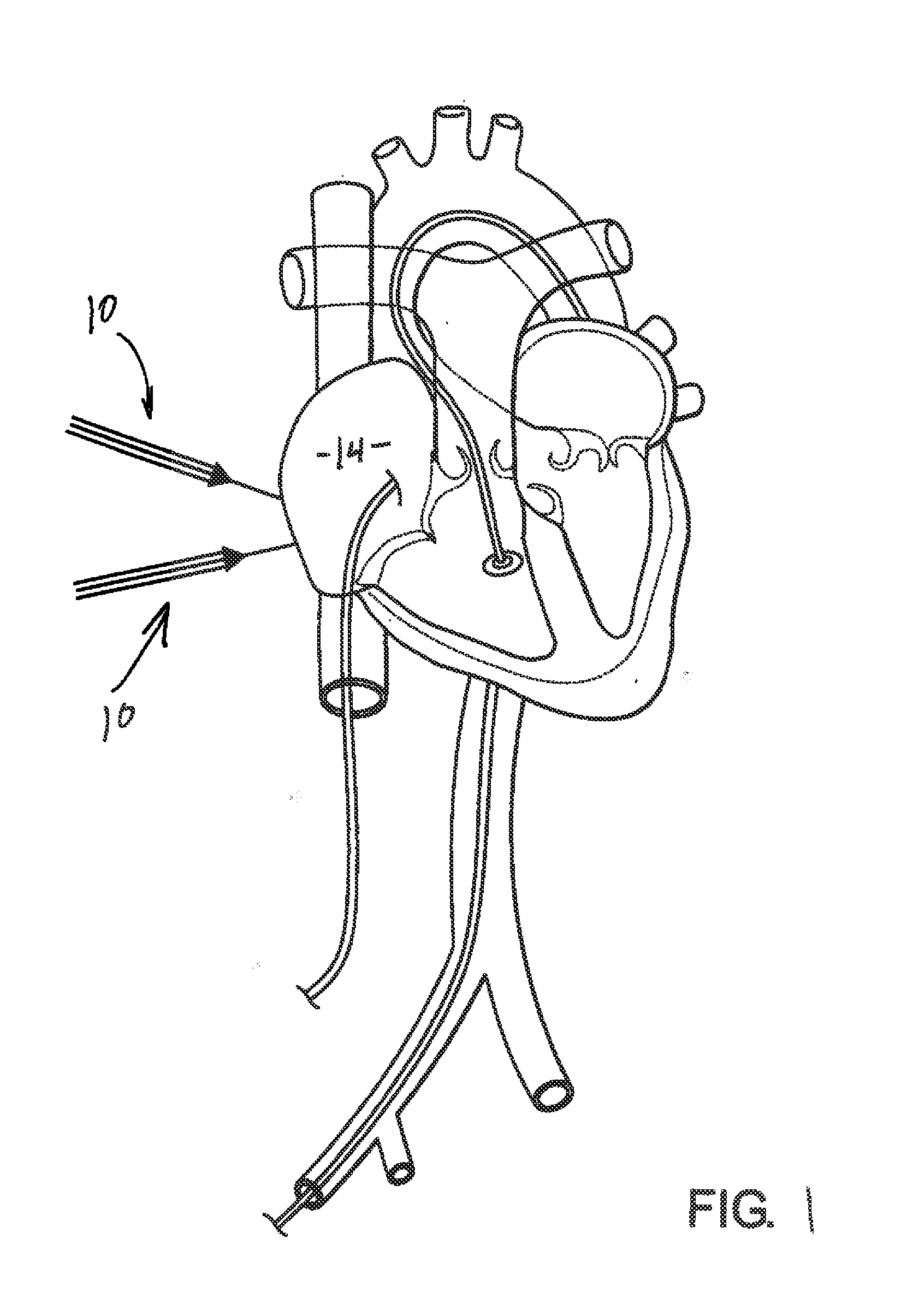
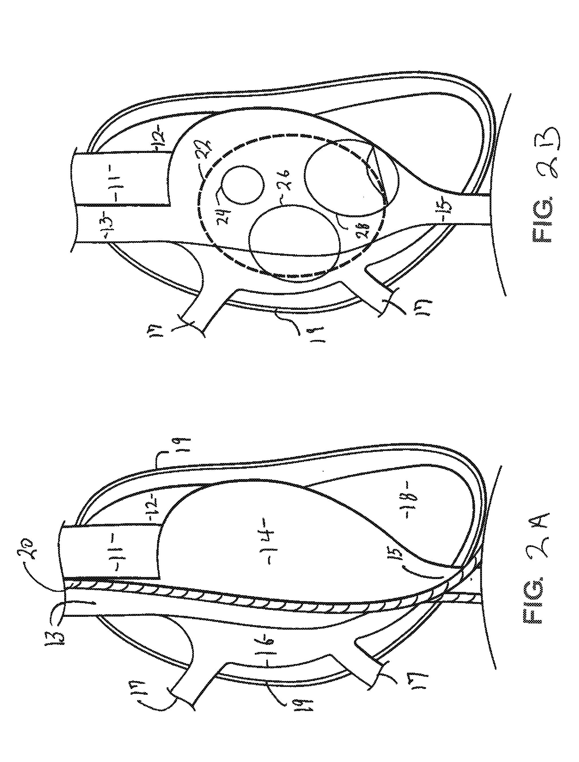
[0062]For purposes of clarity and reference, FIGS. 1, 2A and 2B are schematic representation of the anatomy of the heart. Accordingly, implementing one or more preferred embodiments of the present invention, multiple instruments, including catheters generally indicated as 10, may be concurrently disposed in either the right or left atrium of the heart. As will be set forth in greater detail hereinafter, the instruments 10 pass through the thoracic wall and appropriate ones of intercostal spaces into an interior of a targeted one of the left or right atrium by means of a formed entry...


PUM


Abstract

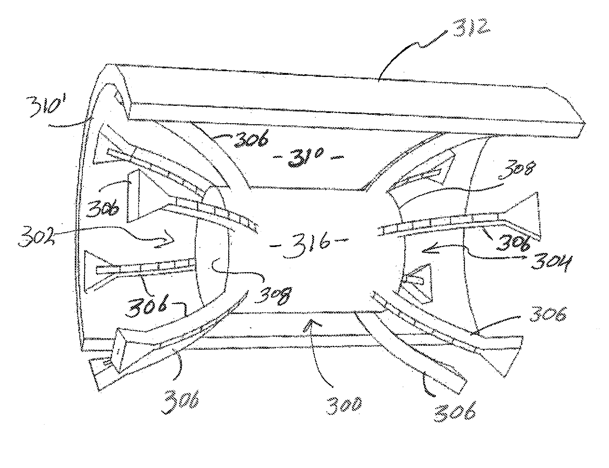
A closure system, assembly and attended method for closing and opening in the tissue of the patient includes first and second closure members each including a biased construction operative for disposition of the closure members into an out of retracted and expanded orientations. A connector is disposed in interconnecting relation with the closure members and structured to establish at least a partial spacing there between. The biased construction and the interconnection of the closure members facilitate con current disposition of the closure members with one another into and through an introductory instrument as well as independent and successive disposition of the first and second closure members into a closing relation to the tissue opening upon exiting the introductory instrument.

Description

CLAIM OF PRIORITY[0001]The present application is a continuation-in-part application of previously filed, now pending application having Ser. No. 13 / 570,347, filed on Aug. 9, 2012, which is a continuation-in-part application of previously filed, now pending application having Ser. No. 13 / 442,230, filed on Apr. 9, 2012, which is based on and a claim of priority is made under 35 U.S.C. Section 119(e) to a provisional patent application that is currently pending in the U.S. Patent and Trademark Office, namely, that having Ser. No. 61 / 574,798 and a filing date of Aug. 9, 2011, and wherein all of the above are incorporated herein in their entirety by reference. The present application is also a continuation-in-part application of previously filed, now pending application having Ser. No. 13 / 838,199, filed on Mar. 15, 2013, which is a continuation-in-part application of previously filed, now pending application, having Ser. No. 13 / 714,989, filed on Dec. 14, 2012, all of which are included ...

Claims


Application Information

Patent Timeline
IPC IPC(8): A61B17/00
CPCA61B17/0057A61B17/12122A61B17/1214A61B17/12168A61B17/12177A61B2017/00575A61B2017/00592A61B2017/00597A61B2017/00623A61B2017/00632A61B2017/12054
Inventor DE CANNIERE, DIDIERDUY, KHANH TRAN
Owner CORQUEST MEDICAL
Learn More