Strontium phosphate microparticle for radiological imaging and therapy

a radioactive strontium phosphate and microparticle technology, applied in the direction of cellulosic plastic layer products, uranium compounds, therapy, etc., can solve the problems of limited selection of radionuclides available for use, lack of flexibility to control dose or radionuclide depending, etc., to reduce time-related degradation of activated radiopharmaceuticals, reduce exposure to medical personnel, and increase radio-opacity

Active Publication Date: 2015-04-30
MO SCI
View PDF1 Cites 7 Cited by
  • Summary
  • Abstract
  • Description
  • Claims
  • Application Information

AI Technical Summary

Benefits of technology

[0088]Another advantage of the invention is the ability to use radioisotopes that have a shorter half-life. For example, Tc-99m (Technetium-99m) cannot be made part of the glass, i.e. the half-life may often be too short to be useful when it is incorporated as part of certain homogeneous glass-radioisotope compositions. Additionally, the ability to use radioisotopes that would otherwise be destroyed or degrade by the glass-melt process. For example, trying to incorporate Technetium or Rhodium into a melt would vaporize the Technetium or Rhodium.
[0089]When melting and stirring is complete, the crucible is removed from the furnace and the melt is quickly quenched to a glass by pouring the melt onto a cold steel plate or into clean, distilled water. This procedure breaks the glass into fragments, which aids and simplifies crushing the glass to a fine powder. The powder is then sized and spheroidized for use.Spheroidizing
[0090]To obtain spheroid microparticles having a diameter in the desired range of micrometres, the glass is processed using varying techniques such as grinding and passing through mesh sieves of the desired size, where the glass particles may be formed into spheroids by passing the sized particles through a gas / oxygen flame where they are melted and a spherical liquid droplet is formed by surface tension. A vibratory feeder located above the gas / oxygen burner slowly vibrates the powder into a vertical tube that guides the falling powder into the flame at a typical rate of 5-25 gm / hr. The flame is directed into a metal container which catches the spheroidized particles as they are expelled from the flame. The droplets are rapidly cooled before they touch any solid object so that, their spherical shape is retained in the solid product.
[0091]After spheroidization, the glass spheres are preferably collected and rescreened based upon size. As a non-limiting example, when the microparticles are intended to be used in the treatment of liver cancer, the fraction less than 30 and greater than 20 micrometres in diameter is recovered since this is a desirable size for use in the human liver. After screening, the −30 / +20 microparticles are examined with an optical microscope and are then washed with a weak acid (HCl, for example), filtered, and washed several times with reagent grade acetone. The washed spheres are then heated in a furnace in air to 500-600° C. for 2-6 hours to destroy any organic material.
[0092]The final step is to examine a representative sample spheres in a scanning electron microscope to evaluate the size range and shape of the spheres. The quantity of undersize spheres is determined along with the concentration of non-spherical particles. The composition of the spheres can be checked by energy dispersive x-ray analysis to confirm that the composition is correct and that there is an absence of chemical contamination.
[0093]The glass microparticles are then ready for phosphate conversion, bonding with radionuclide, and subsequent administration to the patient.

Problems solved by technology

These microspheres are usually provided preloaded with a single radionuclide and so lack the flexibility to control dose or radionuclide depending on the patient's needs.
Further, when radio micro particles are prepared in bulk, off-site by third party providers, the selection of radionuclide available for use may be limited, by the time involved in preparation and delivery.

Method used


Image

  • Strontium phosphate microparticle for radiological imaging and therapy
  • Strontium phosphate microparticle for radiological imaging and therapy
  • Strontium phosphate microparticle for radiological imaging and therapy

Examples


example 1

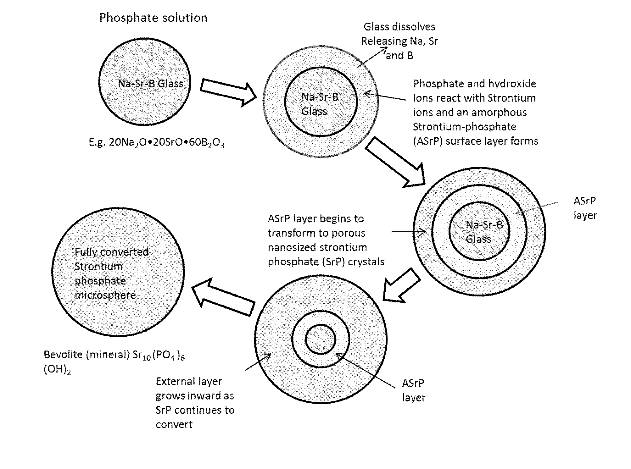
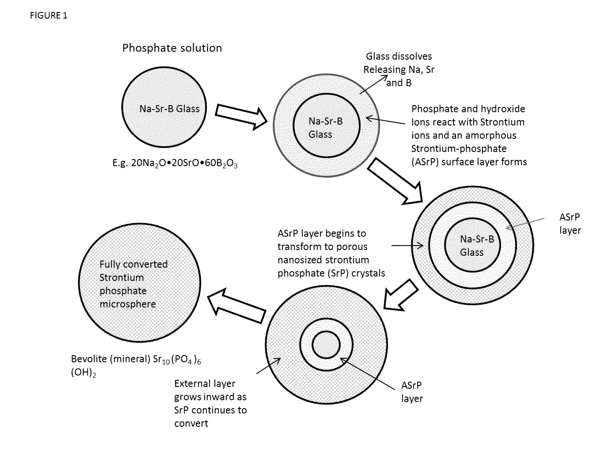
Preparation of Strontium Phosphate Microspheres

[0099]Strontium containing borate glass microspheres were prepared from a batch comprising 20 mol % Na2O, 20 mol % SrO and 60 mol % Ba2O3 as described above. The microspheres ranged in size from 44 to 105 microns.

[0100]The microspheres were reacted with 0.25 molar K2HPO4 solution (pH=12) for 1 h, 6 h, and 72 hrs at 85° C. After rinsing and drying, the surface area was measured by nitrogen absorption using a Micromeretics Tristar 3000 device. Multiple samples were measured and averaged. The measured surface area was 10, 93, and 72 m2 / gm, respectively.

example 2

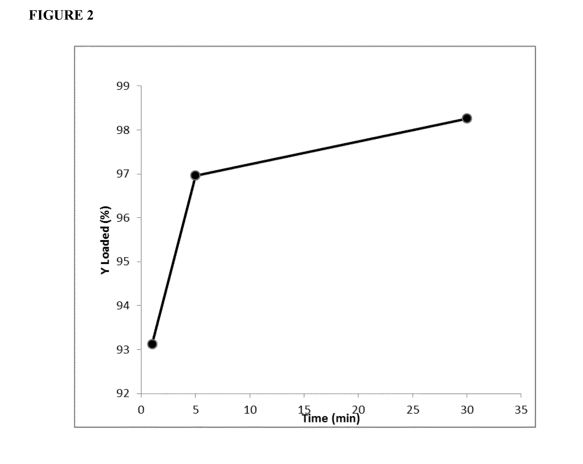
Loading Strontium Phosphate Microspheres with Radioisotopes

[0101]A solution of 5 ug yttrium (Y-90) chloride in 0.5 ml of 0.05N HCl was added to 15 mg of microspheres and incubated for 30 minutes. The amount of yttrium loaded into the microspheres was determined at 1, 5 and 30 minutes in 5 replicates and averaged. Up-take of the yttrium with time is shown in FIG. 1.


PUM

PropertyMeasurementUnit
surface areaaaaaaaaaaa
diameteraaaaaaaaaa
diameteraaaaaaaaaa

Abstract

This invention relates to strontium-phosphate microparticles that incorporate radioisotopes for radiation therapy and imaging.

Description

REFERENCE TO RELATED APPLICATIONS[0001]This application is a continuation-in-part of application Ser. No. 12 / 883,827 filed Sep. 16, 2010 and issued Oct. 21, 2014 as U.S. Pat. No. 8,865,123.FIELD OF THE INVENTION[0002]The field of the present invention is radiomicroparticles for medical therapy and imaging, and particularly radioactive strontium phosphate microparticles for radiological imaging and radioisotope therapy.BACKGROUND[0003]In the treatment of patients with certain kinds of cancer or rheumatoid arthritis, methods are known in which radioactive particles are introduced intravascularly to a tumor site (radioembolization) or locally into the synovial fluid in a joint in order to trap the radioactive particle at a particular site for its radiation effect. Similar methods are used for imaging parts of the body, organs, tumors, and so forth.[0004]According to this technique, a quantity of the radioactive particles are injected into a localized area of the patient to be imaged an...

Claims


Application Information

Patent Timeline
Patent Type & Authority Applications(United States)
IPC IPC(8): A61K51/02C01B25/45A61K51/12
CPCA61K51/02A61K51/1251C01B25/45Y10T428/2982C01P2004/61C01P2006/12C01B25/32C01P2006/44A61K51/1244C01B25/321C01P2004/60
Inventor DAY, DELBERT E.HE, YIYONG
Owner MO SCI
Why Eureka
Try Eureka