High resolution electrophysiology catheter

a high-resolution, electrophysiology technology, applied in the field of high-resolution electrophysiology catheters, can solve the problems of affecting the normal path of depolarization events, disrupting the normal activation of the atria or ventricles, and disrupting the normal propagation of electrical impulses, so as to improve the resolution and fidelity of mapping, and accurately detect tissue contact

Inactive Publication Date: 2015-05-14
BOSTON SCI SCIMED INC
View PDF6 Cites 86 Cited by
  • Summary
  • Abstract
  • Description
  • Claims
  • Application Information

AI Technical Summary

Benefits of technology

[0022]Although the present inventions should not be so limited in their broadest aspects, the use of microelectrodes in the manner described above eliminates detection of the far field electrical activity, thereby increasing the resolution and fidelity of the mapping performed by the medical probe, allowing a user to more precisely measure complex localized electrical activity, and more accurately detecting tissue contact and tissue characterization, including lesion formation assessment.

Problems solved by technology

Sometimes, aberrant conductive pathways develop in heart tissue, which disrupt the normal path of depolarization events.
For example, anatomical obstacles in the atria or ventricles can disrupt the normal propagation of electrical impulses.
These wavelets, called “reentry circuits,” disrupt the normal activation of the atria or ventricles.
The ischemic region, also called a “slow conduction zone,” creates errant, circular propagation patterns, called “circus motion.” The circus motion also disrupts the normal depolarization patterns, thereby disrupting the normal contraction of heart tissue.
Primarily due to the relatively large size of tip electrodes, current catheter designs, such as the type illustrated in FIG. 1, may detect far field electrical activity (i.e., the ambient electrical activity away from the recording electrode(s)), which can negatively affect the detection of local electrical activity.
That is, due to the relatively large size of the tip electrode and the distance from the next ring electrode, the resulting electrical recordings are signal averaged and blurred, and thus not well-defined.
Thus, the electrical activity measured by such catheters does not always provide a physician with enough resolution to accurately identify an ablation site and or provide the physician with an accurate portrayal of the real position of the tip electrode, thereby causing the physician to perform multiple ablations in several areas, or worse yet, to perform ablations in locations other than those that the physician intends.
In addition, many significant aspects of highly localized electrical activity may be lost in the far-field measurement.
For example, the high frequency potentials that are encountered around pulmonary veins or fractioned ECGs associated with atrial fibrillation triggers may be lost.
Also, it may be difficult to determine the nature of the tissue with which the tip electrode is in contact, or whether the tip electrode is in contact with tissue at all, since the far-field measurements recorded by the tip electrode may indicate electrical activity within the myocardial tissue even though the tip electrode is not actually in contact with the endocardial tissue.

Method used


Image

  • High resolution electrophysiology catheter
  • High resolution electrophysiology catheter
  • High resolution electrophysiology catheter

Examples


Embodiment Construction

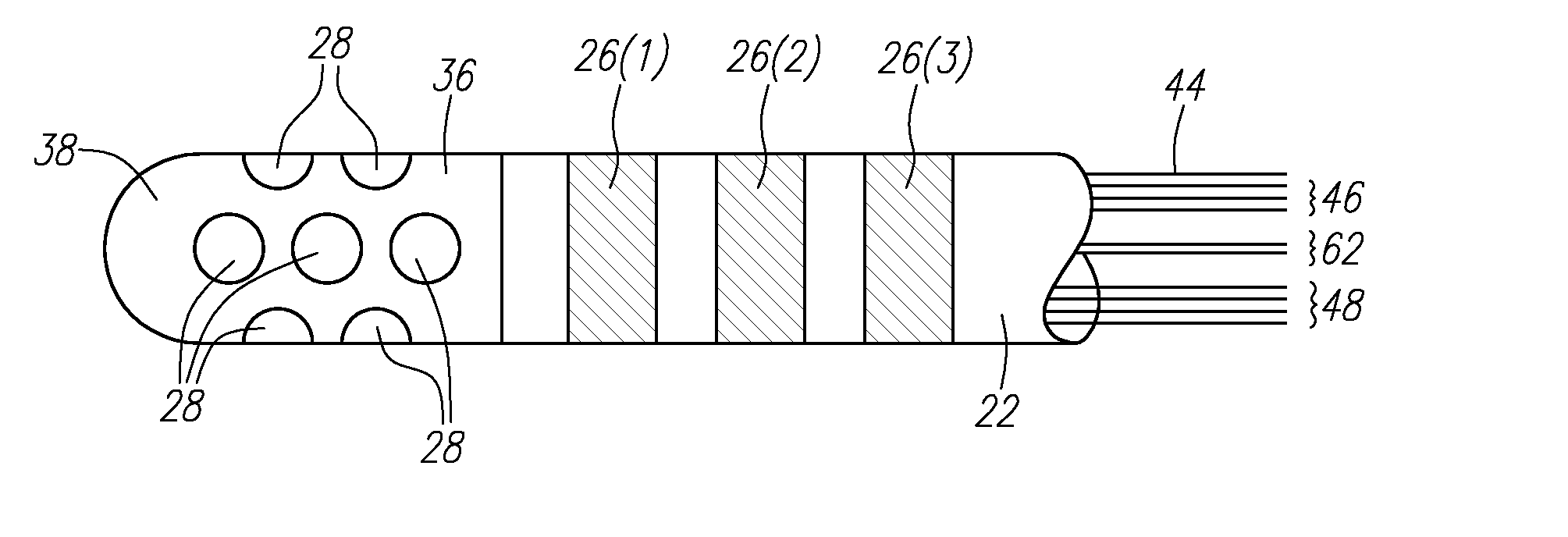
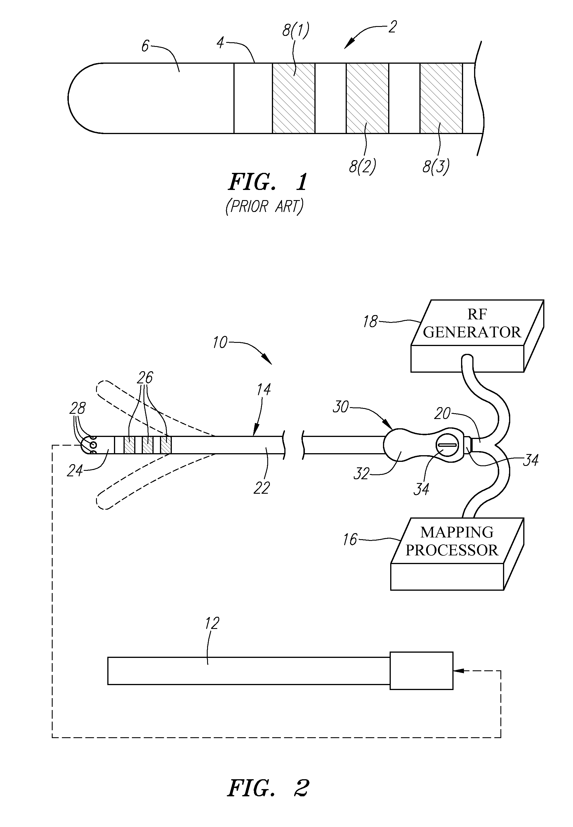
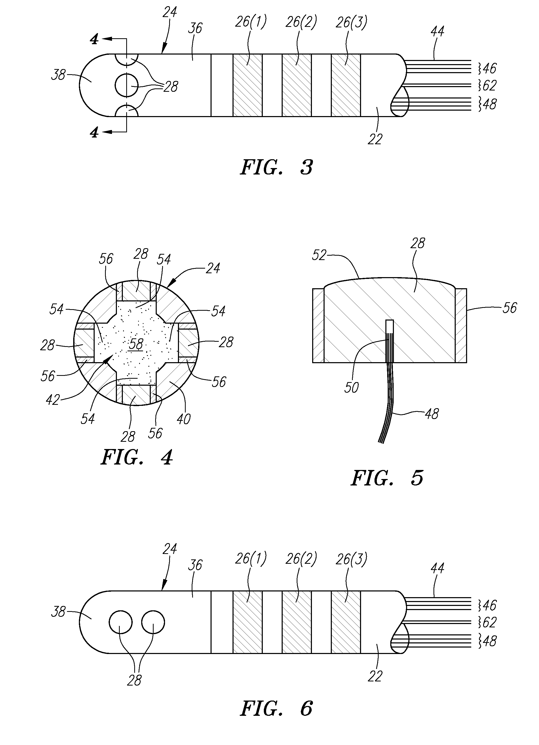
[0041]Referring to FIG. 2, an exemplary electrophysiology system 10 constructed in accordance with the present inventions is shown. The system 10 may be used within body lumens, chambers or cavities of a patient for therapeutic and diagnostic purposes in those instances where access to interior bodily regions is obtained through, for example, the vascular system or alimentary canal and without complex invasive surgical procedures. For example, the system 10 has application in the diagnosis and treatment of arrhythmia conditions within the heart. The system 10 also has application in the treatment of ailments of the gastrointestinal tract, prostrate, brain, gall bladder, uterus, and other regions of the body. As an example, the system 10 will be described hereinafter for use in the heart for mapping and ablating arrhythmia substrates.

[0042]The system 10 generally comprises a conventional guide sheath 12, and an electrophysiology catheter 14 that can be guided through a lumen (not sho...


PUM


Abstract

An electrophysiology medical probes, which may be incorporated into a system and used to perform an electrophysiology procedure, is provided. The medical probe comprises an elongated member (e.g., a flexible elongated member), and a metallic electrode mounted to the distal end of the elongated member. In one embodiment, the metallic electrode is cylindrically shaped and comprises a rigid body. The medical probe further comprises a plurality of microelectrodes (e.g., at least four microelectrodes) embedded within, and electrically insulated from, the metallic electrode, and at least one wire connected to the metallic electrode and the microelectrodes.

Description

RELATED APPLICATION DATA[0001]The present application is a continuation of U.S. patent application Ser. No. 12 / 056,210, filed on Mar. 26, 2008, which claims the benefit under 35 U.S.C. §119 to U.S. Provisional Patent Application Ser. No. 60 / 908,166, filed on Mar. 26, 2007. The foregoing application is incorporated by reference into the present application in its entirety for all purposes.FIELD OF THE INVENTION[0002]The present inventions generally relate to systems and methods for providing therapy to a patient, and more particularly to systems and methods for mapping and ablating tissue within the heart of the patient.BACKGROUND OF THE INVENTION[0003]Physicians make use of catheters today in medical procedures to gain access into interior regions of the body to ablate targeted tissue regions. It is important for the physician to be able to precisely locate the catheter and control its emission of energy within the body during these tissue ablation procedures. For example, in electr...

Claims


Application Information

Patent Timeline
Patent Type & Authority Applications(United States)
IPC IPC(8): A61B18/14A61B5/296
CPCA61B18/1492A61B2018/00577A61B2018/00839A61B2018/00642A61B5/6855A61B2018/1467A61N1/05A61N1/056A61B2018/00791A61B2018/00797A61B5/287A61N1/06A61B5/283
Inventor KOBLISH, JOSEF V.
Owner BOSTON SCI SCIMED INC
Why Eureka
Try Eureka