
Clearing Agent and Mounting Medium for Microscopy
a technology of microscopy and mounting medium, which is applied in the field of preparation of specimens for microscopy, can solve the problems of difficult identification of characteristic features, difficulty in identifying characteristic features, and differences in refractive index within specimens, and achieves the obstruction of detail from observation, and the difficulty of identifying characteristic features
- Summary
- Abstract
- Description
- Claims
- Application Information
AI Technical Summary
Benefits of technology
Problems solved by technology
Method used
Image

Examples
example 1
Preparation of a Clearing Agent and Mounting Solution of the Invention
[0112]4.5 mL of 2,2,2-trichlorethanol (Sigma-Aldrich, 99%, reagent grade) was thoroughly mixed with 1.0 mL of glycerol. To this homogenous solution was added 2.5 mL of a 4% (m / v) aqueous solution of 2,2,2-trichloroacetic acid, at which time cloudiness was observed. The solution was mixed thoroughly until completely clear, about 5 minutes.
example 2
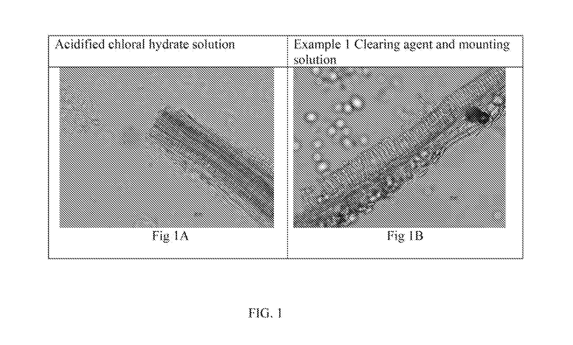
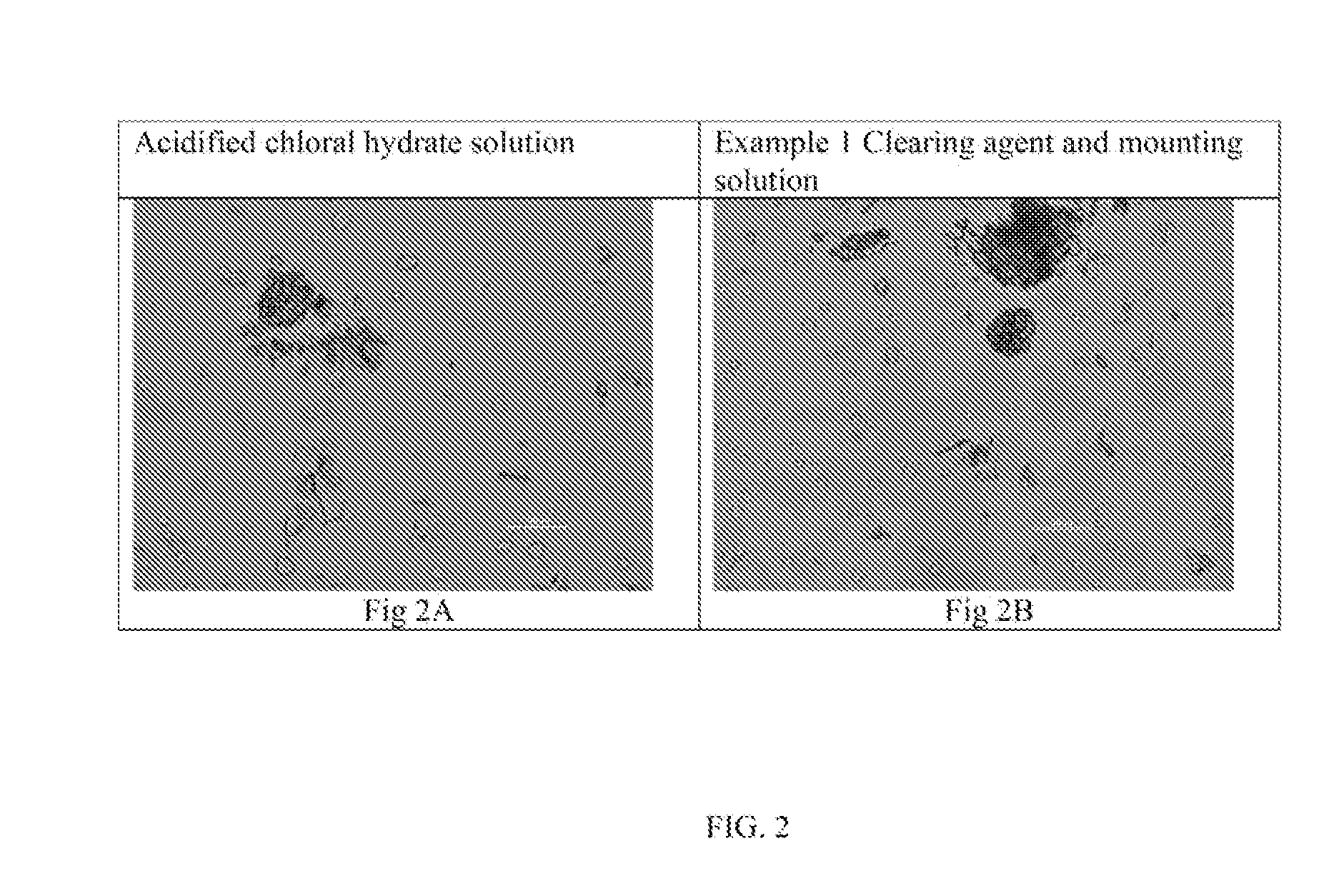
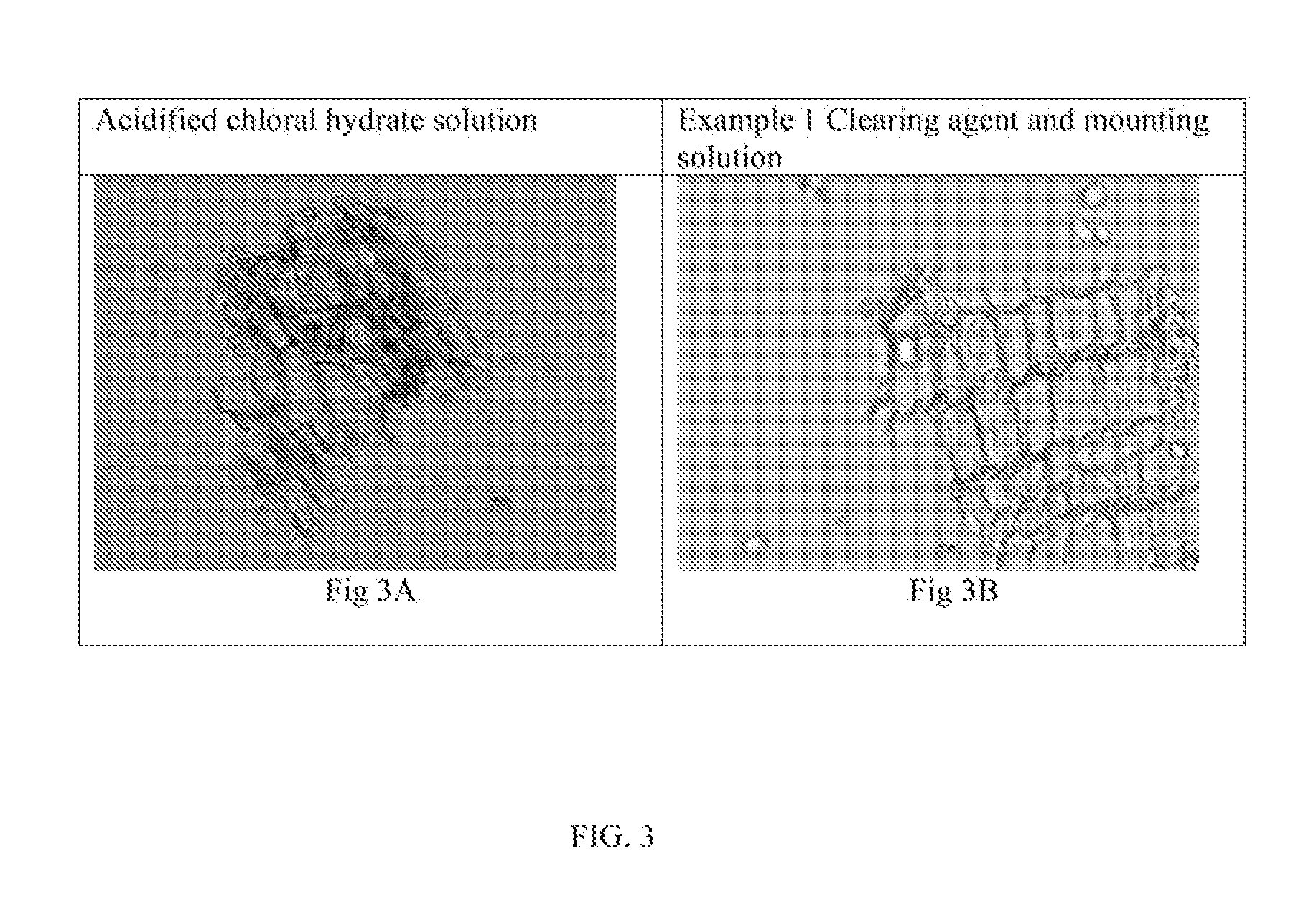
Ginger (Rhizome), Zingiber officinale Roscoe (Zingiberaceae); FIGS. 1-3
[0113]Powdered ginger samples are characterized by numerous fragments of isodiametric thin-walled parenchyma cells containing starch granules; fragments of thin-walled fibers with oblique slit-like pits; fragments of scalariform, reticulate, and spiral vessels, thin-walled cells with suberized radial walls; numerous starch granules with various forms such as simple, flat, oval, oblong with terminal protuberance.
[0114]In ground ginger samples, fragments of parenchyma cells, cell with circular striations (cork cells), fibers accompanied by vessels and abundant starch grains the compact epidermal cells with sharp edges can be observed using the clearing agent and mounting solution of the invention. There was no difference in the structures observed between the inventive and standard clearing agent and mounting solutions.
[0115]FIG. 1 shows a microscopic view of characteristic annular vessel elements and fibers of gin...
example 3
Mate (Leaves), Ilex paraguariensis (Aquifoliacea); FIGS. 4-7
[0118]Ground Ilex leaves are characterized by upper epidermis composed by polygonal cells with unevenly thickened walls. Lower epidermis cells are smaller than those of the upper epidermis, and thinner cell walls, cuticular striations are well marked. Stomata anomocytic (epidermal cells surrounding the guard cell pair are not morphologically distinct from the other epidermal cells). Groups of lignified fibers are visible.
[0119]FIG. 4 shows a microscopic view of fragments of epidermis over leaf veins. The upper epidermis is composed of polygonal cells with unevenly thickened walls. Stomata are absent in the upper epidermis. There are no differences observed using the inventive solution, versus acidified chloral hydrate solution: FIG. 4A, fragment of polygonal cells of the upper epidermis over the vein using acidified chloral hydrate solution; FIG. 4B, fragments of polygonal cells of the upper epidermis over the vein using cl...
PUM
 Abstract
Description
Claims
Application Information