Method of painting microvesicles

a microvesicles and microvesicle technology, applied in the field of microvesicles painting, can solve the problems of determining what is being lost, unable to enrich for enveloped viruses as well as mmv and unable to achieve the effect of mmv enrichment,

Inactive Publication Date: 2015-08-20
VIN DE BONA TRADING CO
View PDF1 Cites 4 Cited by
  • Summary
  • Abstract
  • Description
  • Claims
  • Application Information

AI Technical Summary

Benefits of technology

[0036]Surprisingly, the incubation, or in other words the MP step, works even without having to isolate the MMV first. Hence, there is no need to isolate the MMV from the sample before incubating it with the reactant, in order to still achieve a sufficient MP reaction of the exosomes.
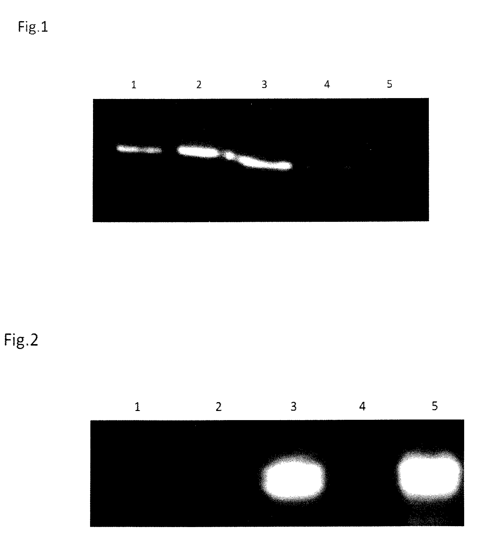
[0145]The fact that the signal in lane 2 of the anti GFP blot (FIG. 2) is much less intense than the signal in lane 3 (and 5) may be explained by the ultracentrifugation step which might have removed not only the excessive GFP-proteins but might also have potentially reduced the amount of exosomes in the supernatant.

Problems solved by technology

However, none of the methods is able to enrich for enveloped viruses as well as MMV, such as exosomes or bigger, simultaneously.
That is because the methods known in the art are either based on purifying everything in the sample by density or size (e.g. filters or ultra-centrifugation) or limited to purifying only the elements that specifically react with the antibody being used, meaning that the purification is limited to molecules that have been previously identified and had antibodies generated for.
Filters are an unknown factor because there are always unknown interactions between the sample elements and the filter materials so it can never be determined what is being lost, especially when the target is novel or unknown, e.g. when researching on new infectious agents or identifying new diagnostic markers.
This approach allows visualisation of membranes, membranes of cells as well as of membrane vesicles, but does not provide the means to tag the vesicles in such a way, that the staining does not allow to physically separate the labelled membrane particles from their surrounding environment.
However these are not suitable to identify new MMV of unknown surface proteins, but limited to isolate or detect those with a known protein or antigen exposed on their surface.
In addition, all of these affinity based methods require the use of antibodies, which is expensive and cumbersome.
All these methods carry potential disadvantages.
Centrifugation processes do not enable the fine separation of membrane vesicles (e.g. exosomes) from cell proteins or certain macromolecular components (DNA, RNA) or macromolecular complexes.
Therefore, these processes do not exclude the presence of unidentified contaminating biological agents, incompatible with therapeutic use in humans.
In addition, these steps are difficult to extrapolate at an industrial scale, particularly when significant volumes are to be treated, or for autologous (i.e. patient by patient) ex vivo applications, in which the process must generally be applied in a confined system.
The use of ultracentrifugation could solve some of the problems, but increases the price of the isolation step significantly, making it unsuitable for a large percentage of laboratories who do not possess the equipment, and makes it unsuitable for high-through-put methods, which is a requirement for an affordable diagnostic tool.
In addition there is—to our knowledge—no method known in the art to isolate and label two different types of vesicles, such as any type of MMV and enveloped virus, simultaneously.
This is costly, time consuming and most importantly would require large amounts of sample, which in most diagnostic scenarios, is not available.

Method used


Image

  • Method of painting microvesicles

Examples


example 1

[0106]Obtaining samples comprising exosomes:

[0107]8×T175 flasks of HEK293T cells were cultured in DMEM until 80-90% confluency. The adherent cells are washed once in 10 ml of PBS, and 18 ml of serum-free DMEM is added into each flask. The cells are incubated in CO2 incubator at 37° C. for approximately 24 hours. After 24 hr incubation, the supernatants (containing exosomes secreted from the cells) are collected.

[0108]By combining the supernatants of two flasks 4×36 ml of cell supernatant were placed in 4×50 ml falcon tubes and centrifuged at 1200 g for 8 minutes at 4° C. 4×35 ml of the supernatant are carefully pipetted up, leaving 1 ml of supernatant and the cell debris in the falcon tube. The supernatant sample is now centrifuged again at 8400 rpm (approx. 12,000 g) for 15 minutes at 4° C. to remove smaller fragments of the cells and cell membranes. After centrifuging, 4×34 ml of the supernatant is taken out and placed into 4 x new tubes. 1 ml of supernatant and any remaining cell...

example 2

[0109]Standard Purification of Exosomes via Ultracentrifugation

[0110]After the intermediate centrifugation step, 4×34 ml of the supernatant were taken out and placed into 4 x new ultracentrifuge tubes. 1 ml of supernatant and any remaining cell debris was left in the used tube. The supernatant samples (within the 4 new ultracentrifuge tubes) are centrifuged at 26,500 rpm (−120,000 g) for 2 hours at 4° C. After centrifugation, the supernatant is discarded and the tube is carefully blotted dry. The remaining pellets (containing exosomes) are resuspended in 2.5 ml of PBS by washing the bottom of the tube. From this 2.5 ml resuspended exosome sample is 5×500 ul samples have been transferred into 5 tubes and labelled as follows:[0111]1) E−: unpainted exosomes, ultracentrifuged (control)[0112]2) E+: painted exosomes, ultracentrifuged (to get rid of unbound gfp-gpi protein)[0113]3) E-MB+: unpainted exosomes, applied over pre-coated beads and subsequently eluted[0114]4) E-MB−: unpainted exo...

example 3

[0117]Generation of His-Tagged GPI-GFP Protein

[0118]The HEK 293 cell line used has been altered to produce the his-tagged and monomeric version of the GPI-GFP protein that is used in the example and to be resistant against hygromycin. In the lab the cells are referred to as “HEK293monogghishyg cells”. They were grown up in a medium which contains 500 ml DMEM high-glucose (Invitrogen 11960 or 11965), 50 ml deactivated FBS (PAA A15-101), 5 ml of 100 mM Sodium Pyruvate (Gibco 11360) and 5 ml of 200 mM L-glutamine (Gibco 25030).

[0119]Finally, hygromycin was added to a final concentration of 50 ug / ml. The cells were grown up in 5-6 T175 flasks to near confluency, then the cell medium was discarded and the cells were washed with 5 ml PBS per flask and then scraped off. The scraped cells from the flasks were collected in 10 ml of PBS each and all flasks rinsed out carefully. The PBS cell suspension was then centrifuged in 15 ml Falcon tubes and centrifuged at 200 g, for 5 mins, at 4° C.

[01...


PUM

PropertyMeasurementUnit
diameteraaaaaaaaaa
diameteraaaaaaaaaa
diameteraaaaaaaaaa

Abstract

The present invention relates to a method to modify and / or to isolate exosomes and other naturally occurring plasma membrane derived microvesicles, by incubation with a reactant, consisting of at least a membrane anchor domain or moiety and a hydrophilic functional domain or moiety. The invention also relates to modification using the same of artificially-prepared lipid bilayer vesicles called liposomes (composed of natural phospholipids) and non-biological or “synthetic” block copolymer membrane mimics which also form vesicles in aqueous solution called polymersomes..

Description

CROSS-REFERENCE TO RELATED APPLICATIONS[0001]This application claims the benefit of priority to U.S. Provisional Application No. 61 / 61 / 712,471 titled “Method of painting microvesicles” filed on 11 Oct. 2013, the entire disclosure of which is hereby incorporated by reference in its entirety for all purposes.FIELD OF THE INVENTION[0002]The present invention relates to a method to modify and / or to isolate exosomes and other naturally occurring plasma membrane derived microvesicles, by incubation with a reactant, consisting of at least a membrane anchor domain or moiety and a hydrophilic functional domain or moiety. The invention also relates to modification using the same of artificially-prepared lipid bilayer vesicles called liposomes (composed of natural phospholipids) and non-biological or “synthetic” block copolymer membrane mimics which also form vesicles in aqueous solution called polymersomes. All of the membrane based vesicles (natural or synthetic) that relate to this membrane...

Claims


Application Information

Patent Timeline
Patent Type & Authority Applications(United States)
IPC IPC(8): G01N1/30G01N33/58
CPCG01N33/582G01N1/30C12Q1/6806G01N33/5432Y10T428/2985C12Q2563/131C12Q2563/161
Inventor DANGERFIELD, JOHNBRANDTNER, EVA MARIATAN, WEE JIN
Owner VIN DE BONA TRADING CO
Why Eureka
Try Eureka