Method for Obtaining Cell and Tissue Specific Biomolecules

a biomolecule and cell technology, applied in the field of biomaterial collection, can solve the problems of destructing the original material being studied, etc., and achieve the effect of increasing the time and cost of cell- and tissue-specific studies

Inactive Publication Date: 2015-09-03
KARSTEN STANISLAV L +1
View PDF6 Cites 1 Cited by
  • Summary
  • Abstract
  • Description
  • Claims
  • Application Information

AI Technical Summary

Benefits of technology

[0021]Aspects of the present invention teach certain benefits in construction and use which give rise to the exemplary advantages described below.
[0022]Currently, isolation of cell-specific biomolecules involves either tissue microdissection performed by various microdissection devices such as laser capture microdissection or tissue dissociation or homogenization and subsequent sorting of the desired cells performed by flow sorting machines. This leads to the destruction of the original material being studied, which is not desirable when samples are rare or difficult to obtain. In addition, the procedure of biomolecule isolation from isolated cells has

Problems solved by technology

This leads to the destruction of the original material being studied, which is not desirable when samples are rare or difficult to obtain.
In addition, th

Method used


Image

  • Method for Obtaining Cell and Tissue Specific Biomolecules
  • Method for Obtaining Cell and Tissue Specific Biomolecules
  • Method for Obtaining Cell and Tissue Specific Biomolecules

Examples


Embodiment Construction

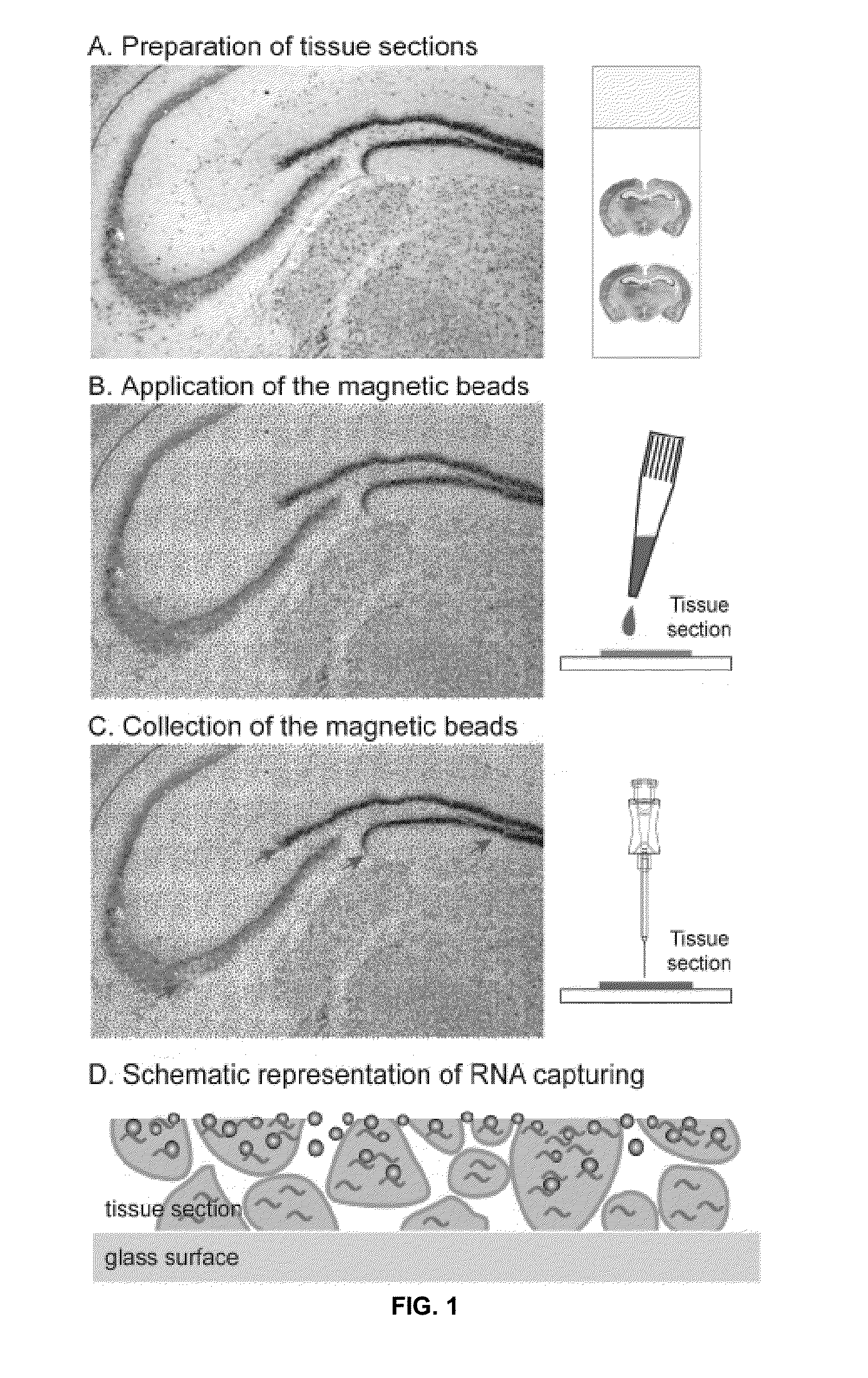
[0043]The above described drawing figures illustrate aspects of the invention in at least one of its exemplary embodiments, which are further defined in detail in the following description. Aspects of the invention and its biomaterial collection methods can now be better understood as set forth below in connection with FIGS. 1-5 disclosing exemplary method steps thereof. Turning now to FIG. 1, the general method steps in one embodiment are shown. First, in FIG. 1A there is shown a schematic view of a representative complex tissue section, in accordance with at least one embodiment; particularly, representing a step showing a tissue (a mouse brain in this particular example) prepared and sectioned. In FIG. 1B, there is shown the application of microbeads to the selected tissue section. Such beads may be magnetic, but not necessarily so; an example of a substantially non-magnetic bead that could be employed according to aspects of the present invention is oligo(dT)25-cellulose beads ...


PUM

PropertyMeasurementUnit
Thicknessaaaaaaaaaa
Diameteraaaaaaaaaa
Densityaaaaaaaaaa

Abstract

Methods for tissue microdissection- or dissociation-free means of collecting cell-specific biomolecules from complex heterogeneous tissue sources including native tissues, fresh frozen, fixed, and archived specimens. The tissue section having a plurality of beads with immobilized capture probes on their surface evenly distributed over all cell types of the tissue is arranged. Alternatively the tissue section being coated with a thin layer of resin containing incorporated or covalently bound immobilizing capture probes is arranged. The beads or resin are incubated directly on the tissue section permitting capturing of the biomolecules by the capture probes. After incubation, the tissue section is visualized and the cells or area of interest are identified. Next, the beads or resin samples, located in these cells or areas of interest, are collected and transferred to a sample tube where biomolecules are separated from the beads or resin using standard methods and are then used in downstream applications.

Description

RELATED APPLICATIONS[0001]Not applicable.INCORPORATION BY REFERENCE[0002]Applicant(s) hereby incorporate herein by reference any and all patents and published patent applications cited or referred to in this application.BACKGROUND OF THE INVENTION[0003]1. Field of the Invention:[0004]Aspects of this invention relate generally to biomaterial collection, and more particularly to compositions and methods for obtaining cell and tissue samples.[0005]2. Description of Related Art:[0006]The field of the invention is biomaterial collection, more specifically, the present invention relates to direct collection of cell and region specific biomolecules including DNA, RNA, and proteins as an alternative to tissue microdissection and flow sorting. The biomaterial collection compositions and methods presented herein can be used in conjunction with the capillary-based cell and tissue acquisition system (“CTAS”) as disclosed in WO / 2008 / 021202, which is incorporated herein by reference in its entire...

Claims


Application Information

Patent Timeline
IPC IPC(8): G01N33/543C12Q1/68G01N1/30
CPCG01N33/54333C12Q1/6806G01N1/30
Inventor KARSTEN, STANISLAV L.KUDO, LILI C.
Owner KARSTEN STANISLAV L
Why Eureka
Try Eureka