Apparatus and Method for Assessment of Interstitial Tissue

a technology of interstitial tissue and optical coherence tomography, which is applied in the field of apparatus and method for optical coherence tomography for assessment of interstitial tissue, can solve the problems of reducing the linearity of the scan, unable to accurately detect the interference pattern, and unable to achieve the effect of reducing the difficulty of each technique, so as to achieve minimal tissue disruption, high-resolution oct, and minimal invasive oct imaging

Inactive Publication Date: 2016-01-14
PHYSICAL SCI
View PDF8 Cites 3 Cited by
  • Summary
  • Abstract
  • Description
  • Claims
  • Application Information

AI Technical Summary

Benefits of technology

[0011]One advantage of the invention is that is allows for high-resolution OCT imaging of interstitial tissue by correcting for the nonlinearity of the manual scans and for the distortion of the image caused by tissue noncompliance when the probe is passed through it. Another advantage of the invention is that it provides minimally invasive OCT imaging a minimal tissue disruption due to the operator controlling the position of an optical probe within the interstitial tissue.
[0012]Another advantage of the invention is that it does not require probes that are rotated at a high speed and / or moved axially at a high speed to generate high fidelity OCT images. Another advantage is that the low speed scanning enables the recording of high yield co-registered OCT / spectroscopy images. The reasonable integration times (e.g., tens to hundreds of ms) can be used for each imaging voxel, and thus to collect sufficient photons and generate such images. The elimination of the high speed rotation / movement requirements can also allow the use low cost disposable probes.
[0013]Another advantage of the invention is that is allows for tissue mapping over relatively long distances (e.g., up to several centimeters), due to the fact that a scanning engine is no longer needed. The interventional radiologist passes the probe through the investigated tissue mass, and the OCT image of the entire trajectory of the probe can be recorded in real-time and conveyed to the operator. Another advantage of the invention is that it can capture a high-resolution OCT image independent of scanning speed. Another advantage of the invention is ease of repeating a procedure because manual positioning of the probe allows for the procedure to be repeated several times without removing the probe from the tissue.
[0014]Another advantage of the invention is that it allows for fluorescence imaging or spectroscopy imaging synchronously with OCT imaging due to the elimination of the need for high speed movement to capture an OCT image.
[0015]In one aspect, the invention involves a method of imaging a sample. The method involves inserting a guidance needle and an optical probe into an investigation site of the sample, the optical probe being positioned within the guidance needle. The method also involves establishing, using a position sensor, a reference location of the optical probe at a first spatial position at the investigation site relative to the guidance needle. The method also involves capturing a first optical coherence tomography (OCT) A-line with the optical probe at the first spatial position when the optical probe is moved relative to the reference location. The method also involves detecting, using the position sensor, a spatial location of the optical probe relative to the reference location during movement of the optical probe within the sample. The method also involves capturing an OCT A-line with the optical probe at a second spatial position if the reference location and the spatial location are separated by greater than a predetermined threshold value. The method also involves if the second OCT A-line is captured, determining whether the second OCT A-line is identical to the first OCT A-line and discarding the second OCT A-line if it is identical to the first OCT A-line and storing the second OCT A-line and the first OCT A-line if the second OCT A-line and the first OCT A-line are not identical.
[0016]In some embodiments, the method involves detecting, using the position sensor, a second spatial location of the optical probe relative to the reference location when the optical probe is moved relative to the reference location. The method also involves capturing an OCT A-line with the optical probe in a third spatial position if the second spatial location and the first spatial location are separated by greater than the predetermined threshold value. If the third OCT A-line is captured, determining whether the third OCT A-line is identical to the second OCT A-line then discarding the third OCT A-line if it is identical to the second OCT A-line, and storing the second OCT A-line and the first OCT A-line if the second OCT A-line and the first OCT A-line are not identical.

Problems solved by technology

The combination of reflected light from the sample arm and reference light from the reference arm can yield an interference pattern when the interferometer arms are substantially matched within the coherence length of the light source.
However, when the imaging has to be done with a needle size probe that passes through several mm to several cm of an interstitial sample (e.g., tissue), the generation of high linearity scan can require the use of rotary or axial movement of the probe within the tissue, which becomes problematic due to tissue friction.
Each of these techniques can prove difficult when imaging interstitial tissue.
High-speed and / or rotational movement of an imaging probe in interstitial tissue can cause tissue morbidity by, for example, catching and / or dislocating the tissue, and thus also degrading the linearity of the scan.
However, the size of the scan is limited to a few mm or less, and therefore is not practical for imaging large size areas within the tissue.
However, a protective tube typically cannot be pushed to penetrate the tissue.
In addition, it increases the overall diameter of a probe, and thus cannot be passed through be 19 gauge or smaller diameter biopsy guidance needles.
Scanning linearity however still remains a serious issue.
Hand-held OCT imaging devices can suffer from inaccurate imaging due to high nonlinearity of the manual scan.
However, this approach is computationally intensive and is not realistic for real-time correction of images.

Method used


Image

  • Apparatus and Method for Assessment of Interstitial Tissue
  • Apparatus and Method for Assessment of Interstitial Tissue
  • Apparatus and Method for Assessment of Interstitial Tissue

Examples


Embodiment Construction

[0030]Generally, an apparatus including an optical probe capable of taking an OCT image is inserted into interstitial tissue. The optical probe is manually moved (e.g., by a technician or a robot). When the optical probe is moved more than approximately 5 microns from a reference point (e.g., a guidance needle or a handle), an OCT reflectivity profile or A-line is taken. Each OCT A-line is analyzed by a data processor to determine whether or not it is a repeat of the previous A-line. Repeated A-lines are discarded. The data process presents an aggregate OCT image of the interstitial tissue based on each non-discarded OCT A-line.

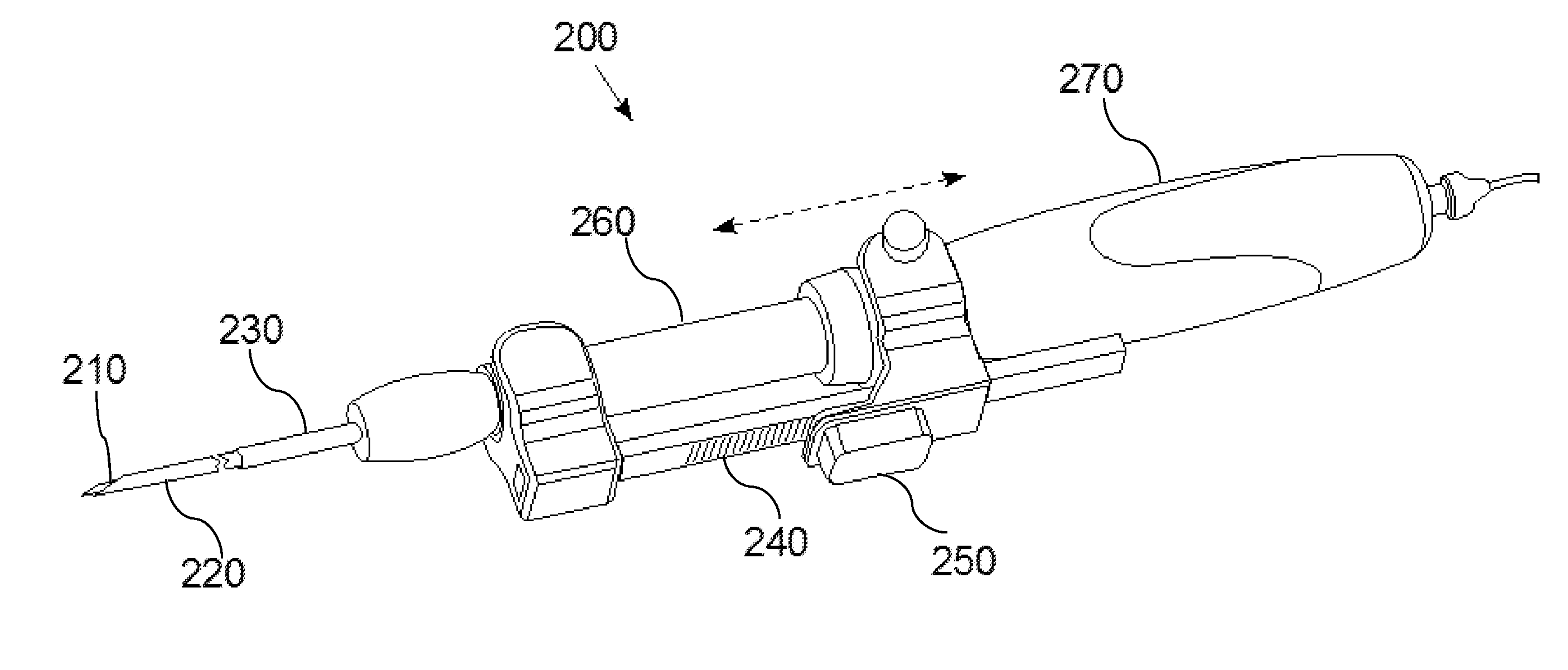
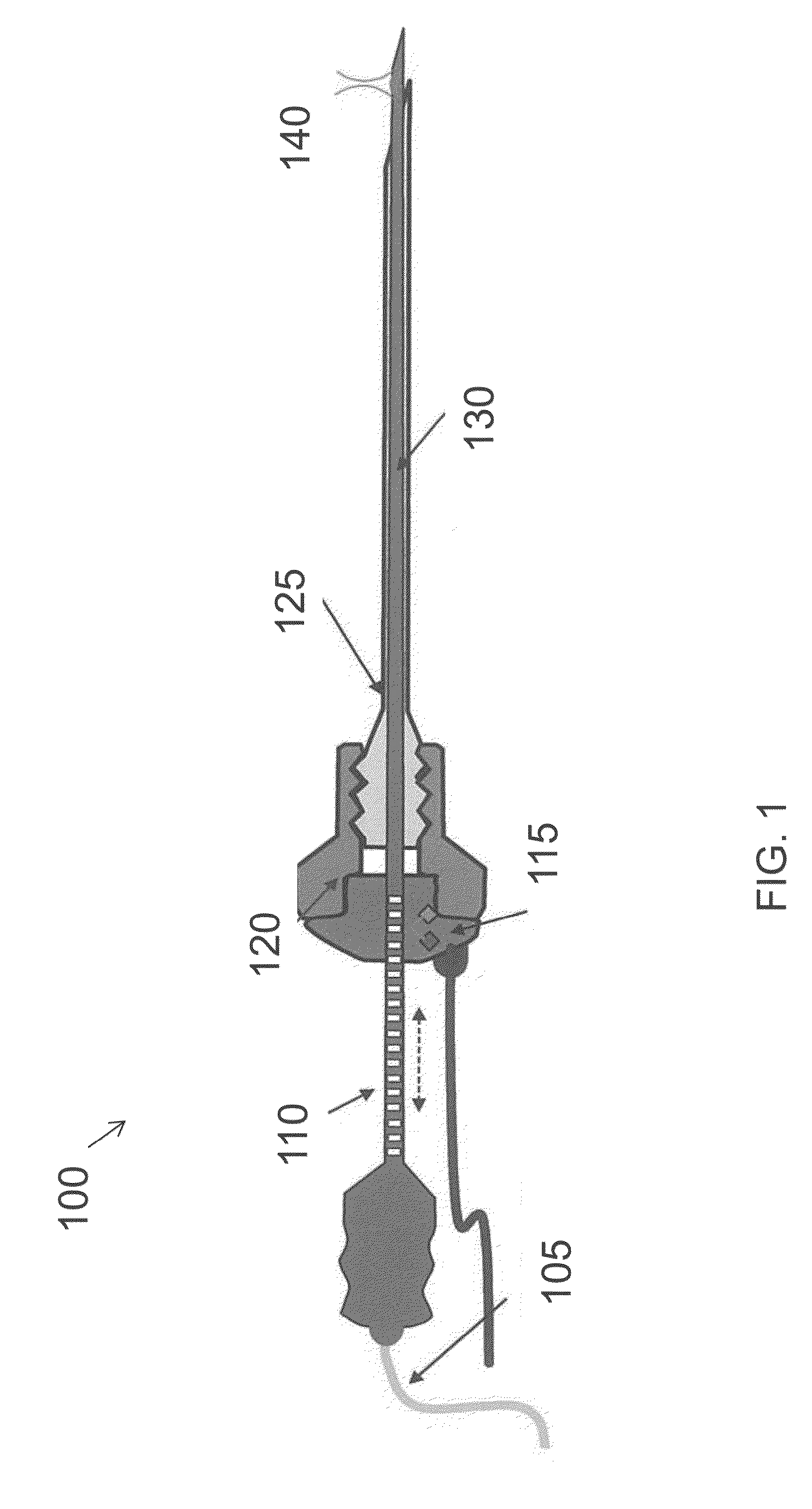
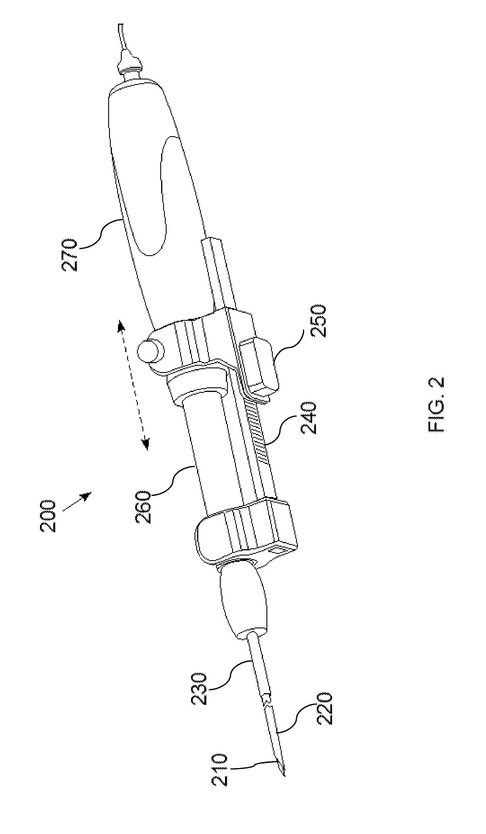
[0031]FIG. 1 is a diagram of a hand-held optical imaging device 100, according to an illustrative embodiment of the invention. The hand-held optical imaging device 100 includes an optical fiber 105, an optical scale 110, a position sensor 115 (e.g., optical encoder), a connector 120 (e.g., male luerlock connector), a guidance needle 125, and an optical probe ...


PUM


Abstract

A handheld optical coherence tomography imaging and tissue sampling system and method of imaging and sampling a tissue is disclosed. The method includes inserting a catheter probe into a biopsy needle. The biopsy needle can be attached to a hand-held scanning and sampling device. The biopsy needle is maneuvered to an investigation site. A three-dimensional image of the tissue at the investigation site is captured with the catheter probe.

Description

CROSS REFERENCE TO RELATED APPLICATIONS[0001]This application claims the benefit of and priority to U.S. Provisional Application No. 62 / 022,497, filed Jul. 9, 2014, which is owned by the assignee of the instant application and the disclosure of which is hereby incorporated herein by reference in its entirety.STATEMENT OF GOVERNMENT INTEREST[0002]The subject matter described herein was developed in connection with funding provided by the National Institute of Health (NIH) under Grant No. 5R44CA117218-04 and NIH contract No. HHSN26120140006C. The Federal government has rights in the technology.FIELD OF THE INVENTION[0003]The invention relates generally to an apparatus and method for optical coherence tomography (“OCT”) imaging for assessment of interstitial tissue. More particularly, OCT images of the interstitial tissue are taken as a needle including an optical probe is moved within the tissue.BACKGROUND[0004]Optical coherence tomography (OCT) can be viewed as an optical analog to u...

Claims


Application Information

Patent Timeline
Patent Type & Authority Applications(United States)
IPC IPC(8): A61B5/00A61B5/06
CPCA61B5/0066A61B5/0075A61B5/6848A61B5/065A61B5/742A61B5/0084A61B5/0071
Inventor IFTIMIA, NICUSOR V.CHANG, WHANWOOK
Owner PHYSICAL SCI
Why Eureka
Try Eureka