
Ultrasound imaging sheath and associated method for guided percutaneous trans-catheter therapy

Inactive Publication Date: 2016-01-14
GUIDED INTERVENTIONS
View PDF1 Cites 19 Cited by
  • Summary
  • Abstract
  • Description
  • Claims
  • Application Information

AI Technical Summary

Benefits of technology

The present invention is a catheter with an integrated ultrasound imaging system that allows for high-quality 2D and 3D images to be generated without the need for a separate imaging catheter. This results in cost savings, reduced procedure time, and improved image quality. Additionally, the invention contemplates a mounting a circular ultrasound imaging array on the outside of the sheath at the distal end, which provides the largest possible aperture and best possible image quality and penetration. Furthermore, the invention also contemplates 3D imaging, which makes instrument orientation easier and shortens the learning curve.

Problems solved by technology

However, today's ultrasound imaging catheters do not provide simultaneous guidance relative to the intervention or therapy if the imaging catheter is exchanged for the treatment catheter.
Therefore, the image guidance cannot be obtained simultaneously to the therapeutic action.
However, this requires an additional puncture for the imaging catheter.
Currently, percutaneous valve repair procedures utilize Trans Esophageal Echocardiography (TEE) imaging for guidance due to the lack of high quality ICE imaging and 3D ICE imaging.
The current ICE imaging is limited to 2 dimensional imaging with rather limited image quality.
In addition the long axis imaging format makes orientation difficult which requires a significant learning curve for electronic ICE imaging.
Also cable management from the imaging sensor(s) to the ultrasound instrument is challenging.
Atrial fibrillation is a common problem with high healthcare consumption and increased morbidity and mortality.
It is difficult though to provide such accurate positioning of a transducer or RF electrode within a beating heart.
Unfortunately a fixed, complete lesion shape does not completely fit all anatomic variations.
Also, the risk of collateral damage is increased since these lesion shapes are rather fixed (i.e. balloon shapes) and therewith do not avoid energy deposition into collateral structures.
In theory, such a procedure could bring about shrinkage of the annulus and repair mitral insufficiency.
It is difficult to provide such accurate positioning of a transducer within a beating heart.
Although it is possible to momentarily halt the heartbeat, perform the procedure and then restart the heart, this adds considerable risk to the procedure.
Moreover, localized heating of the annulus by a transducer in contact with the annulus introduces the further risk of damage to the epithelial cells overlying the annulus with attendant risk of thrombus formation after the procedure.
However, also this approach involves potential collateral damage because of the difficulty of limiting catheter movement and therewith unwanted energy deposition superior and inferior to the mitral annulus.
Besides this collateral energy deposition there is always the risk of damaging the mitral leaflets and chordae tendinae by unintentional energy deposition.
Also, since the energy is directed from the inside of the heart outward there is always a potential for collateral damage in neighboring organs or structures, for example, AV node damage or atrio-esophageal fistulae.

Method used


Image

  • Ultrasound imaging sheath and associated method for guided percutaneous trans-catheter therapy
  • Ultrasound imaging sheath and associated method for guided percutaneous trans-catheter therapy
  • Ultrasound imaging sheath and associated method for guided percutaneous trans-catheter therapy

Examples


Embodiment Construction

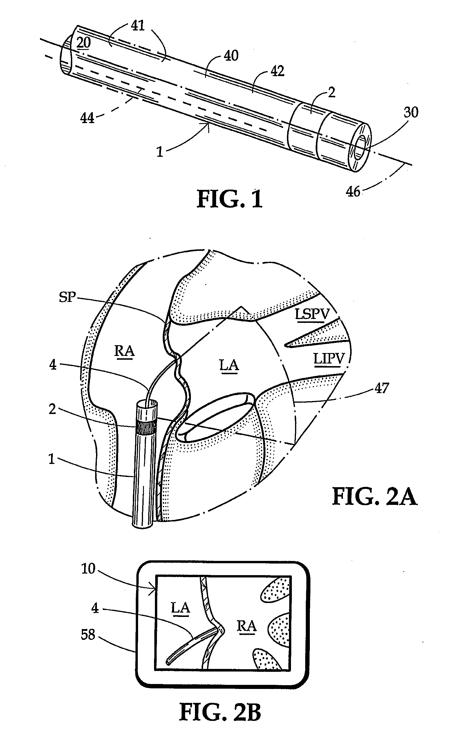
[0050]Apparatus according to one embodiment of the invention includes a sheath 1 (FIG. 1) generally in the form of an elongated tube having a proximal end 20, a distal end 30 and a proximal-to-distal axis. As used in this disclosure with reference to elongated elements for insertion into the body, the term “distal” refers to the end which is inserted into the body first, i.e., the leading end during advancement of the element into the body, whereas the term “proximal” refers to the opposite end.

[0051]Sheath 1 has an interior bore or lumen (not separately designated) extending between its proximal end 20 and its distal end 30. Desirably, sheath 1 has a relatively stiff proximal wall section 41 extending from its proximal end 20 to a juncture 40, and a relatively limber distal wall section or sheath end portion 42 extending from the juncture 40 to the distal end or tip 30. One or more pull wires 44 (only one shown) are slideably mounted in the proximal wall section 41 and connected to...


PUM


Abstract

Intra-organ ultrasound images are obtained by integrating ultrasound array configurations at the distal region of a sheath or guiding catheter integral to any catheter based intervention. A dual mode ablation / imaging circular ultrasound array is used to create circular or partial circular lesions. The sites of the individual lesion segments are identified in an ultrasound 2D image. In the case of PV isolation the process of ablating individual segments identified in the ultrasound image is repeated until a circumferential, continuous lesion has been achieved and PV isolation has been confirmed with the coaxial loop sensing catheter which also serves as a guide wire.

Description

CROSS-REFERENCE TO RELATED APPLICATIONS[0001]This application claims the benefit of U.S. Provisional Application No. 61 / 770,810 filed Feb. 28, 2013 and the benefit of U.S. Provisional Application No. 61 / 770,818 filed Feb. 28, 2013.FIELD OF THE INVENTION[0002]The present invention relates in part to integrated ultrasound imaging with a catheter delivery sheath as used for electrophysiology (EP), interventional cardiology and interventional radiology procedures.[0003]The present invention also relates to percutaneous catheter based treatments of various diseases as, for example, Atrial Fibrillation (AF), GERD, urinary tract disease, valve disease and lung tumors in mammalian subjects.BACKGROUND OF THE INVENTION[0004]Ultrasound imaging is well established to guide interventional procedures. Ultrasound imaging has the advantage that real time guidance with morphological information (unlike with fluro guidance which does not provide morphological information) is obtained without radiatio...

Claims


Application Information

Patent Timeline
IPC IPC(8): A61N7/02A61B5/055A61B8/00A61B8/08A61B8/12
CPCA61N7/022A61N2007/0052A61B8/12A61B8/445A61B8/4488A61B8/461A61B8/483A61B5/055A61B2018/00351A61B2018/00375A61B2018/00023A61B2018/00577A61B2018/00553A61B2018/00517A61N2007/0004A61B8/0841
Inventor WARNKING, REINHARD, J.
Owner GUIDED INTERVENTIONS
Learn More