Nanopore sensor for enzyme-mediated protein translocation

a technology of enzyme-mediated protein translocation and nanopore sensor, which is applied in the field of single-molecule protein analysis, can solve the problems of difficult drive translocation via applied voltage, and achieve the effect of improving the “threading” of proteins

Active Publication Date: 2016-02-04
RGT UNIV OF CALIFORNIA
View PDF2 Cites 9 Cited by
  • Summary
  • Abstract
  • Description
  • Claims
  • Application Information

AI Technical Summary

Benefits of technology

[0022]In one embodiment of the present invention, the step of measuring current changes comprises measuring current changes for states of (i) open channel in the nanopore, (ii), capture of the nondenatured protein by the nanopore, and (iii) passage of a protein from (ii) through the nanopore. In a preferred embodiment, the measuring comprises detecting differences between states (i), (ii) and (iii). In another preferred embodiment, the measuring comprises measuring differences during state (iii) caused by amino acid structure of the protein passing through the nanopore. In another preferred embodiment, the nanopore is defined by a pore protein. In a yet more preferred embodiment, the pore protein is α-hemolysin. In another preferred embodiment, the NTP driven unfoldase is attached to the pore protein. In a more preferred embodiment, the NTP driven unfoldase is an AAA+ enzyme. In a yet more preferred embodiment, the AAA+ enzyme is ClpX. In another embodiment, the circuit comprises a patch clamp amplifier applying a constant voltage between the cis chamber and the trans chamber.
[0023]In certain aspects of the present invention, the nanopore is defined by α-hemolysin or another multimeric pore protein. The pore protein does not need to be functionalized; that is it may be used as the protein exits in its native environment; it does not need to have any molecular structures added to it to attach or bind the protein being translocated. That is, it may be generic for translocation of any protein sequence, and does not specifically bind to or recognize the protein to be translocated. The protein to be translocated is also preferably in a native form. It may, in certain embodiments of the present invention, have attached to it a molecular structure for improving “threading” of the protein through the nanopore. In certain aspects of the present invention, the protein to be translocated comprises an exogeneous sequence comprising a targeting domain for the protein translocase. The targeting domain may comprise at least about 10-30 negatively charged amino acids, e.g. 30-100 glutamate or aspartate residues, or other negatively charged synthetic monomers, e.g. dextran sulfate, located at an amino or carboxy terminus of the protein to be translocated.

Problems solved by technology

However, unlike nucleic acids, proteins are generally not uniformly charged (making it difficult to drive translocation via an applied voltage) and they also fold into complex, large, and stable structures that cannot transverse a nanopore's aperture.

Method used


Image

  • Nanopore sensor for enzyme-mediated protein translocation
  • Nanopore sensor for enzyme-mediated protein translocation
  • Nanopore sensor for enzyme-mediated protein translocation

Examples


example 1

Construction of a Nanopore Device for Monitoring Protein Translocation

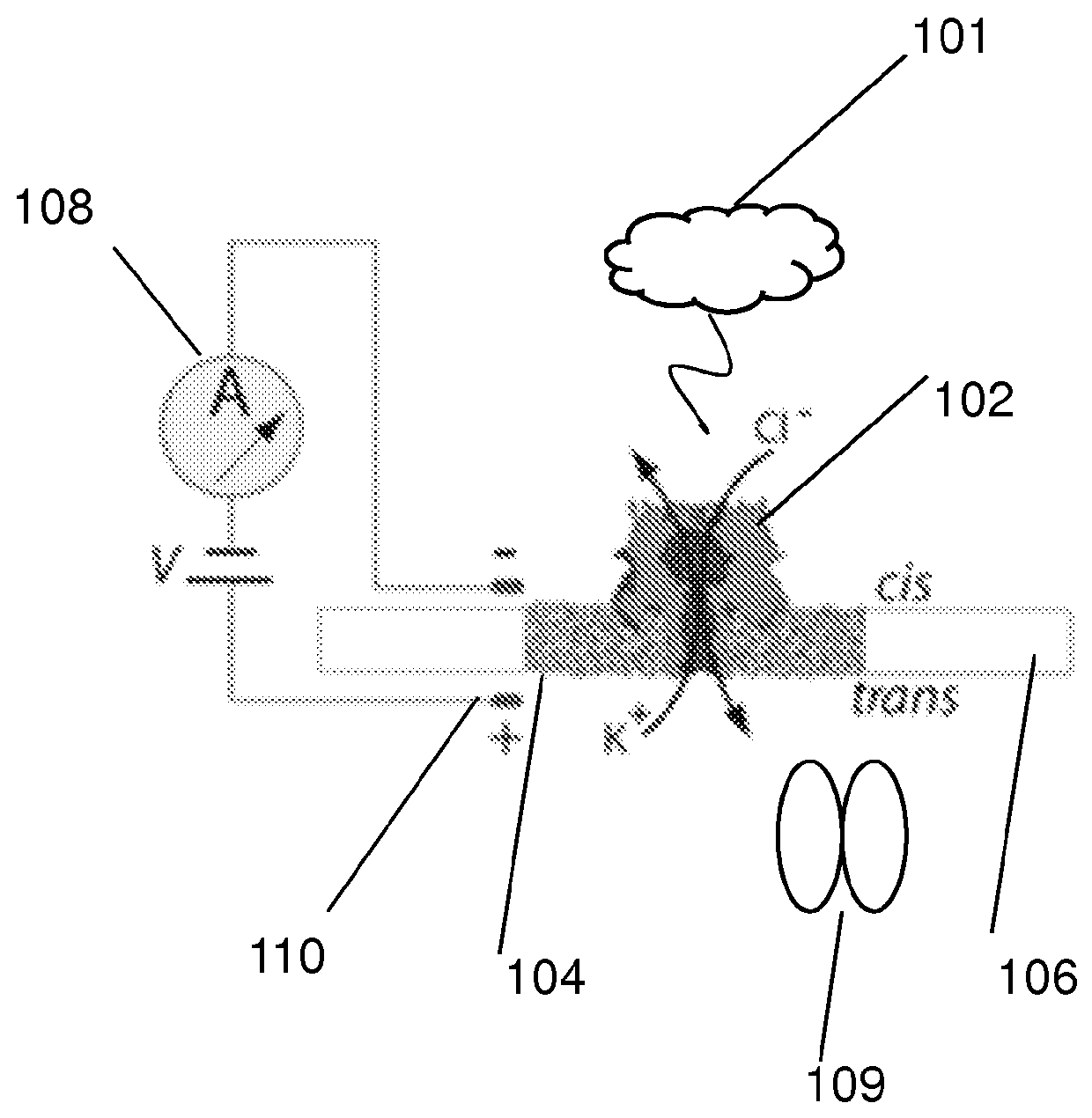
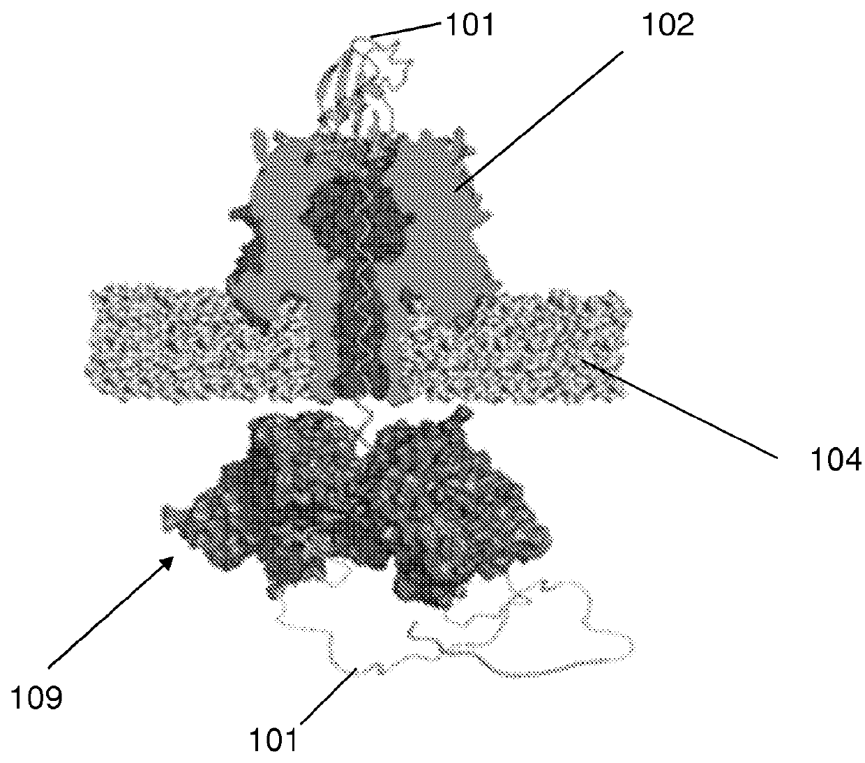
[0114]The nanopore device of this example is diagrammed in FIG. 1A, which shows diagrammatic drawing of a nanopore sensor with a single α-HL pore embedded in a lipid bilayer separating two Teflon® PTFE polymer wells each containing 100 μl of 0.2 M KCl solution (30° C.). Voltage is applied between the wells (trans side+180 mV), causing ionic current flow through the channel. Current diminishes in the presence of a captured protein molecule.

[0115]Briefly, for each experiment a single α-HL nanopore was inserted into a 30 μm diameter lipid bilayer that separates two wells (termed cis and trans) that each contained 100 μl of PD buffer (pH7.6). A covalently-linked trimer of an N-terminal truncated ClpX variant (ClpX-ΔN3) was used for all ClpX nanopore experiments. The ClpX-ΔN3 BLR expression strain was obtained from Andreas Martin (UC Berkeley). ClpX protein expression was induced at an OD 600 of ˜1 by addition of 0.5 m...

example 2

Engineering Protein S1, S2-35 and S2-148 for Translocation

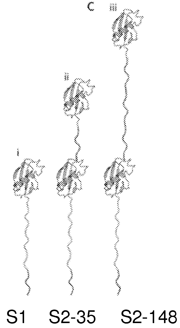
[0118]These proteins are schematically illustrated in FIG. 1C, which shows (i) S1, a protein bearing a single N-terminal Smt3-domain coupled to a 65-amino-acid-long charged flexible segment capped at its carboxy-terminus with the 11 amino acid ClpX-targeting domain (ssrA tag); (ii) S2-35, similar to S1 but appended at its N-terminus by a 35 amino acid linker and a second Smt3 domain; (iii) S2-148, identical to S2-35 except for an extended 148 amino acid linker between the Smt3 domains.

[0119]For our initial experiments, we used a modified version of the ubiquitin-like protein Smt3. Smt3 is comprised of ˜100 amino acids arranged into four β-strands and a single α-helix. We further engineer Smt3 into S1. To construct substrate protein S1, DNA encoding the 76 amino acid tail (GGSSGGSGGSGSSGDGGSSGGSGGSGSSGDGGSSGGSGGDGSSGDGGSDGDSDGSDGD GDSDGDDAANDENYALAA) (SEQ ID NO: 1) was constructed by polymerase chain reaction (PCR) and cloned...

example 3

Detection of Translocation of Protein S1

[0124]A representative ionic current trace for capture and translocation of protein S1 in the presence of ClpX and ATP is shown in FIG. 2A. FIG. 2A shows the ionic current traces during S1 translocation. (i) Open channel current through the α-HL nanopore under standard conditions (˜34±2 pA, RMS noise 1.2±0.1 pA). (ii) Capture of the S1 substrate. Upon protein capture, the ionic current drops to ˜14 pA (˜0.7 pA RMS noise). (iii) ClpX-mediated ramping state. The ionic current decreases to below 10 pA and is characterized by one or more gradual amplitude transitions. This pattern is only observed in the presence of ClpX and ATP (trans compartment). (iv) Smt3 domain unfolding and translocation through the nanopore (˜3.8 pA, 1.7 pA RMS noise). (v) Return to open channel current upon completion of substrate translocation to the trans compartment, From the open channel current of ˜34±2 pA (FIG. 2A, i), S1 capture resulted in a current drop to ˜14 pA ...


PUM

PropertyMeasurementUnit
ionic current state dwell timesaaaaaaaaaa
internal diameteraaaaaaaaaa
sizeaaaaaaaaaa

Abstract

Described herein is a device and method for translocating a protein through a nanopore and monitoring electronic changes caused by different amino acids in the protein. The device comprises a nanopore in a membrane, an amplifier for providing a voltage between the cis side and trans side of the membrane, and an NTP driven unfoldase which processed the protein to be translocated. The exemplified unfoldase is the ClpX unfoldase from E. coli.

Description

CROSS-REFERENCE TO RELATED APPLICATIONS[0001]This is a US Provisional Patent Application. U.S. Provisional Patent Application 61 / 599,754, filed Feb. 16, 2012 by Jeffrey Nivala, entitled “Unfolding and Translocation of Proteins Through a Nanopore Sensor and Methods of Use,” SC2012-400, is related and hereby incorporated by reference for all purposes.STATEMENT OF GOVERNMENTAL SUPPORT[0002]This invention was made with government support under contract R01HG006321 awarded by the National Institutes of Health. The government has certain rights in this invention.REFERENCE TO SEQUENCE LISTING, COMPUTER PROGRAM, OR COMPACT DISK[0003]A sequence listing will be submitted upon filing of a utility patent application.BACKGROUND OF THE INVENTION[0004]1. Field of the Invention[0005]The present invention relates to the field of single molecule protein analysis and also to the field of nanopore analysis.[0006]2. Related Art[0007]Presented below is background information on certain aspects of the pre...

Claims


Application Information

Patent Timeline
Patent Type & Authority Applications(United States)
IPC IPC(8): C12M1/00
CPCC12M47/06B82Y15/00G01N33/48721C12Q1/48G01N33/54366
Inventor NIVALA, JEFFREY MMARKS, DOUGLAS BAKESON, MARK A
Owner RGT UNIV OF CALIFORNIA
Why Eureka
Try Eureka