Three dimensional tissue imaging system and method

a tissue imaging and three-dimensional technology, applied in the field of three-dimensional tissue imaging systems and methods, can solve the problems of not being able to display or quantify the morphological disorganization characteristics obtained from the skin below, 20-30% of early melanomas going undetected, and not being able to achieve the effect of reducing sampling errors, improving the standard of care for biopsies, and increasing surgery efficiency

Active Publication Date: 2016-03-03
MELA SCIENCES
View PDF8 Cites 24 Cited by
  • Summary
  • Abstract
  • Description
  • Claims
  • Application Information

AI Technical Summary

Benefits of technology

[0013]One possible indication for use of the 3D Optical Spectral Imaging technology disclosed herein may be for Image Guided Tissue Sampling, the intent of which is to sample the most disorganized area of the lesion, thus reducing sampling error. A second possible indication for use of the Optical Spectral Imaging technology may be for Image Guided Tissue Sectioning for dermatopathologists, the intent of which is to select the most disorganized area of the lesion to be used for evaluation by the pathologist under the microscope. A third possible indication for use of the Optical Spectral Imaging technology may be for Image Guided Biopsies, the intent of which is to increase surgery efficiency by viewing the 3-dimensional volume and construct of the lesion for margin guidance. As a result, the present invention is intended to improve the standard of care for biopsies, increase dermatopathology sectioning efficiency and accuracy, as well as aid in the surgeon's excision quality of removed tissue.

Problems solved by technology

This limited information results in 20-30% of early melanomas going undetected.
Additional imaging tools are available to the market, but none display or quantify the morphological disorganization characteristics as obtained from below the surface of the skin.
While the excisional biopsy and sauceration shave biopsy demonstrated near 100% accuracy, punch technique was only 86.5% accurate.
The paper reveals, “Due to the inherent intralesional heterogeneity of cutaneous melanoma, small punch biopsies may not always obtain representative specimens and may subject the patient to significant risk of misdiagnosis.”
Further misdiagnosis occurs among pathologists who either over diagnose or under diagnose a lesion.
This technique results in additional sections being ordered by the dermatologist if he or she believes the diagnosis is not correct, and perhaps the area of interest was not reviewed by the pathologist due to ineffective sectioning.
Currently, no imaging device is known that offers the capability to guide sectioning as performed by a pathologist and his or her grossers, and this lack of image guidance in determining which areas of the lesion to section, in addition to subjective evaluation under the microscope, puts patients at undue risk.
Unfortunately, ultrasound is unreliable on pigmented lesions and is difficult to read.
These images however are not transformed into a 3-dimensional volumetric rendering of the lesion, and require interpretation by the user on the areas between each vertical layer image.

Method used


Image

  • Three dimensional tissue imaging system and method
  • Three dimensional tissue imaging system and method
  • Three dimensional tissue imaging system and method

Examples


first example implementation

[0038

[0039]Obtaining Multi-Spectral Images

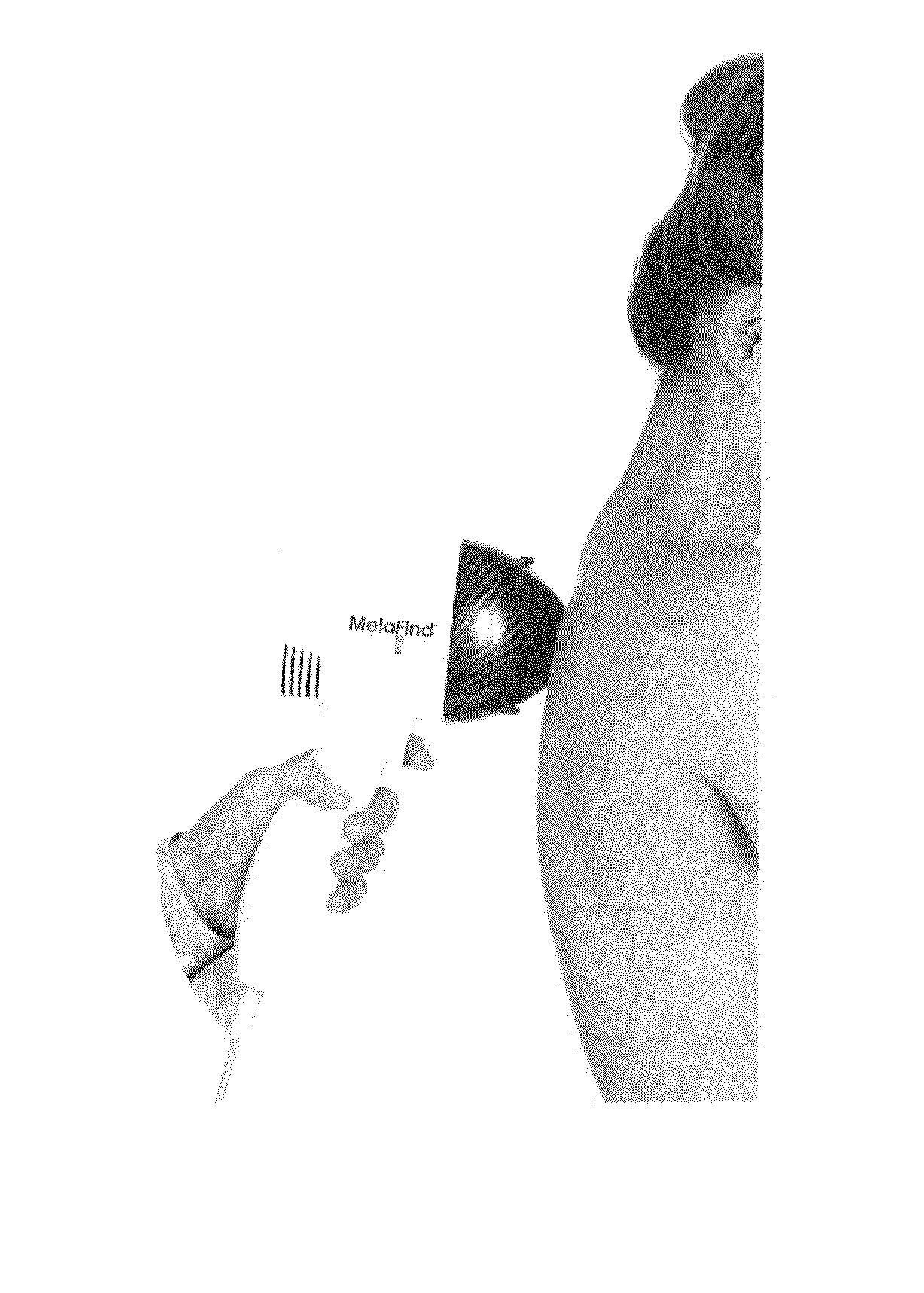
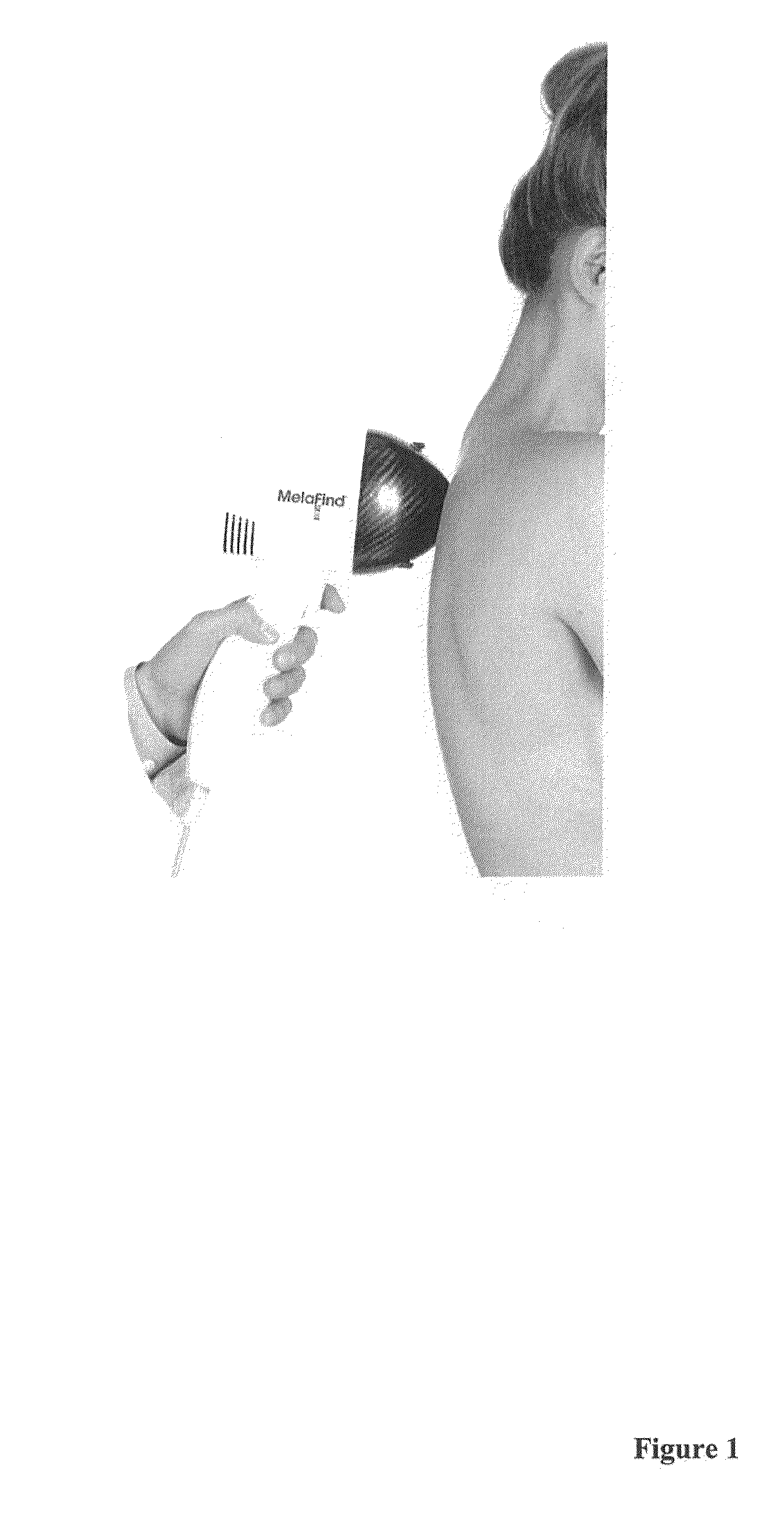
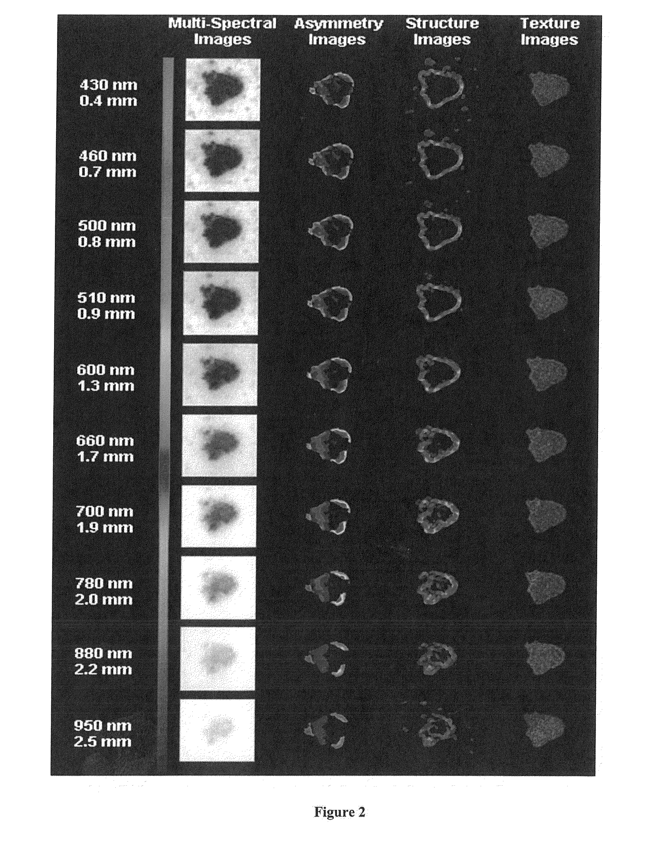
[0040]The inventive system first obtains multi-spectral images. This can be achieved using the hardware and some of the software used with the MelaFind computer-controlled multi-spectral imaging and analysis system, in which ten distinct spectral bands are used to illuminate a skin lesion with wavelengths distributed over visible to near-infrared ranges, from 430 nm (blue) through 950 nm (near infrared spectrum). Appendix Image 1 illustrates the MelaFind optical scanner being held by a medical professional or technician against a region of interest on the back of a patient. Appendix Image 2 shows the wavelengths used to obtain data from various depths below the surface of the skin and examples of the images that can be created using the existing MelaFind system.

[0041]In use, the imaging assembly illuminates the skin with light through a thin layer of alcohol, via an illumination circuit containing light-emitting diodes (i.e. LED) composed to...


PUM


Abstract

Multispectral digitized images are provided by utilizing 2 or more spectral bands, providing images and information regarding a pigmented or other differentiated tissue sample at differing depths. The images are transformed to enhance morphological patterns characteristic of melanoma, such as the statistical properties of the pigment network, image texture or homogeneity, and the lesion reflectance, in order to provide information about lesion morphology at different depths. The images can also be processed and transformed to extract features such as blood volume saturation, oxygen saturation, or other characteristics that can be identified based on differential reflectance or absorption of light.

Description

CROSS-REFERENCE TO RELATED APPLICATIONS[0001]This application is related to, and claims the benefit of, U.S. Provisional Patent Application Ser. No. 62 / 042,858, filed Aug. 28, 2014, which is incorporated herein by reference.FIELD OF THE INVENTION[0002]The present invention relates to systems and methods for optical spectral imaging of tissue in three dimensions using multiple spectrums of light.BACKGROUND OF THE INVENTION[0003]Conventionally, lesion information is obtained via surface visual analysis by the dermatologist. This limited information results in 20-30% of early melanomas going undetected. The estimated total direct cost associated with the treatment of melanoma in 2010 in the United States due to lack of early detection was $2.36 billion, with 65% of total cost going to continued care and another 20% of total cost going to the last year of life.[0004]To see below the topmost layer of skin, a dermatoscope with a lighted magnifier may be used, but the image is limited to t...

Claims


Application Information

Patent Timeline
Patent Type & Authority Applications(United States)
IPC IPC(8): A61B5/00G06T7/00G06T15/08
CPCA61B5/0077G06T15/08G06T7/0012A61B5/0075A61B5/742A61B2560/0475A61B5/444G06T2207/30024G06T2207/30088G06T2207/10152A61B5/748G06T17/10G06T2210/41
Inventor DEBERNARDIS, FRANK, A.TUCKER, NATALIE, J.REITER, AUSTIN, D.PALASARA, MINHAZ
Owner MELA SCIENCES
Why Eureka
Try Eureka