Detection of Early-Stage Pancreatic Adenocarcinoma

a pancreatic cancer and early-stage technology, applied in the field of anti-pancreatic cancer antibodies, can solve the problems of difficult detection of small tumors, difficult differentiation of pancreatic cancer from focal pancreatitis, and difficult diagnosis of pancreatic cancer at an early stage of tumor growth, etc., and achieve high specificity and sensitivity

Inactive Publication Date: 2016-03-10
IMMUNOMEDICS INC
View PDF0 Cites 0 Cited by
  • Summary
  • Abstract
  • Description
  • Claims
  • Application Information

AI Technical Summary

Benefits of technology

The patent text describes using anti-pancreatic cancer antibodies, such as the hPAM4 antibody, to detect and diagnose pancreatic cancer at early stages with high sensitivity and specificity. The antibodies can label at least 70% of well-differentiated, moderately-differentiated, and poorly-differentiated pancreatic cancer, and more than 90% of invasive pancreatic adenocarcinomas. The antibodies can also detect 85% or more of precursor lesions.

Problems solved by technology

Thus, diagnosing pancreatic cancer at an early stage of tumor growth is often difficult and requires extensive diagnostic work-up, often times including exploratory surgery.
However, reliable detection of small tumors, as well as differentiation of pancreatic cancer from focal pancreatitis, is difficult.
This is significant, since late detection of pancreatic cancer results in low survival rate.
Current treatment procedures available for pancreatic cancer have not led to a cure, or to a substantially improved survival time.
However, due to a large tumor burden, only 10% to 25% of patients are candidates for “curative resection.” For those patients undergoing a surgical treatment, the five-year survival rate is still poor, averaging only about 10%.
However, CA19.9 levels were found to be elevated in a number of other malignant and benign conditions, and currently the assay cannot be used for diagnosis.
However, previously reported antibodies against pancreatic cancer antigens have not been successfully employed to date for the effective therapy or early detection and / or diagnosis of pancreatic cancer.

Method used


Image

  • Detection of Early-Stage Pancreatic Adenocarcinoma
  • Detection of Early-Stage Pancreatic Adenocarcinoma
  • Detection of Early-Stage Pancreatic Adenocarcinoma

Examples


example 1

Humanized PAM4 MAb

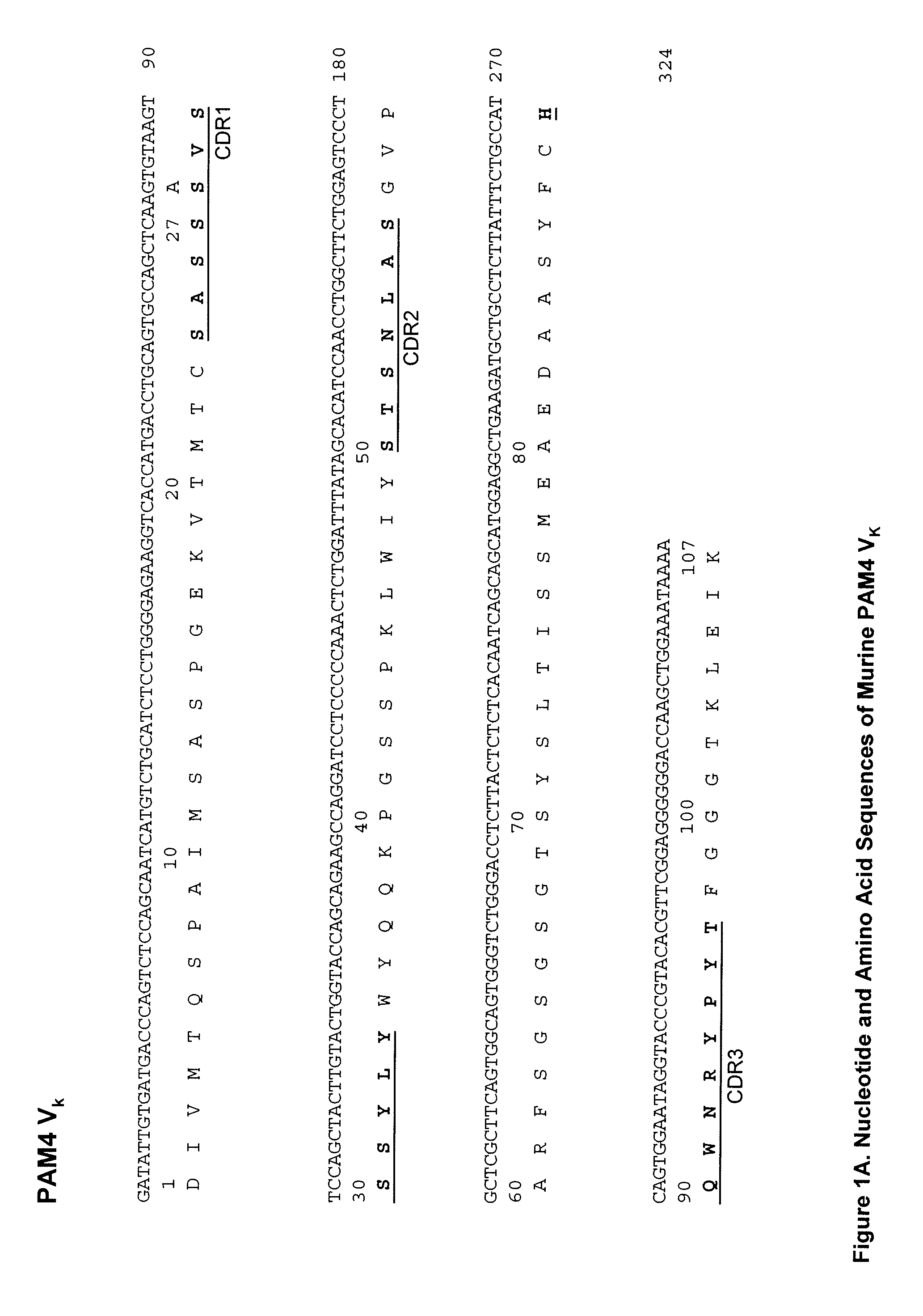
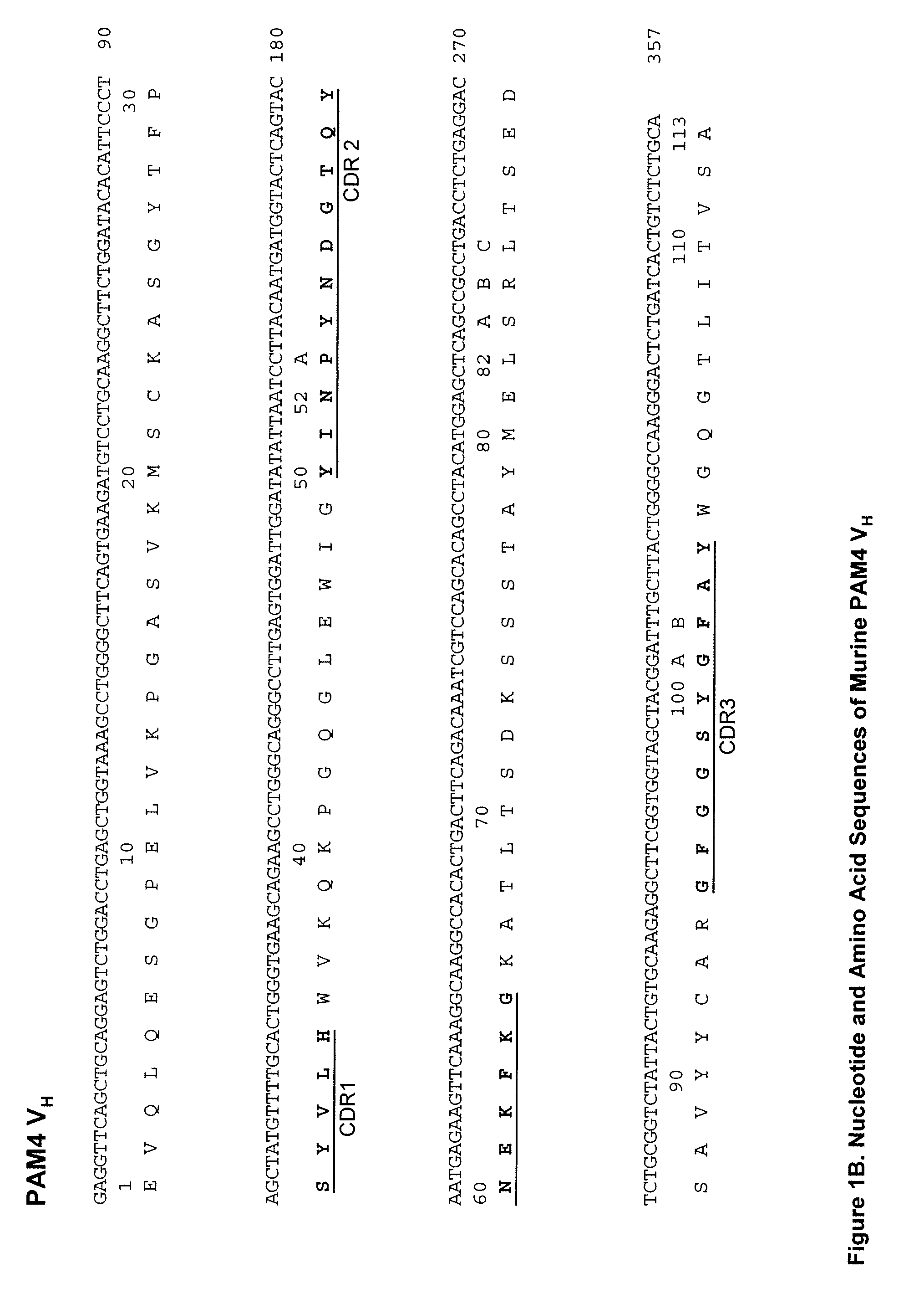
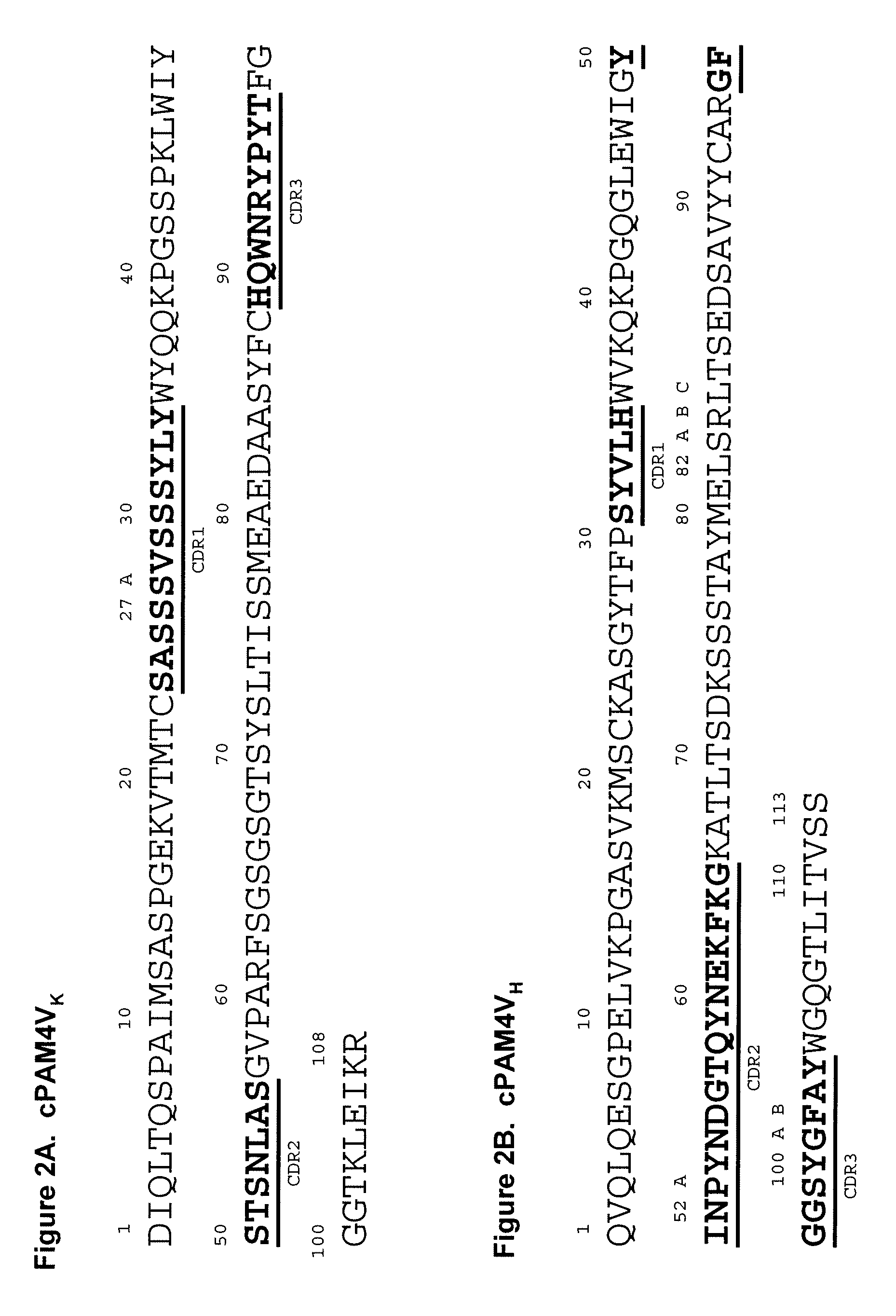
[0244]In preferred embodiments, the claimed methods and compositions utilize the antibody hPAM4 which is a humanized IgG of the murine PAM4 MAb raised against pancreatic cancer mucin. Humanization of the murine PAM4 sequences was utilized to reduce the human antimouse antibody (HAMA) response. To produce the humanized PAM4, murine complementarity determining regions (CDR) were transferred from heavy and light variable chains of the mouse immunoglobulin into human framework region (FR) antibody sequences, followed by the replacement of some human FR residues with their murine counterparts. Humanized monoclonal antibodies are suitable for use in in vitro and in vivo diagnostic and therapeutic methods.

[0245]Comparison of the variable region framework sequences of the murine PAM4 MAb (FIGS. 1A and 1B) to known human antibodies in the Kabat database showed that the FRs of PAM4 VK and VH exhibited the highest degree of sequence homology to that of the human antibodies Wa...

example 2

Immunohistochemistry Staining Studies

[0258]Immunohistochemistry on normal adult tissues showed that the PAM4 reactive epitope was restricted to the gastrointestinal tract where staining was weak, yet positive (Table 2). Normal pancreatic tissue, including ducts, ductules, acini, and islet cells, were negative for staining A PAM4 based enzyme immunoassay with tissue homogenates as antigens generally supported the immunohistology data (Table 3). The PAM4 epitope was absent from normal pancreas and other non-gastrointestinal tissues. In neoplastic tissues, PAM4 was reactive with twenty one out of twenty five (85%) pancreatic cancers (Table 4 and Table 5) and ten out of twenty six colon cancers, but only limited reactivity with tumors of the stomach, lung, breast, ovary, prostate, liver or kidney (Table 5). PAM4 reactivity appeared to correlate with the stage of tumor differentiation, with a greater percentage of staining observed in well differentiated pancreatic cancers than in modera...

example 3

In Vivo Biodistribution and Tumor Targeting of Radiolabeled PAM4

[0260]Initial biodistribution studies of PAM4 were carried out in a series of four different xenografted human pancreatic tumors covering the range of expected differentiation. Each of the four tumor lines employed, AsPc1, BxPc3, Hs766T and CaPan1, exhibited concentrations of 131I-PAM4 within the tumors (range: 21%-48% ID / g on day three) that were significantly (P<0.01-0.001) higher than concomitantly administered nonspecific, isotype-matched Ag8 antibody (range: 3.6%-9.3% ID / g on day three). The biodistribution data were used to estimate potential radiation doses to the tumor of 12,230; 10,684; 6,835; and 15,843 cGy / mCi of injected dose to AsPc1, BxPc3, Hs766T and CaPan1, respectively. With an actual maximum tolerated dose (MTD) of 0.7 mCi, PAM4 could provide substantial rad dose to each of the xenografted tumor models. In each tumor line the blood levels of radiolabeled PAM4 were significantly (P<0.01-0.001) lower tha...


PUM


Abstract

Described herein are compositions and methods of use of anti-pancreatic cancer antibodies or fragments thereof, such as murine, chimeric, humanized or human PAM4 antibodies. The subject antibodies show a number of novel and useful diagnostic characteristics, such as binding with high specificity to pancreatic and other cancers, but not to normal pancreatic tissues and binding to a high percentage of early stage pancreatic cancers. In preferred embodiments, the antibodies bind to pancreatic cancer mucins. The antibodies and fragments are of use for the detection and diagnosis of early stage pancreatic cancer. In preferred embodiments, the anti-pancreatic cancer antibodies can be used for immunoassay of serum samples, wherein the immunoassay can detect a marker for early stage pancreatic cancer in serum. More preferably, the serum is extracted with an organic phase, such as butanol, before immunoassay.

Description

RELATED APPLICATIONS[0001]This application is a divisional of U.S. patent application Ser. No. 13 / 010,129, filed Jan. 20, 2011, which was a continuation-in-part of U.S. patent application Ser. No. 12 / 537,803 (now issued U.S. Pat. No. 8,491,896), filed Aug. 7, 2009, which was a continuation-in-part of U.S. patent application Ser. No. 11 / 849,791 (now abandoned), filed Sep. 4, 2007, which was a divisional of U.S. patent application Ser. No. 10 / 461,885 (now issued U.S. Pat. No. 7,282,567), filed Jun. 16, 2003, which claimed the benefit under 35 U.S.C. 119(e) of U.S. Provisional Patent Application 60 / 388,314, filed Jun. 14, 2002. U.S. patent application Ser. No. 12 / 537,803 claimed the benefit under 35 U.S.C. 119(e) of Provisional U.S. Patent Application Ser. Nos. 61 / 087,463, filed Aug. 8, 2008, and 61 / 144,227, filed Jan. 13, 2009. U.S. patent application Ser. No. 13 / 010,129 claimed the benefit under 35 U.S.C. 119(e) of U.S. Provisional Patent Application 61 / 297,303, filed Jan. 22, 2010; ...

Claims


Application Information

Patent Timeline
Patent Type & Authority Applications(United States)
IPC IPC(8): G01N33/574
CPCG01N2333/4725G01N33/57438A61K51/0406A61K51/0491A61K51/08A61K51/088A61K51/109A61K2039/505A61K2039/507B82Y5/00B82Y15/00C07K16/303C07K16/3092C07K16/44C07K2317/24C07K2317/31C07K2317/54C07K2317/55C07K2317/565G01N33/57488A61K47/6897A61K49/0058A61K51/1018
Inventor GOLD, DAVID V.GOLDENBERG, DAVID M.
Owner IMMUNOMEDICS INC
Why Eureka
Try Eureka