
Systems and methods for monitoring muscle rehabilitation

a monitoring system and muscle technology, applied in the field of systems and methods for monitoring muscle rehabilitation, can solve the problems of spinal stabilization system dysfunction, dysfunction of these subsystems, and disruption of the feedback loop, and achieve the effect of accurate temporal averaging

Active Publication Date: 2016-04-21
MAINSTAY MEDICAL
View PDF7 Cites 70 Cited by
  • Summary
  • Abstract
  • Description
  • Claims
  • Application Information

AI Technical Summary

Benefits of technology

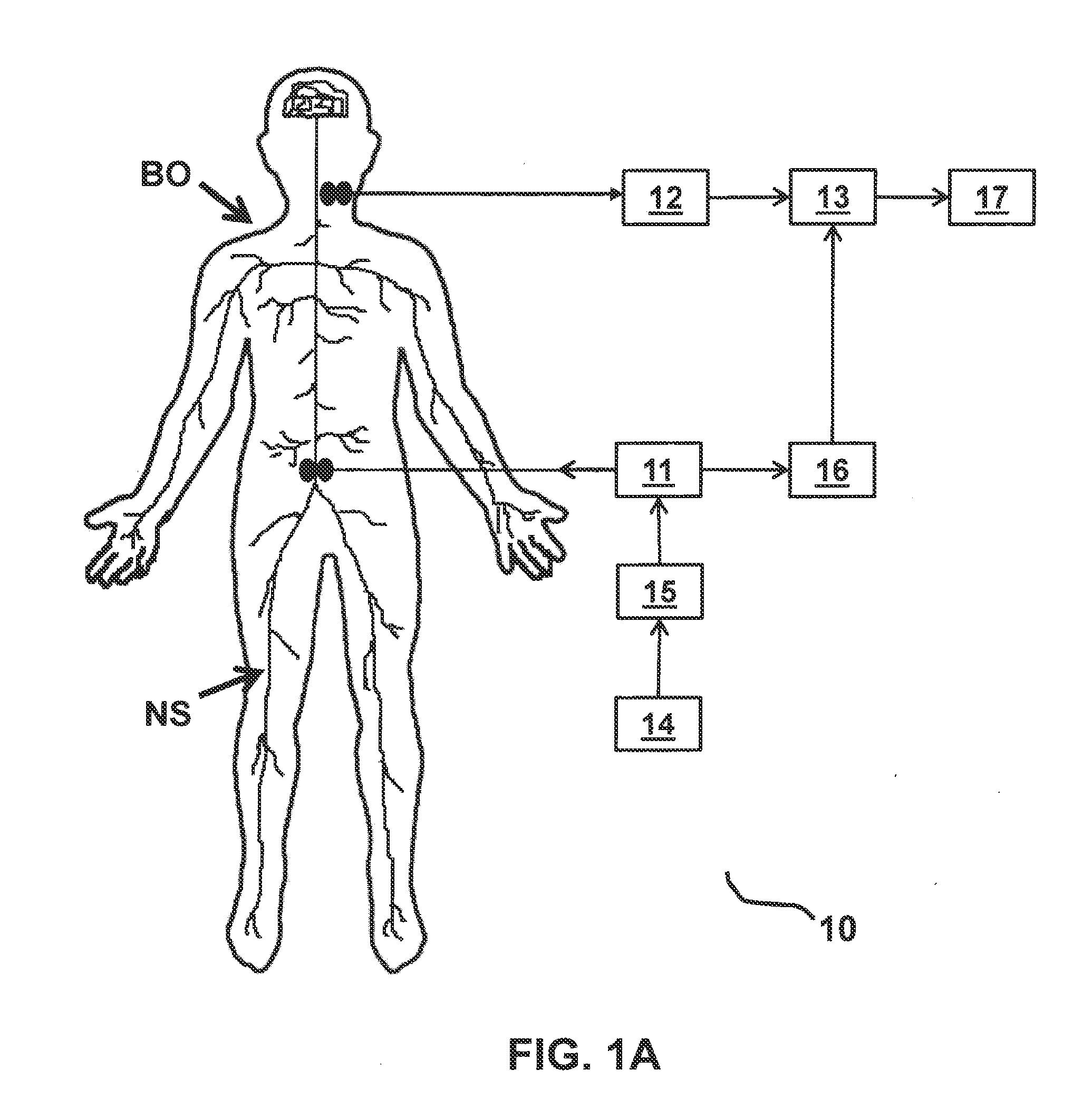
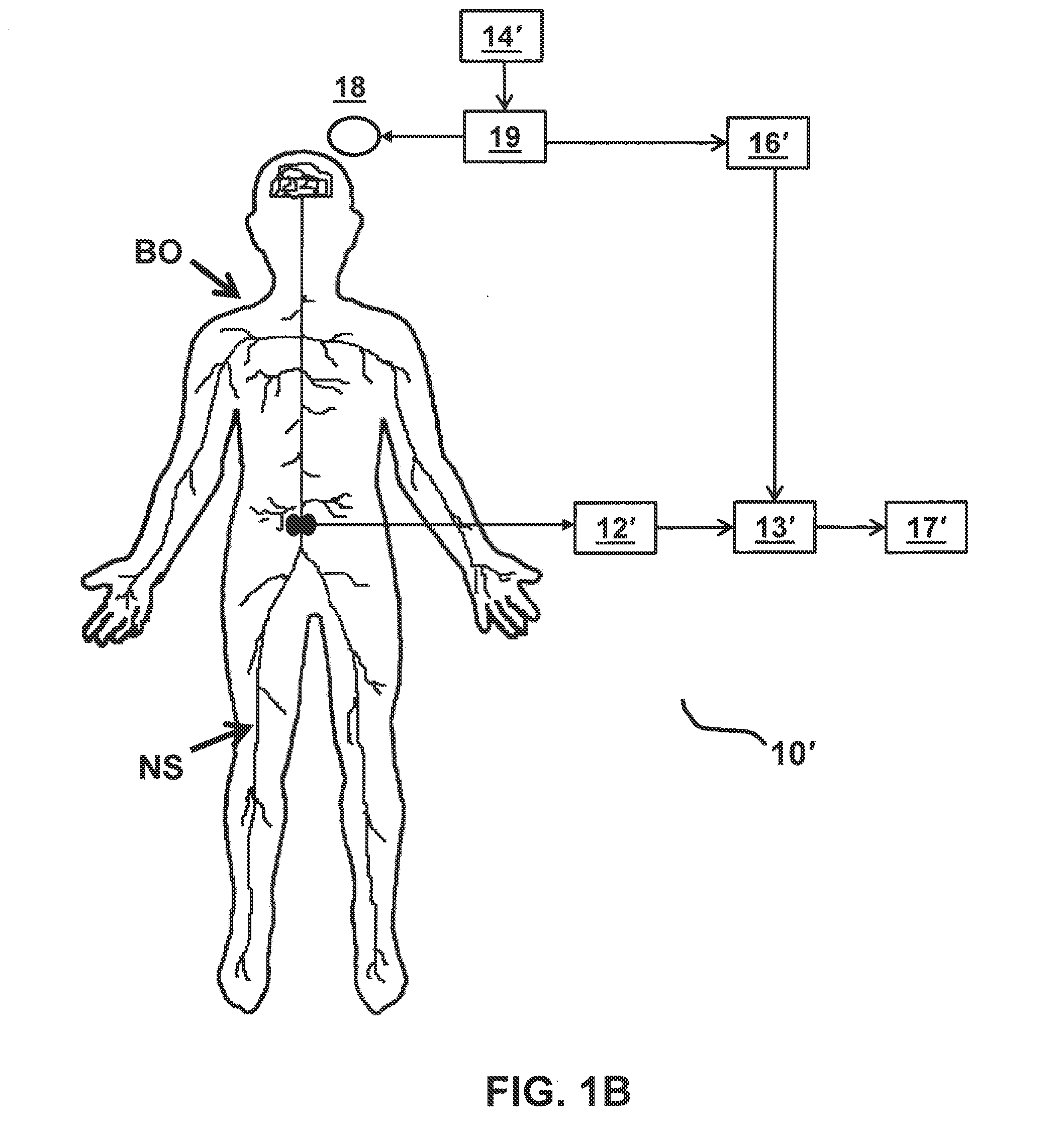
The present invention provides a system and method for monitoring the progress of rehabilitation of a muscle by measuring the response of the body to stimulations. The system includes an implantable device with a recording electrode and a processor, a transcranial magnetic stimulation device, a trigger detector, an external receiver, and a user interface. The system can automatically apply a stimulation protocol to the body and record the response signal generated by the body in response to the stimulation protocol. The recorded response signal can be processed and displayed on a computer screen for further analysis. The system can also adjust the stimulation protocol based on the recorded response signal and provide feedback on the rehabilitation process. The technical effects of the invention include improved accuracy in measuring muscle response to stimulations and more efficient and effective rehabilitation protocols.

Problems solved by technology

It is applicant's realization that lower back pain often results from dysfunction of these subsystems and disruption of the feedback loop.
It is believed that in some patients with back pain, the spinal stabilization system is dysfunctional.
However, ligament injury, soft tissue fatigue, viscoelastic creep, and other connective tissue injuries may cause mechanoreceptors to produce corrupted signals about vertebral position, motion, or loads, leading to an inappropriate muscle response.
In addition, muscles themselves may be injured, fatigued, atrophied, or lose their strength, thus aggravating dysfunction of the spinal stabilization system.
Moreover, muscles may disrupt the spinal stabilization system by going into spasm, contracting when they should remain inactive, developing trigger points, or contracting out of sequence with other muscles.
Such muscle dysfunction may cause muscle spindle mechanoreceptors to send abnormal signals to the motor cortex, which further may compromise normal muscle activation patterns via the feedback loops.
Through such mechanisms, disruptions to the spinal stabilization system can result in spine instability, which can lead to low back pain.
In particular, spine instability can result in the generation of high loads on spinal structures when the spine moves beyond its neutral zone.
High loads can lead to inflammation, disc degeneration, facet joint degeneration, and muscle fatigue.
Since the endplates and annulus have a rich nerve supply, it is believed that abnormally high loads on such structures, resulting from spine instability, may be a common cause of pain.
Load transmission to the facet joints also may increase with degenerative disc disease, leading to facet arthritis and facet joint pain.
In general terms, stimulation at a rate significantly higher than the fusion frequency has minimal effect on the strength or nature of contraction and may, in fact, have an adverse impact on fatigue of the muscle.
Stimulation at a frequency higher than necessary to achieve the desired (e.g., maximum) force is energy inefficient, which is an important consideration for an implantable device.
In patients with spinal cord injury, the sensory nervous system is usually damaged as well as the motor system, and thus the afflicted person loses proprioception of what the muscle and limbs are doing.
One of the challenges of stimulation therapies such as NMES and TMS is monitoring to ensure the stimulation device is positioned properly, applying appropriate levels of stimulation, and resulting in a positive therapeutic effect.
Monitoring can be especially challenging for deep muscles such as the deep fascicles of the lumbar multifidus, which are too deeply positioned for contractions to be reliably observed visually.
A related challenge of NMES and TMS for rehabilitation of skeletal muscles is to diagnose when the therapy has been successful and may be discontinued.
This is particularly important with patients who cannot communicate, e.g., young children, or patients who do not want to communicate, e.g., malingerers who may be motivated for the therapy to not be successful as it would result in loss of worker's compensation insurance.

Method used


Image

  • Systems and methods for monitoring muscle rehabilitation
  • Systems and methods for monitoring muscle rehabilitation
  • Systems and methods for monitoring muscle rehabilitation

Examples


Embodiment Construction

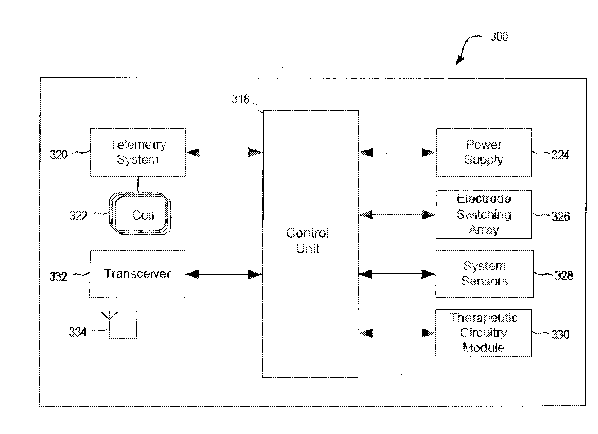
[0051]A system and a method for monitoring stimulation therapy of skeletal muscles are described herein. Increasingly, neuromuscular electrical stimulation (NMES) and trans-cranial magnetic stimulation (TMS) are being utilized to treat and rehabilitate muscles. It is believed that such therapies may achieve positive outcomes by improving the functionality of feedback loops between muscles and the nervous system controlling them. NMES may be applied to a patient through an implanted NMES apparatus, such as through the implantable pulse generator (IPG) described in U.S. Pat. Nos. 8,428,728 and 8,606,358 to Sachs and U.S. Application Publication No. 2011 / 0224665 to Crosby, each of which is incorporated by reference herein. Alternatively, TMS may be applied to a patient externally using electromagnetic coils positioned on or near a patient's skull. While it is known that such techniques deliver a stimulating electrical current or voltage to patients which results in muscle contraction t...


PUM


Abstract

System and method for rehabilitating a muscle and monitoring such rehabilitation are provided, the system including a user input receiver for receiving stimulation parameter inputs from a user, a stimulator for generating stimulations to be applied to a patient's body based on the stimulation parameter inputs, a signal receiver to receive, detect, and record a signal containing an evoked potential generated by the body in response to the stimulations, a signal processor for processing the recorded signal, for example, by amplifying, filtering, digitizing and temporal averaging the recorded signal, a trigger detector to alert the signal processor module when stimulations are generated to enable synchronization of the response signal with the stimulus for accurate temporal averaging, and an output display for providing data representative of the evoked potential to the user.

Description

I. CROSS-REFERENCE TO RELATED APPLICATIONS[0001]This application claims the benefit of priority of U.S. Provisional Patent Application No. 62 / 064,924, filed Oct. 16, 2014, the entire contents of which are incorporated herein by reference.II. FIELD OF THE INVENTION[0002]This application generally relates to assessment of the physiological state of a muscle subject to therapeutic stimulation. In particular, this application is directed to a system and method for monitoring progress of muscular rehabilitation.III. BACKGROUND OF THE INVENTION[0003]Back pain in the lower, or lumbar, region of the back is common. In many cases, the cause of back pain is unknown. The human back is a complicated structure including bones, muscles, ligaments, tendons, nerves and other structures, which together form the spinal stabilization system. The spinal stabilization system may be conceptualized to include three subsystems: 1) the spinal column, which provides intrinsic mechanical stability; 2) the spi...

Claims


Application Information

Patent Timeline
IPC IPC(8): A61N2/00A61N2/02A61N1/372A61N1/05A61N1/36
CPCA61N1/36135A61N1/37211A61N2/02A61N1/36067A61N1/0551A61N1/36003A61N2/004A61B5/4836A61N1/36071A61N1/36139A61B5/24A61B5/389A61B5/686A61N2/006A61B5/4519A61B2505/09A61B2562/043
Inventor CROSBY, PETER A.JAAX, KRISTEN N.RAWAT, PRASHANT B.
Owner MAINSTAY MEDICAL
Learn More