Quantitative molar concentration detection of specific apolipoprotein-containing particles present in bodily fluids by using capillary electrophoresis

a technology of apolipoprotein and molar concentration, which is applied in the direction of fluorescence/phosphorescence, instruments, biocide, etc., can solve the problems of increasing the distribution profile of the individual's lipoprotein subclass may be indicative of a health risk, and the difference in the amount of cholesterol in the particle may also correlate with the risk of cardiovascular disease. , to achieve the effect of fast, accurate and detailed resolution

Inactive Publication Date: 2016-04-21
HELENA LAB
View PDF1 Cites 3 Cited by
  • Summary
  • Abstract
  • Description
  • Claims
  • Application Information

AI Technical Summary

Benefits of technology

[0017]The methods and system described herein provide for the simultaneous separation and detection of lipoprotein particles present in a biological sample. The advantage of the methods and system of the present invention include the fast,

Problems solved by technology

It is well-established that the lipoprotein subclass distribution profile of an individual may be indicative of a health risk.
For example, apoB is a constituent of VLDL and LDL particles, which are associated with increased risk of cardiovascular disease.
Moreover, differences in the amount of cholesterol in a particle may also correlate with the risk of cardiovascular disease.
While NMR can be used to detect HDL, VLDL, IDL and LDL, it cannot be used to detect Lp(a) particles.
Likewise, the applicability of NMR to lipoprotein characterization is limited by the low sample throughput and cost of equipment.
However, such techniques are labor intensive and not automatable.
The most accurate method for isolating lipoprotein is ultracentrifugation; but, this method is very time consuming and expensive and therefore not suitable for large-scale population studies.
These methods are limited by the absence of effective and scalable methods to calculate lipid particle concentration.
This

Method used


Image

  • Quantitative molar concentration detection of specific apolipoprotein-containing particles present in bodily fluids by using capillary electrophoresis
  • Quantitative molar concentration detection of specific apolipoprotein-containing particles present in bodily fluids by using capillary electrophoresis
  • Quantitative molar concentration detection of specific apolipoprotein-containing particles present in bodily fluids by using capillary electrophoresis

Examples


example 1

Optical Apparatus for use in CE-ITP-LIF Systems

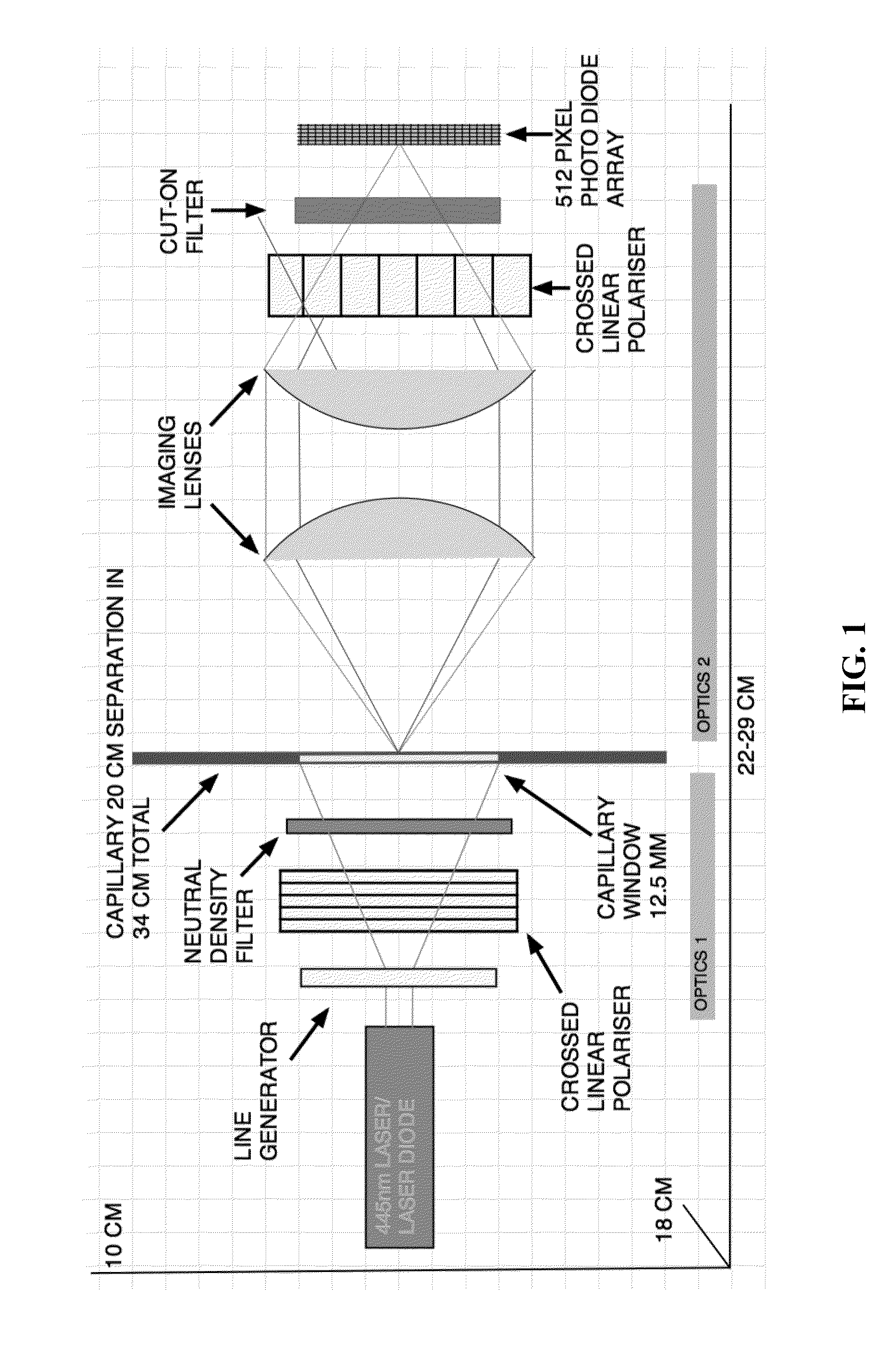
[0122]A schematic of an optical apparatus comprising two optical zones for use in a CE-ITP-LIF system is shown in FIG. 1. Optics zone 1 comprises an optical rail on which are arranged a 445 nm or other specific wavelength laser or laser diode. Light from these sources is focused through a series of optical components comprising, but not limited to, a line generator, a crossed linear polarizer, and a neutral density filter. Light from optics zone 1 is focused onto a 12.5 mm area of a 100 μM internal diameter fused silica capillary (˜365 μM o.d.) in which a 20 mm viewing window has been created by thermal removal of the polyamide sheath. The light then passes through the sample that is being separated by ITP and excites the fluorescent label attached to each analyte molecule (e.g., a lipoprotein and / or lipid particle). Emitted light energy, at a wavelength specific to the fluorescent label is then focused onto a 512 pixel photo diode arra...

example 2

Replicate Lipoprotein Profiles of a Single Biological Sample

[0139]To test the reproducibility of the CE-ITP-LIF system, several replicate biological samples from a single patient were evaluated. As a control experiment, the non-specific lipophilic dye CF was run on the ITP system in the absence of a biological sample. FIG. 6A shows an electropherogram of the control experiment with a peak corresponding to CF (migration time=0.7999), area under peak=2.345). Next, lipoprotein particles in replicate biological samples from patient 8 were labeled with CF and run with a standard CF sample. FIG. 6B is an electropherogram showing the lipoprotein profile of each replicate sample tested. The lipid profile remains constant even after CF has degraded (FIG. 6C).

example 3

Lipoprotein Particle Spiking Results in a Marked Increase in the Corresponding Detected Lipoprotein Peak Height

[0140]The lipoprotein profile of a biological sample stained with NBD-ceramide generates several peaks corresponding to individual serum lipoproteins (FIGS. 6A-6B). To validate the identity of each individual lipoprotein peak, biological samples were spiked with known amounts of purified lipoprotein. To validate peaks corresponding to HDL and LDL, native samples from patient 8 were spiked with purified HDL and LDL, respectively. The lipid profile of the HDL spiked sample (FIG. 7A, top) and the LDL spiked sample (FIG. 7B, top) were aligned with the lipid profile generated by the native sample (FIG. 7A, bottom; FIG. 7, bottom). As shown in FIG. 7A, there was a marked increase in the peak height and area under the peak in the HDL spiked sample compared to the native sample. FIG. 7B shows the same relationship between the LDL spiked sample compared to the native sample. FIG. 7C...


PUM

PropertyMeasurementUnit
Concentrationaaaaaaaaaa
Molar densityaaaaaaaaaa
Therapeuticaaaaaaaaaa

Abstract

A method for determining the molar concentration of specific lipoprotein particles present in a bodily fluid is presented. Multipixel Capillary Isotachophoresis Laser Induced Fluorescence is applied to fluorescently-labeled lipoproteins or immunologically-labeled apolipoproteins, facilitating quantification of lipoproteins and/or lipid particles and/or their associated apolipoproteins in a sample. The measurements are used to predict the risks of developing, progressing in severity of diseases related to lipoprotein particles, including cardiovascular and metabolic disorders.

Description

[0001]This application claims the benefit of U.S. Provisional Patent Application Ser. No. 62 / 066,573, filed Oct. 21, 2014 and U.S. Provisional Patent Application Ser. No. 62 / 147,665 filed Apr. 15, 2015, which are hereby incorporated by reference in their entirety.FIELD OF THE INVENTION[0002]The present invention is relates to a method and a system for determining the molar concentration and / or particle number of a lipoprotein present in a biological sample. The invention also teaches a method for assessing a cardiovascular risk in a subject.BACKGROUND OF THE INVENTION[0003]Lipoproteins are biological assemblies comprising an outer layer of protein and phospholipids and a core of neutral lipids including cholesterol esters and triacylglycerols. Lipoproteins include very low-density lipoprotein (VLDL), low-density lipoprotein (LDL), intermediate-density lipoprotein (IDL), high-density lipoprotein (HDL), chylomicrons, and lipoprotein(a) (Lp(a)) particles. Each lipoprotein particle is f...

Claims


Application Information

Patent Timeline
IPC IPC(8): G01N33/92G01N27/447G01N21/64
CPCG01N33/92G01N21/6428G01N2021/6439G01N2800/32G01N2800/50G01N27/447
Inventor GUADAGNO, PHILIPBELLIN, ERIN GRACE SUMMERSDEVANUR, DEEPIKAHASSARD, STUART
Owner HELENA LAB
Why Eureka
Try Eureka