Catheter for detection of ultrasound and photoacoustic signals and ultrasound/photoacoustic image acquisition system using the same

a technology of ultrasound and photoacoustic signal, which is applied in the field of catheters for acquiring photoacoustic signal and ultrasound catheters for detecting ultrasound and photoacoustic signals, can solve the problems of inability to accurately identify the physiological components of the lesion, the inability to accurately identify the plaque causing acute myocardial infarction, and the inability to manufacture endoscopic catheters and other problems, to achieve the effect of accurate diagnosis of disease in the inner portion, easy to

Inactive Publication Date: 2016-04-28
POSTECH ACAD IND FOUND
View PDF6 Cites 8 Cited by
  • Summary
  • Abstract
  • Description
  • Claims
  • Application Information

AI Technical Summary

Benefits of technology

[0011]In an ultrasound / photoacoustic image acquisition system according to the present invention, by combining ultrasound diagnosis and a photoacoustic effect, it is possible to obtain various advantages and functional effects of an ultrasound diagnosis method for an inner portion of a blood vessel of the related art and to obtain various advantages of acquisition of physiological information, good contrast between light and dark, spatial resolution, and the like due to the photoacoustic effect. As a result, in comparison with methods or apparatus of the related art, it is possible to more accurately diagnose disease in the inner portion of the blood vessel based on the ultrasound image and the photoacoustic image.
[0012]In addition, in the ultrasound / photoacoustic image acquisition system according to the present invention, the variable wavelength pulse laser, the lens optical fiber, the miniaturized ultrasound transducer, and the endoscopic catheter are used without addition of complicated peripheral devices, so that the ultrasound / photoacoustic image acquisition system can be easily manufactured. In addition, the lens optical fiber integrated with the lens is used, so that it is possible to overcome a complication of a structure and a limitation in size caused by internal optical members of an existing endoscope, to relatively reduce the size of the catheter, and to obtain a high-resolution image.
[0013]In addition, due to a difference in structure from the catheter of the related art, the size of the endoscopic catheter according to the present invention is remarkably reduced. In particularly, in the catheter according to the present invention, the lens optical fiber is used for transmitting laser, and the lens optical fiber focuses the emitted light in a predetermined direction by the lens. In this manner, if the lens optical fiber is used, the lens of the optical fiber end adjusts the direction and focus of the light, and thus, there is no need for an additional mirror. In addition, without an additional lens for condensing the light, a high-resolution photoacoustic image can be obtained.
[0014]Due to these features of the lens optical fiber, it is possible to solve the problem of the related art in that the size of the catheter is increased by the optical members, and it is possible to obtain a high-resolution image.

Problems solved by technology

Therefore, since physiological information of inner tissue of the blood vessel cannot be identified, there is a problem in that it is difficult to accurately determine physiological components of the lesion.
In particular, there is a problem in that a vulnerable plaque causing acute myocardial infarction cannot be accurately identified by using only the ultrasound image.
However, in the related art, there is a problem in that an apparatus for acquiring the photoacoustic image cannot be easily manufactured as an endoscopic catheter because mirrors, lenses, light sources, and other optical members has complicated structures and large sizes.
In addition, in the related art, in the case of using the apparatus for acquiring the photoacoustic image, peripheral devices for obtaining a high-resolution image are complicated, so that it is difficult to manufacture a miniaturized apparatus.

Method used


Image

  • Catheter for detection of ultrasound and photoacoustic signals and ultrasound/photoacoustic image acquisition system using the same
  • Catheter for detection of ultrasound and photoacoustic signals and ultrasound/photoacoustic image acquisition system using the same
  • Catheter for detection of ultrasound and photoacoustic signals and ultrasound/photoacoustic image acquisition system using the same

Examples


Embodiment Construction

[0032]A catheter according to the present invention and an ultrasound / photoacoustic image acquisition system can simplify a structure, minimize a size, and improve resolution by using a lens optical fiber and can more accurately diagnose lesion in a blood vessel by simultaneously acquiring an ultrasound image and a photoacoustic image.

[0033]Hereinafter, a configuration and operations of the ultrasound / photoacoustic image acquisition system using a catheter for detection of ultrasound and photoacoustic signals according to the present invention will be described in detail.

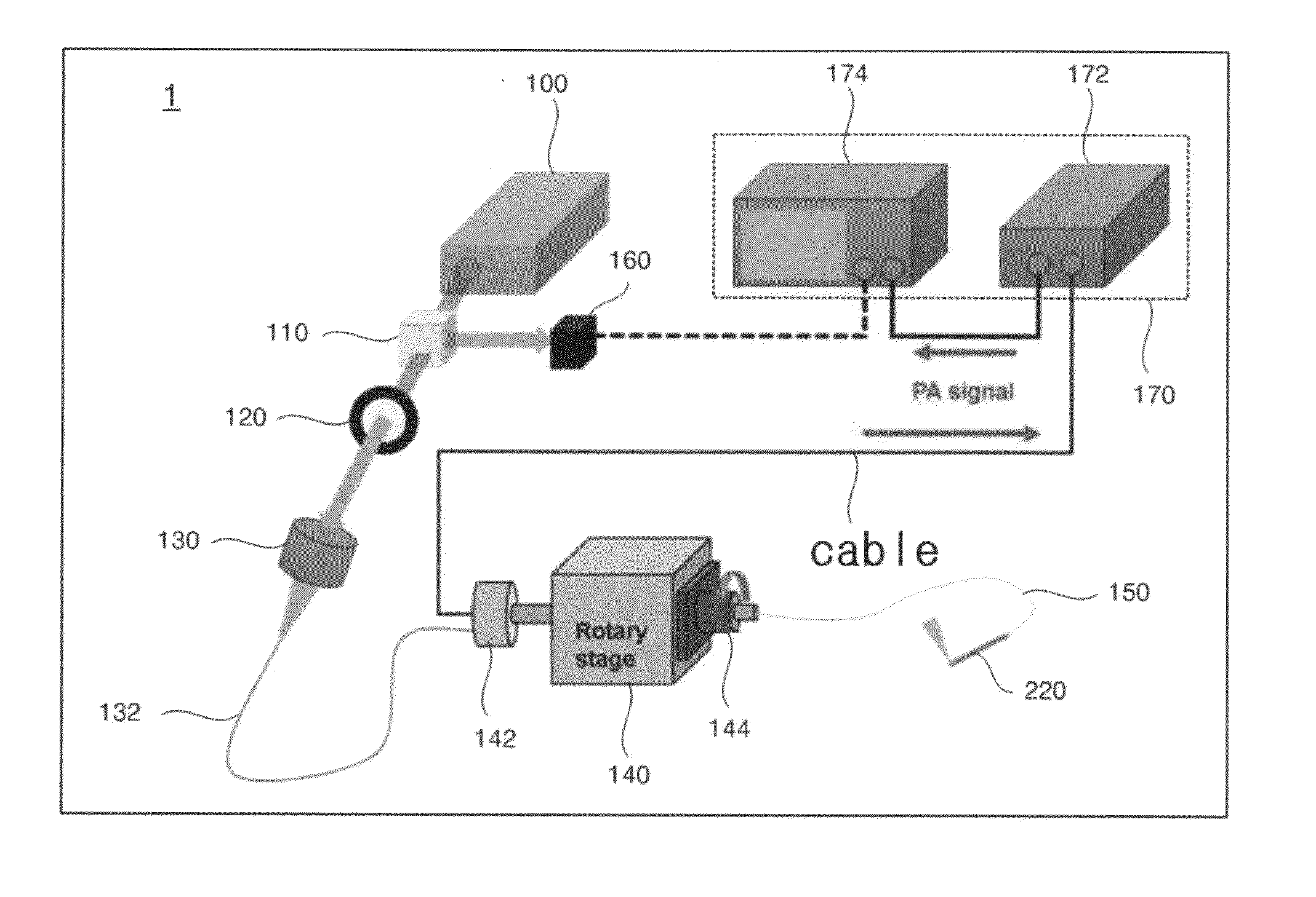
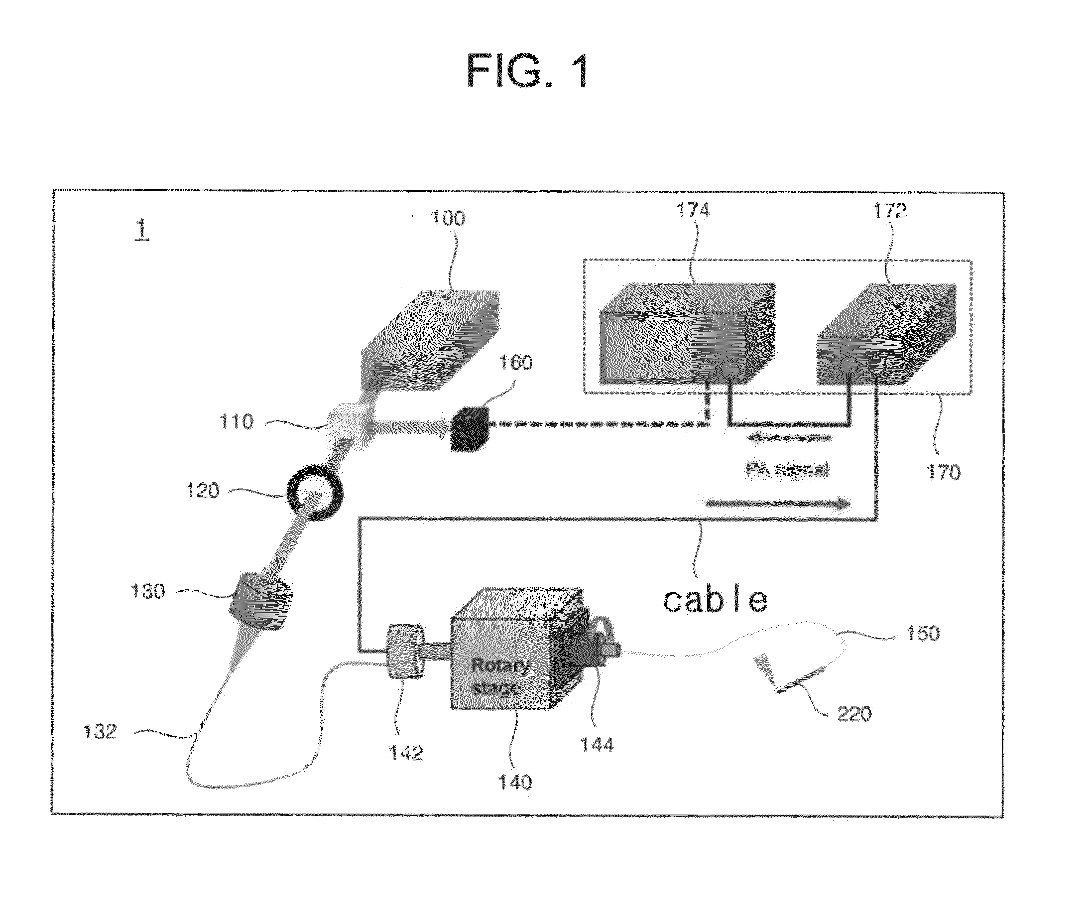
[0034]FIG. 1 is a diagram illustrating overall configuration of the ultrasound / photoacoustic image acquisition system using the catheter for detection of ultrasound and photoacoustic signals according to the present invention. Referring to FIG. 1, the ultrasound / photoacoustic image acquisition system 1 according to the present invention is configured to include a light source 100, a beam splitter 110, a lens 120, a ...


PUM


Abstract

Provided are a catheter for detection of ultrasound and photoacoustic signals and an image acquisition system using the catheter. The catheter for detection of ultrasound and photoacoustic signals includes a lens optical fiber where a lens is attached to an end of an optical fiber, an ultrasound transducer, a catheter main body where the lens optical fiber and the cable are disposed to pass through an inner portion thereof, a catheter head which is connected to an end of the catheter main body, and a membrane which surrounds a surface of the catheter head.
The catheter is configured so as to irradiate condensed light by using the lens optical fiber and to receive the photoacoustic signal or to receive and transmit the ultrasound signal. Accordingly, it is possible to simultaneously acquire an ultrasound image and a high-resolution photoacoustic image.

Description

CROSS-REFERENCE TO RELATED PATENT APPLICATION[0001]This application claims the benefit of Korean Patent Application No. 10-2014-0144082, filed on Oct. 23, 2014, in the Korean Intellectual Property Office, the disclosure of which is incorporated herein in its entirety by reference.BACKGROUND OF THE INVENTION[0002]1. Field of the Invention[0003]The present invention relates to a catheter for detection of ultrasound and photoacoustic signals and, more particularly, to an endoscopic catheter capable of simultaneously an ultrasound signal and a photoacoustic signal and an image acquisition system capable of acquiring a superimposed image of ultrasound and photoacoustic images by using the endoscopic catheter.[0004]2. Description of the Prior Art[0005]In the related art, a blood vessel disease diagnosis apparatus using ultrasound inside a blood vessel displays a current position and an ultrasound image of a cross section of an inner wall of the blood vessel by using the ultrasound. By usi...

Claims


Application Information

Patent Timeline
Patent Type & Authority Applications(United States)
IPC IPC(8): A61B8/00A61B5/00A61B8/08A61B8/12
CPCA61B8/4416A61B8/12A61B8/4483A61B8/5261A61B8/445A61B5/0095A61B5/02007A61B5/6852A61B1/00096A61B1/05A61B1/07
Inventor KIM, CHUL HONGPARK, SUNG JOCHOI, CHANG HOON
Owner POSTECH ACAD IND FOUND
Why Eureka
Try Eureka