Methods for phenotyping of intact whole tissues

a whole tissue and tissue phenotyping technology, applied in the field of tissue preparation and characterization, can solve the problem of lack of a whole-organ imaging method

Active Publication Date: 2016-05-05
CALIFORNIA INST OF TECH
View PDF2 Cites 8 Cited by
  • Summary
  • Abstract
  • Description
  • Claims
  • Application Information

AI Technical Summary

Benefits of technology

[0133]The various methods and techniques described above provide a number of ways to carry out the invention. Of course, it is to be understood that not necessarily all objectives or advantages described can be achieved in accordance with any particular embodiment described herein. Thus, for example, those skilled in the art will recognize that the methods can be performed in a manner that achieves or optimizes one advantage or group of advantages as taught herein without necessarily achieving other objectives or advantages as taught or suggested herein. A variety of alternatives are mentioned herein. It is to be understood that some preferred embodiments specifically include one, another, or several features, while others specifically exclude one, another, or several features, while still others mitigate a particular feature by inclusion of one, another, or several advantageous features.

Problems solved by technology

At the root of this knowledge gap is the absence of a method that enables whole-organ imaging.

Method used


Image

  • Methods for phenotyping of intact whole tissues
  • Methods for phenotyping of intact whole tissues
  • Methods for phenotyping of intact whole tissues

Examples


example 1

Results

Method for Passive Clearing and Immunostaining of Whole Organs

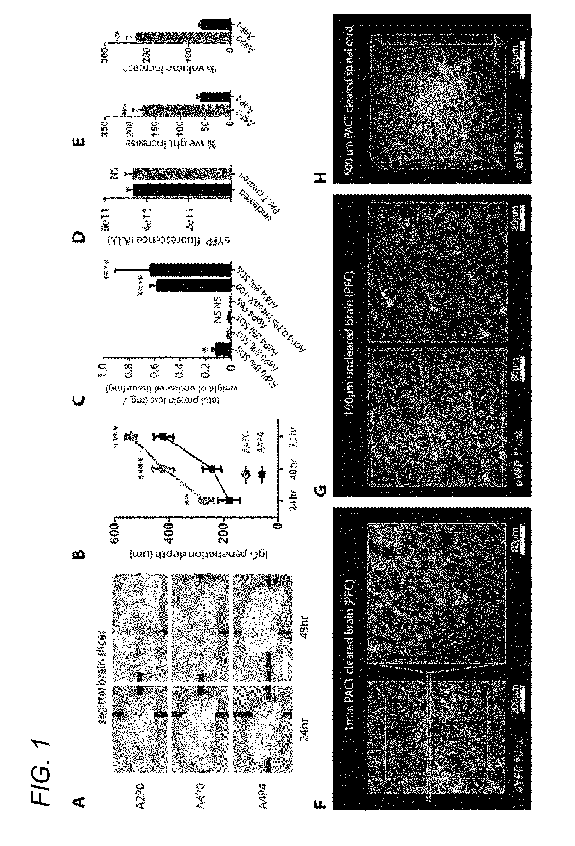
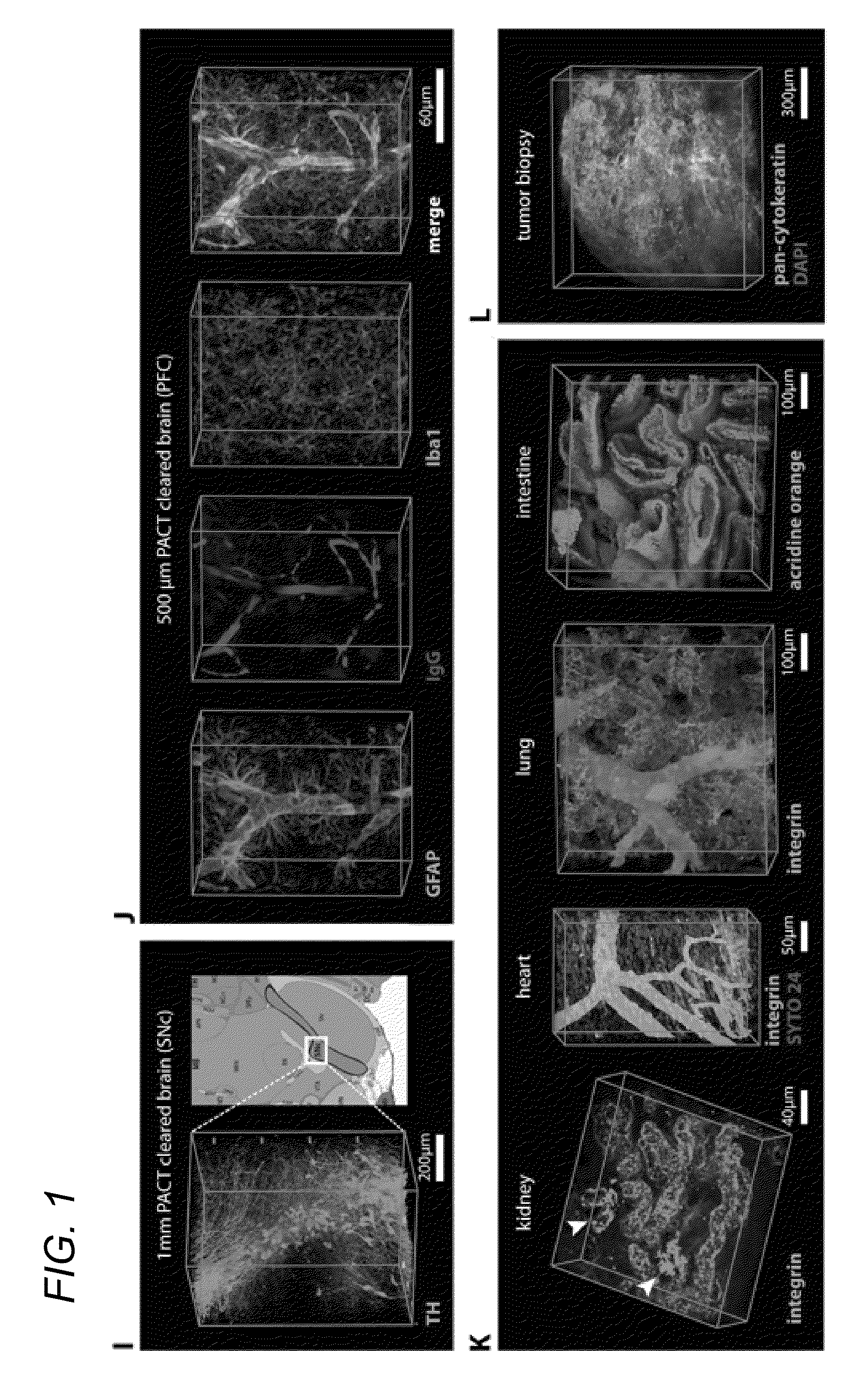
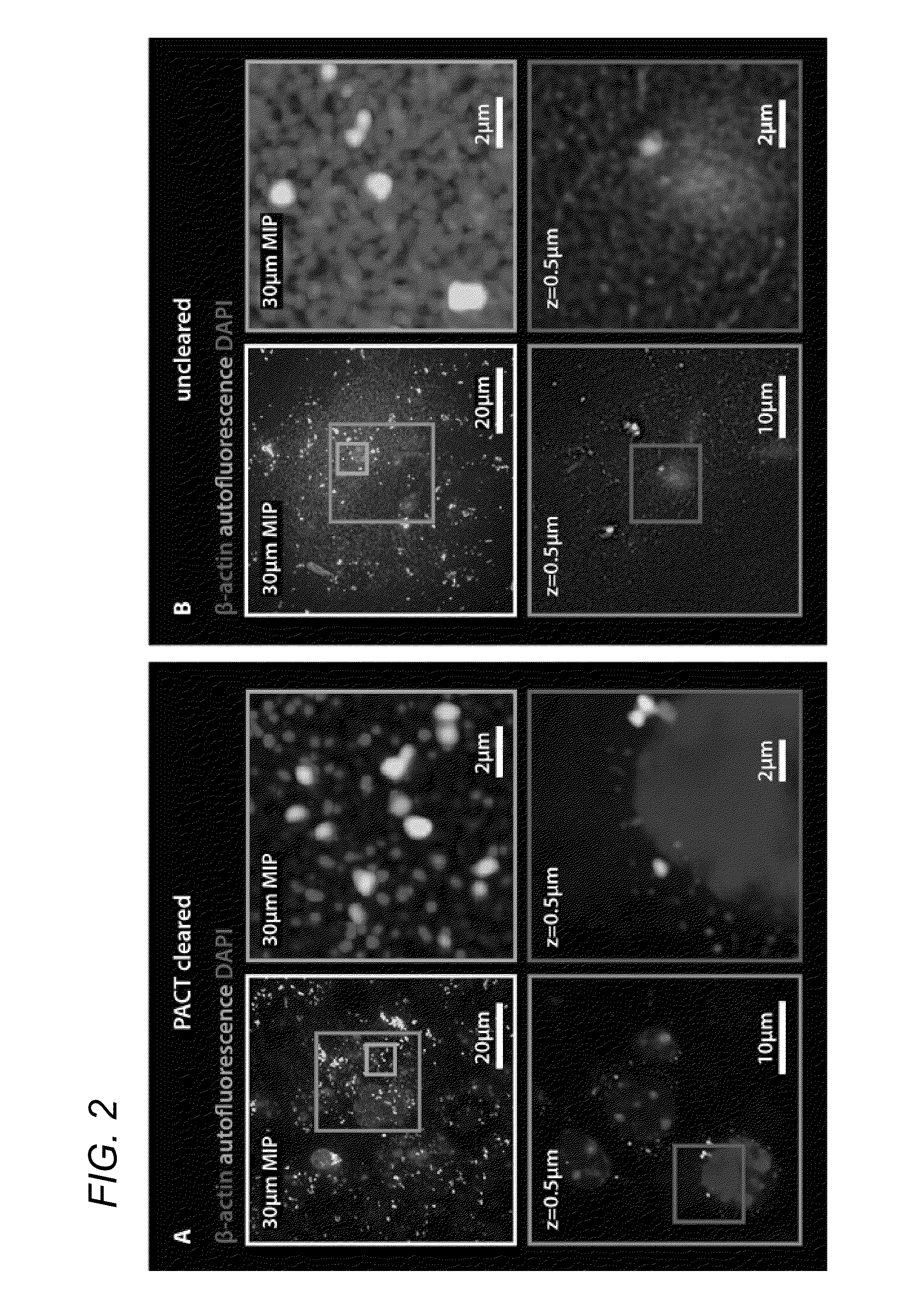
[0052]In some embodiments, thick tissue is rendered optically transparent for imaging in three main steps. First, tissue is cross-linked and hybridized to hydrogel monomers to stabilize biomacromolecules. Second, tissue lipids are extracted from the tissue-hydrogel matrix with ionic detergent(s). Third, cleared tissue is embedded in RIMS for imaging, or for long-term storage. Although wholebody clearing was the primary goal, it was recognized that the processing of small or particularly fragile specimens and organs would best be accomplished by a mild, passive clearing protocol. PACT was developed for rendering rodent whole organs, their 1-3 mm thick sections, including brain, spinal cord, kidney, heart, lung, and intestine, or human tissue biopsies transparent. The clearing speed depends in part on the rate of lipid solvation by detergent micelles, and the rate of diffusion of detergent micelles in tissue (Hoffman...

example 2

Discussion

[0066]In some embodiments, the invention teaches PARS, a method that renders intact whole-organisms transparent for imaging with single cell resolution while preserving fluorescent and protein-based signals and tissue architecture. The starting point, the CLARITY method (Chung et al., 2013) provided scientists with a brain-processing platform for elucidating the 3D cellular arrangement and connectome in toto. Numerous laboratories have previously reported on new clearing reagents in the decade before CLARITY, however many of these reagents were highly application- or tissue-specific (summarized in Table 1). In contrast, CLARITY introduced two broadly applicable techniques pertaining to tissue preservation (hydrogel embedding) and clearing efficiency (electrophoretic tissue clearing, ETC), both of which could be incorporated into the design, or redesign, of other clearing procedures.

[0067]Traditionally, making tissue transparent was a process that demanded solvent incubatio...

example 3

Methods

PACT Clearing

[0085]4% paraformaldehyde (PFA)-fixed tissue sections were incubated at 4° C. overnight in the hydrogel monomer solution A4P0 (4% acrylamide in PBS) supplemented with 0.25% photoinitiator 2,2′-Azobis[2-(2-imidazolin-2-yl)propane]dihydrochloride (VA-044, Wako Chemicals USA, Inc.). A4P0-infused samples were degassed with nitrogen for 1-5 minutes and then incubated for 2-3 hours at 37° C. to initiate tissue-hydrogel hybridization. After removing excess hydrogel via brief PBS washes, tissue-hydrogel matrices were transferred into 50 mL conical tubes containing 8% SDS in 0.1M PBS (pH 7.5), and depending on tissue size, were incubated for 2-5 days at 37° C. with shaking. For immunostaining, 1-3 mm thick PACT-processed samples were washed in PBS with 4-5 buffer changes over the course of a day and then transferred to buffer containing small-molecule dyes or primary antibodies followed by fluorescently-conjugated secondary antibody (1:200-400, in PBS containing 2% normal...


PUM

PropertyMeasurementUnit
depthaaaaaaaaaa
imaging stack depthaaaaaaaaaa
depthaaaaaaaaaa

Abstract

In various embodiments, the present application teaches methods and compositions for tissue clearing in which whole organs and bodies are rendered macromolecule-permeable and optically-transparent, thereby exposing their cellular structure with intact connectivity. In some embodiments, the present application teaches PACT, a protocol for passive tissue clearing and immunostaining of intact organs. In other embodiments, the present application teaches RIMS, a refractive index matching media for imaging thick tissue. In yet other embodiments, the application teaches PARS, a method for whole-body clearing and immunolabeling.

Description

CROSS REFERENCE TO RELATED APPLICATIONS[0001]This application is a divisional of U.S. application Ser. No. 14 / 447,607, filed Jul. 30, 2014, currently pending, which claims priority under 35 U.S.C. §119(e) from U.S. Provisional Application No. 61 / 880,401, filed Sep. 20, 2013, now expired, and U.S. Provisional Application No. 61 / 992,103, filed May 12, 2014, now expired, the contents of all of which are hereby incorporated herein by reference in their entirety.FIELD OF THE INVENTION[0002]The present invention generally relates to the field of tissue preparation and characterization.BACKGROUND[0003]The following description includes information that may be useful in understanding the present invention. It is not an admission that any of the information provided herein is prior art or relevant to the presently claimed invention.[0004]Understanding the structure-function relationships at cellular, circuit, and organ-wide scale requires 3D anatomical and phenotypical maps, currently unavai...

Claims


Application Information

Patent Timeline
Patent Type & Authority Applications(United States)
IPC IPC(8): G01N1/30
CPCG01N2001/302G01N1/30G01N33/57492G01N2001/305
Inventor GRADINARU, VIVIANAYANG, BIN
Owner CALIFORNIA INST OF TECH
Why Eureka
Try Eureka