
Processes for producing stable exosome formulations

a technology of exosome and formulation, which is applied in the direction of drug composition, inorganic non-active ingredients, skeletal/connective tissue cells, etc., can solve the problems of increasing the number of heart attack survivors and the overall global economic burden of ischemic heart diseas

Inactive Publication Date: 2016-06-09
CAPRICOR
View PDF4 Cites 21 Cited by
  • Summary
  • Abstract
  • Description
  • Claims
  • Application Information

AI Technical Summary

Benefits of technology

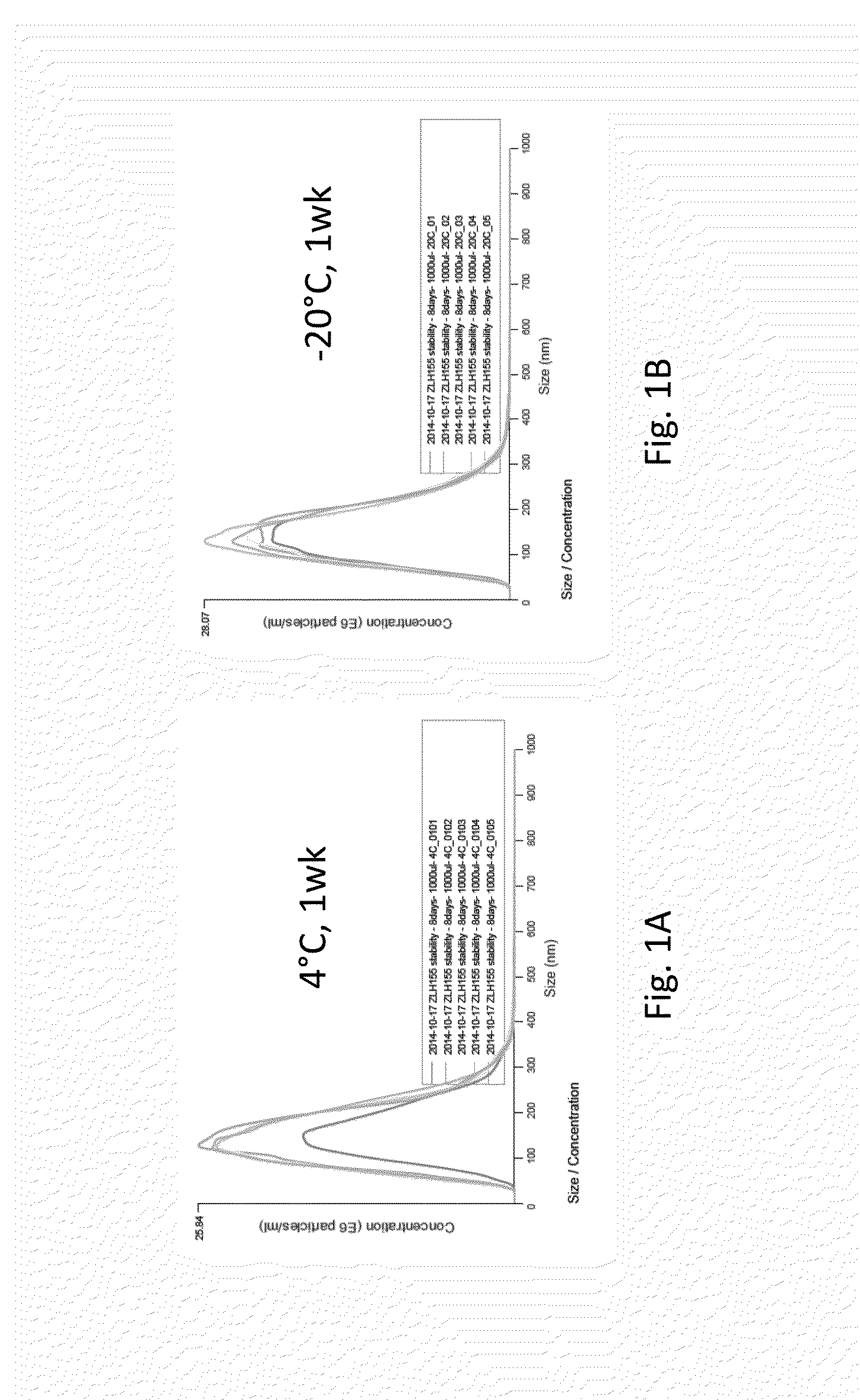
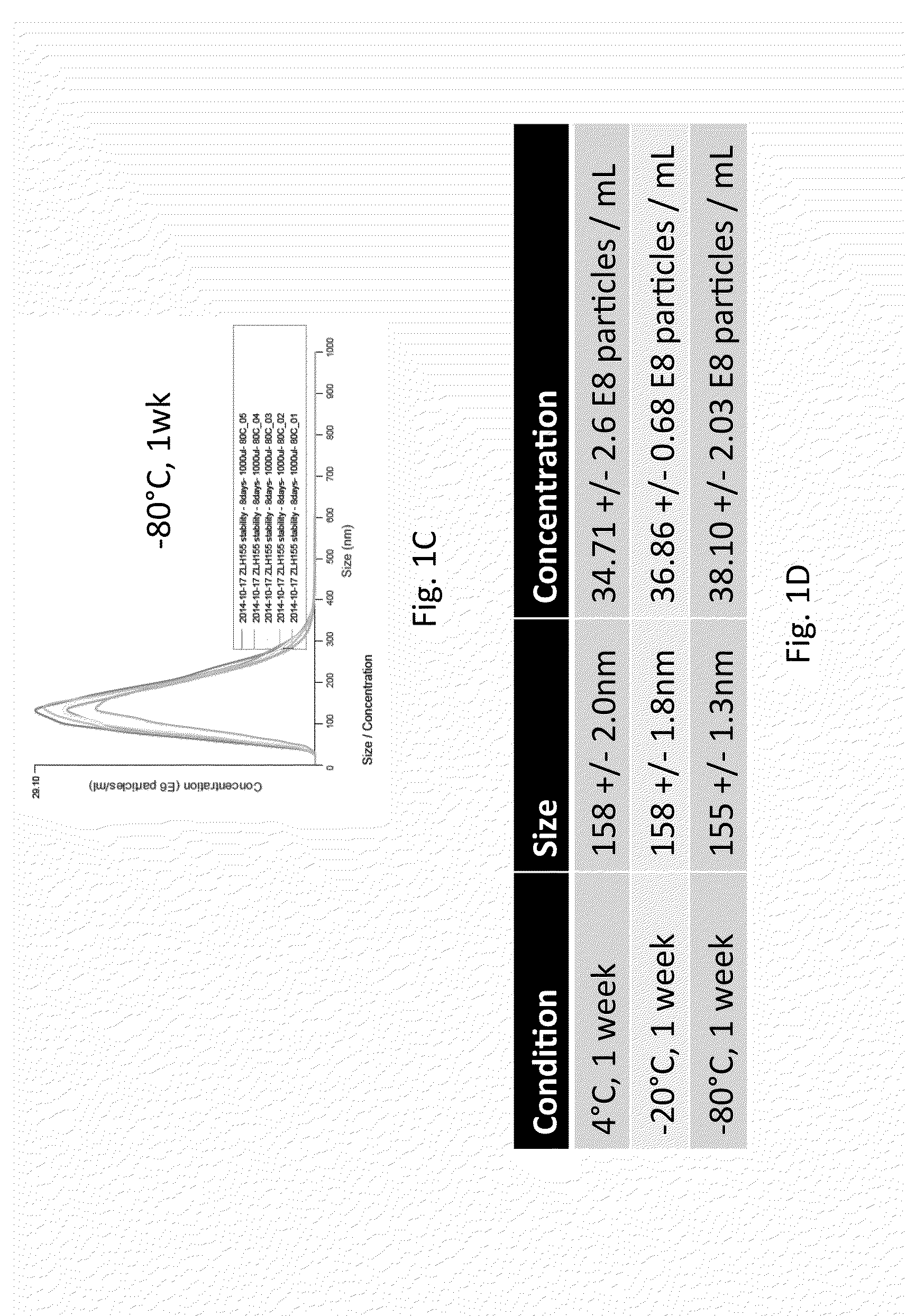
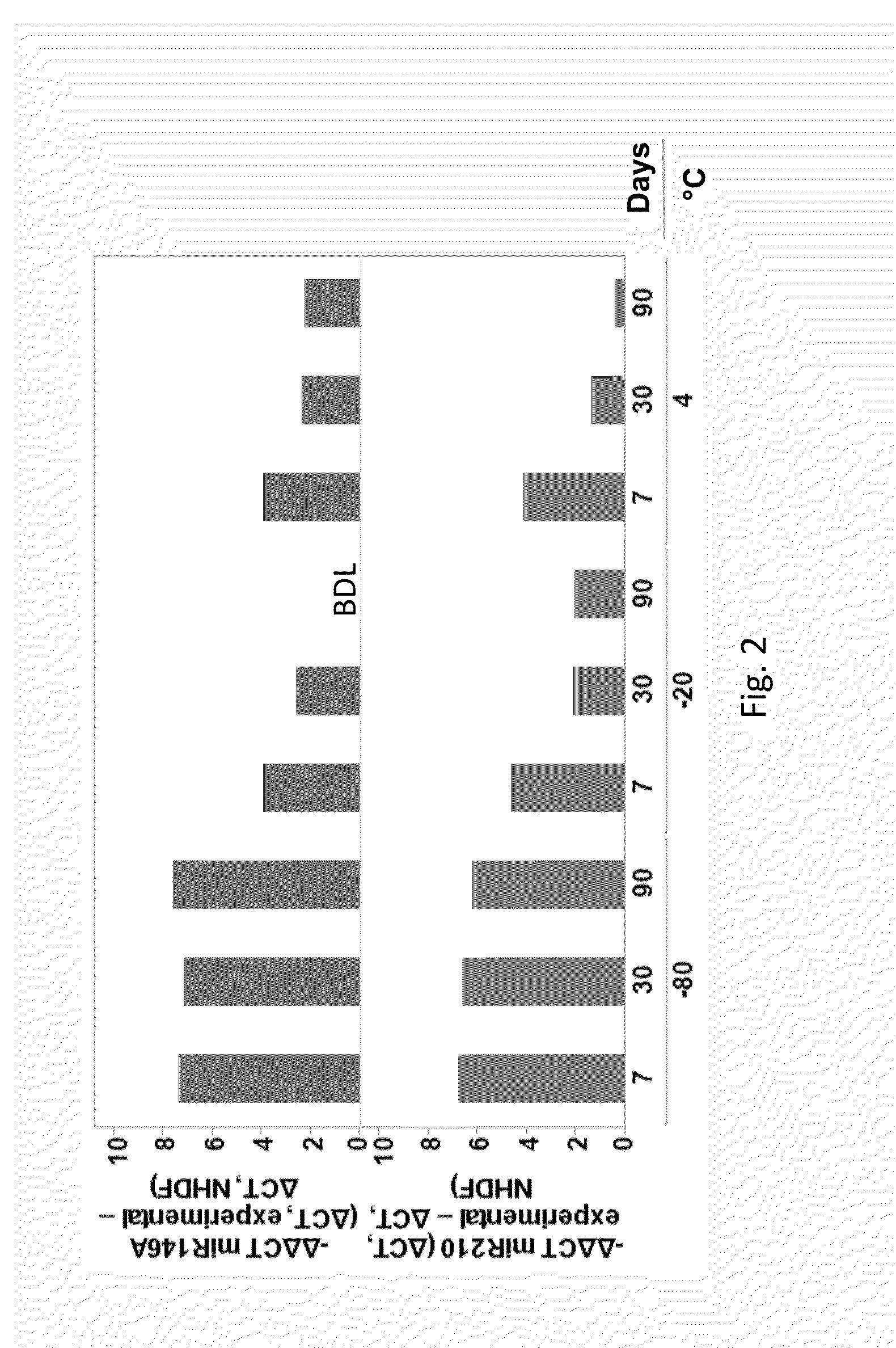
This patent describes methods for creating stable exosome formulations for use in humans. These formulations contain at least 106 exosomes that maintain the levels of certain molecules, such as miR-210 RNA and miR-146A. The exosomes are able to maintain biological activity even after being lyophilized (freeze-dried) or stored at 4°C for 30 days. The patent also describes a stable liquid formulation of cardiosphere-derived cell (CDC) exosomes that maintain miR-210 RNA and miR-146A levels for at least 75% of the starting formulation. This stable formulation can be generated from CDCs through ultrafiltration and diafiltration. Overall, the patent provides technical means for creating stable and biologically active exosome formulations for therapeutic use.

Problems solved by technology

However, the result is an increasing number of heart attack survivors and disability years due to nonfatal ischemic heart disease, which contributes greatly to the overall global economic burden of ischemic heart disease.

Method used


Image

  • Processes for producing stable exosome formulations
  • Processes for producing stable exosome formulations
  • Processes for producing stable exosome formulations

Examples


example 1

Exosome Preparation

[0109]Immediately upon receipt, hearts were grossly dissected and cut into biopsy-sized pieces of about 25 mg each (500 μmπl×500 μπl×500 μπl; though in some embodiments, other sizes are used), referred to as explants. Human hearts were cut using an automated tissue slicer (Zimmer® Dermatome) and automated tissue chopper (Mcllwain™ Tissue Chopper, Ted Pella, Inc.) as previously described (see e.g. United States Patent US20150216905 A1). Explants were then processed as previously described (see e.g., Smith et al. 2007 and U.S. patent application Ser. No. 11 / 666,685, filed Apr. 21, 2007 and Ser. No. 13 / 412,051, filed Mar. 5, 2012, the entireties of each of which are incorporated by reference herein).

[0110]In order to generate allogeneic CDCs, explants were plated on CELLBIND® CellSTACK® vessels (Corning Life Sciences). After 1-2 weeks, cellular outgrowth emerging from the explants became confluent. These explant derived cells (EDCs) were harvested using IX TrypLE™ (I...

example 2

Exosome Isolation

[0112]Exosomes were filtered using a 0.45 μm to remove cellular debris and then isolated by ultrafiltration based on size (2 kda to 30 kda), polyethylene glycol precipitation or Exoquick (SBI, Mountain View, Calif.). In certain situations, isolated exosomes were filter sterilized with a 0.22 μm microbial exclusion filter. Exosomes were formulated using several diafiltrations to replace the buffer to an acceptable infusion solution (e.g. PLASMALYTE, RINGERS solutions).

example 3

Exosome Analysis

[0113]Exosomal protein was assessed using DC assay (Bio-Rad, Hercules, Calif.). Exosome particle size and concentration was assessed using Brownian motion and the Nanosight tracking analysis (Malvern Instruments Ltd, Malvern UK). RNA was isolated using miRNeasy micro kit (Qiagen, Valencia, Calif.) and quantified using either the Nanodrop, Qubit or AATI fragment analyzer (Advance Analytics, Ankeny, Iowa). Reverse transcription and qPCR reactions were conducted using TaqMan miR probes (ThermoFisher Scientific, Grand Island, N.Y.).


PUM

PropertyMeasurementUnit
Temperatureaaaaaaaaaa
Fractionaaaaaaaaaa
Fractionaaaaaaaaaa

Abstract

The invention encompasses methods for generating stable exosome formulations and encompasses stable exosome formulations. The exosome formulations encompass stable liquid exosome formulations and stable lyophilized exosome formulations. In some embodiments, the exosome formulations can be generated by ultrafiltration and diafiltration. The exosome formulations can be suitable for administration to a human.

Description

BACKGROUND OF THE INVENTION[0001]Exosomes are cell-derived vesicles. Hong et al., PLoS ONE 9(8): e103310. doi:10.1371 / journal.pone.0103310. They are found in biological fluids, such as urine, plasma, and ascites. Id. Exosomes are generated by inward budding of endosomal multivesicular bodies. Id. The cargo of exosomes includes proteins / glycoproteins expressed on the cell membrane as well as molecules and soluble factors present in the cytosol of parental cells. Id. Exosomes normally have diameters ranging from 40-100 nm. Zhang et al., Oncology Letters 8: 1701-1706, 2014. Exosomes contain special proteins, lipids, RNA and micro-RNAs. Id.[0002]Exosomes produced from cardiosphere-derived cells enhance angiogenesis and promote cardiomyocyte survival and proliferation. Ibrahim et al., 2014 May 8; 2(5):606-19, which is hereby incorporated by reference. Exosomes produced from cardiosphere-derived cells are enriched in miR146a.[0003]The leading cause of death in the US remains heart disease...

Claims


Application Information

Patent Timeline
IPC IPC(8): A61K35/34C12N15/113
CPCA61K35/34C12N15/113C12N2320/30C12N2330/10C12N2310/141A61K9/08A61K9/19A61K47/02A61P9/00A61P43/00C12N5/0657C12N2320/32C12N2533/52C12N2500/02
Inventor KREKE, MICHELLESMITH, RACHELHANSCOME, PETERPECK, KIELIBRAHIM, AHMED
Owner CAPRICOR
Learn More