Spectroscopy for gunshot residue analysis

a technology of spectroscopy and gunshot residue, applied in the direction of material analysis through optical means, instruments, weapons, etc., can solve the problems of difficult recovery, detection and preservation of discharge samples, inability to perform sem analysis of tape collection substrates, and relatively expensive instruments and time-consuming analyses, etc., to achieve more sensitive detection, time and cost-effective, and more labor.

Active Publication Date: 2016-08-18
THE RES FOUND OF STATE UNIV OF NEW YORK
View PDF2 Cites 5 Cited by
  • Summary
  • Abstract
  • Description
  • Claims
  • Application Information

AI Technical Summary

Benefits of technology

[0010]A spectroscopic method of detecting inorganic gunshot residue particles provides several improvements over current elemental analyses used for GSR detection. This method is more labor, time and cost effective. Furthermore, the method of the present invention will allow for more sensitive detection of inorganic gunshot residue particles. Additionally, detection of explosive particles (2,4-DNT) will be useful in the fields of home land security and counter-terrorism.
[0011]A novel approach for GSR detection via microscopic-attenuated total reflectance (ATR) Fourier transform infrared (FT-IR) spectroscopic imaging was investigated by applicants. ATR-FT-IR analysis of GSR is nondestructive, with analysis times competitive to current methodologies, and offers a molecular “fingerprint” of the detected GSR. The vibrational signatures collected from the tape substrate and GSR particles (both OGSR and IGSR) are easily discernible by the naked eye. Furthermore, this vibrational “fingerprint” targets a wider range of chemicals compared to current methodologies, increasing the selectivity of the method. The optics of ATR-FT-IR imaging provides pseudo-immersion analysis. The high refractive index (4.0) of the germanium ATR crystal increases the numerical aperture of the optics, enhancing spatial resolution by a factor of 4, without the use of a synchrotron light source (“ATR Accessories An Overview,”PerkinElmer Life and Analytical Sciences (2004), which is hereby incorporated by reference in its entirety). Analysis is performed in ambient conditions and may detect GSR regardless of debris collection. Exhaustive research applying micro-ATR-FT-IR chemical imaging (mapping) to the fields of bio-medical (Chan et al., Appl. Spectrosc. 59:149 (2005); Anastassopoulou et al., Vibrational Spectroscopy 51:270 (2009); Kazarian et al., Biochimica et Biophysica Acta (BBA)—Biomembranes 1758:858 (2006); Kazarian et al., Analyst 138:1940 (2013), which are hereby incorporated by reference in their entirety) and forensic research (Dirwono et al., Forensic Sci. Int'l 199:6 (2010); Ng et al., Anal. and Bioanal. Chem. 394:2039 (2009); Spring et al., Anal. and Bioanal. Chem. 392:37 (2008), which are hereby incorporated by reference in their entirety) have been reported. GSR detection via micro-ATR-FT-IR chemical imaging has not previously been investigated, although it has been suggested (Ng et al., Anal. and Bioanal. Chem. 394:2039 (2009); Ricci et al., 6402:64020J (2006), which are hereby incorporated by reference in their entirety).

Problems solved by technology

Recovery, detection, and preservation of these discharge samples is often challenging.
Unfortunately, the collection of debris and fibers from certain surfaces, is known to inhibit SEM analysis of tape collection substrates, as these analytes are not electronically conductive (Mastruko, Forensic Sci.
Other pitfalls for the technique include relatively expensive instrumentation and time consuming analyses (Romolo et al., Forensic Sci.
However, the advent of heavy metal free (HMF) ammunition reduces the probability of detecting “characteristic” IGSR particles and weakens the specificity (rate of true negatives) of the technique for the identification of GSR.
Although, LA-ICPMS was reported to be capable of analyzing a 12.8 mm2 area in approximately 66 minutes, it unfortunately required expensive instrumentation and was also dependent upon detection of heavy metals.
However, these methods have been limited to chemical characterization of GSR.

Method used


Image

  • Spectroscopy for gunshot residue analysis
  • Spectroscopy for gunshot residue analysis
  • Spectroscopy for gunshot residue analysis

Examples


example 1

GSR Collection

[0060]GSR samples were generated by discharging 0.38 in. caliber Winchester brand “0.38 special” ammunition from a Smith and Wesson Model 10 Revolver and 0.40 in. Federal brand “S&W” full metal jacketed ammunition from a Smith and Wesson M&P 40 firearm. The firearms were discharged approximately 0.3 m away from a cloth collection substrate. For real world applications of this method, the cloth substrate mimics the clothing of a shooting victim or suspect. 3M brand double-sided PSA tape (common office tape) was utilized for tape lifting. Pieces of tape were adhered to a 1×1 in. glass microscope slide. PSA tape is composed of an acrylic polymer adhesive layer, supported on a hydrocarbon backbone (Toyama et al., J. of Applied Polymer Sci. 17:3495 (1973), which is hereby incorporated by reference in its entirety). The available side of the adhesive tape was pressed against the cloth discharge substrate to collect the GSR particles. Several macroscopic (>500 μm diameter) GS...

example 2

ATR-FT-IR Microscope

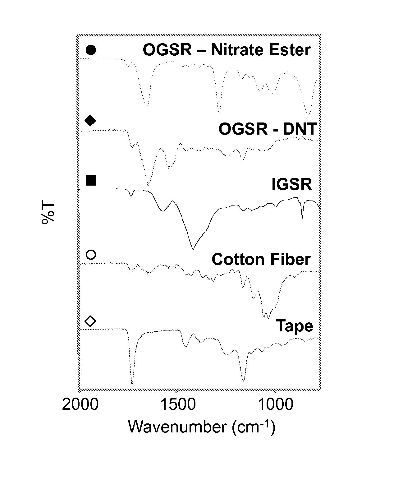
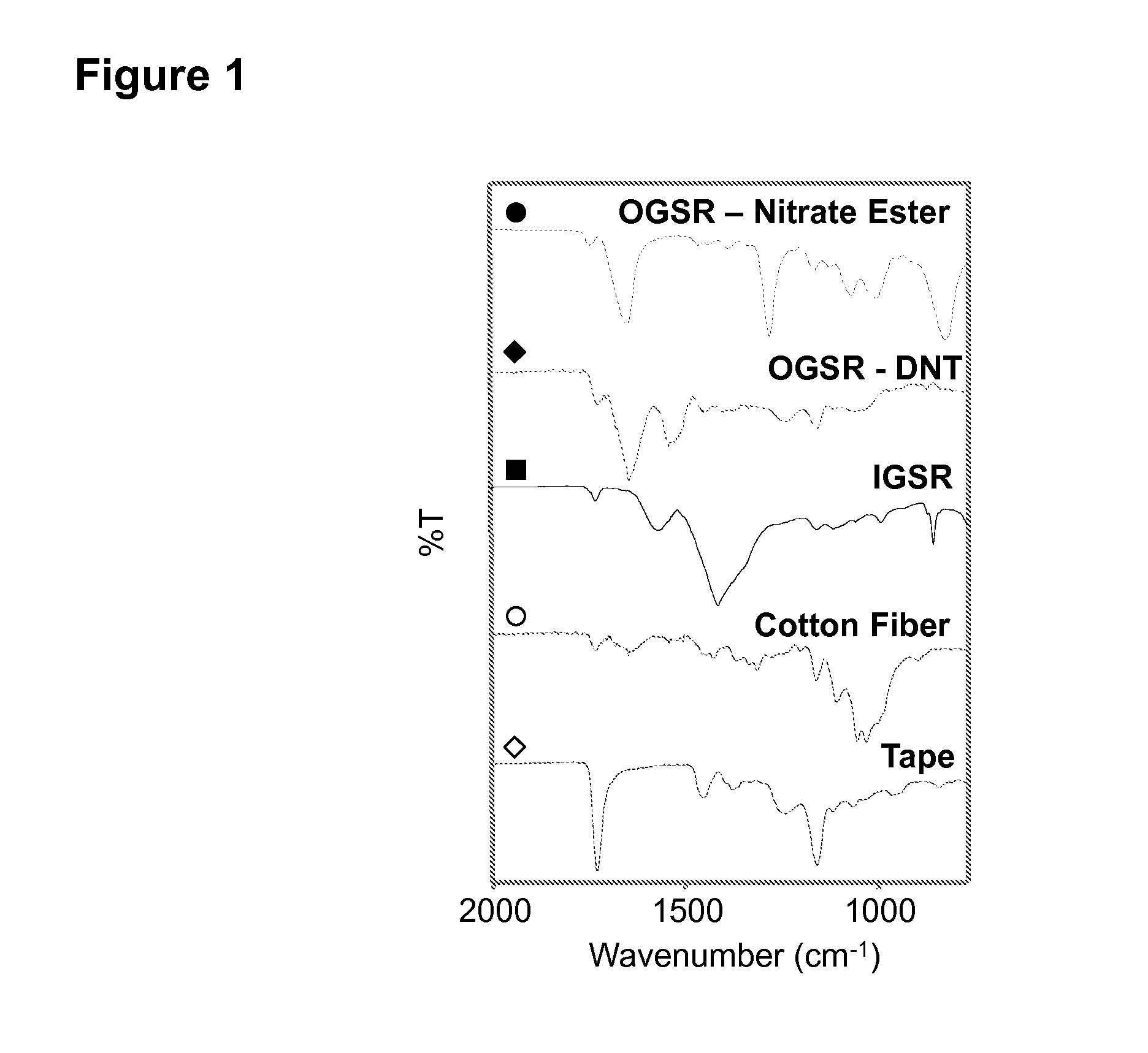
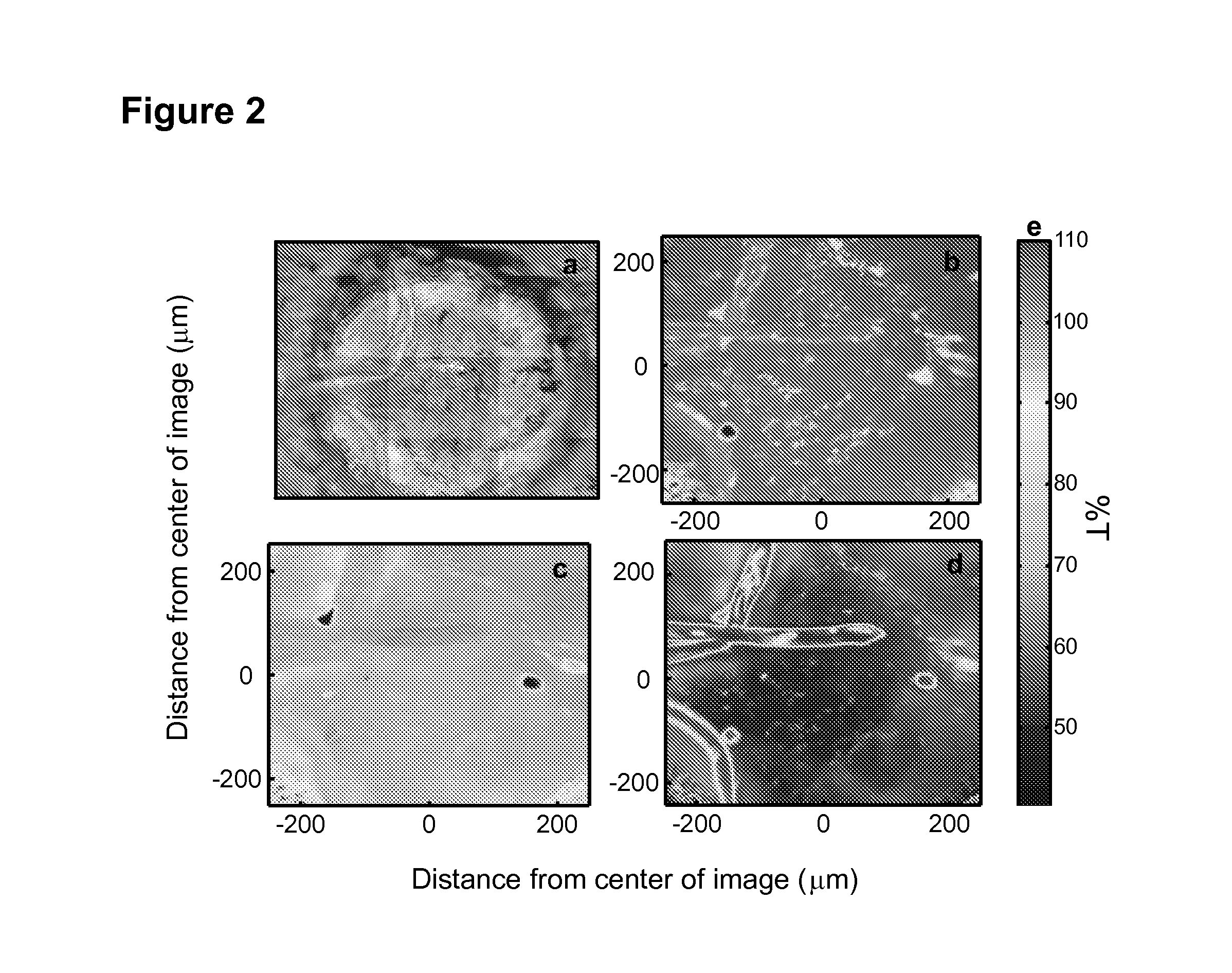
[0061]Micro-ATR-FT-IR imaging was performed with a Perkin Elmer Spotlight 400 IR Microscope and Spectrum 10 software. The 1×1 in. glass microscope slides with the adhered tape and GSR samples were placed under the micro-ATR objective. The micro-ATR objective utilized to collect the IR images was composed of a germanium crystal with a 600 μm tip. The tip of the Ge crystal was kept in constant contact with the surface of the samples due to a spring mechanism. This mechanism allowed the crystal to navigate the three-dimensional sample surface. The depth of penetration of the evanescent wave was 0.65 μm. 500 μm2 areas of the tape substrate were mapped individually. A mercury cadmium telluride (MCT) detector array allowed for the collection of an ATR-FT-IR spectrum every 1.56 μm (pixel size). 320 spectra were collected in both the X and Y directions, and, therefore, each IR image is the representation of 102,400 pixels, each corresponding to one ATR-FT-IR spectrum. Th...

example 3

Scanning Electron Microscope

[0062]For SEM analysis, OGSR particles with diameters of approximately 500 μm were manually deposited on adhesive carbon discs obtained from Ted Pella, Inc. The discs were mounted on SEM stubs. Police typically use these adhesive discs for GSR collection and detection, since they are conductive, stable under an electron beam and in a vacuum. All of these properties are required for SEM analysis. SEM / EDS analysis was performed by Evans Analytical Group, Inc. A Hitachi 3400-N Variable Pressure SEM with INCA software (Oxford Instruments, Inc.) was used for the acquisition of electron images and collection of elemental spectra (EDS) from several OGSR particles. A magnification of 100× was used to obtain the SEM images. An accelerating voltage of 20 kV was utilized. EDS spectra were collected over a range of 0 to 10 keV.


PUM

PropertyMeasurementUnit
sizeaaaaaaaaaa
sizeaaaaaaaaaa
sizeaaaaaaaaaa

Abstract

The present invention relates to a method of detecting inorganic gunshot residue (GSR) particles. The method includes providing a sample comprising gunshot residue, subjecting the sample to spectroscopic analysis to produce a spectroscopic signature for the sample, and identifying inorganic gunshot residue particles based on the spectroscopic signature for the sample. Also disclosed is a method of detecting gunshot residue particles. The method includes providing a sample comprising gunshot residue, subjecting the sample to spectroscopic analysis to produce a spectroscopic signature for the sample, where the spectroscopic signature spans a range of wavenumbers, creating one or more spectroscopic maps from the spectroscopic signature for the sample, where each different spectroscopic map is for a different wavenumber, and identifying gunshot residue particles based on the one or more spectroscopic maps for the sample.

Description

[0001]This application claims benefit of U.S. Provisional Patent Application Ser. No. 61 / 887,059, filed Oct. 4, 2013, which is hereby incorporated by reference in its entirety.FIELD OF THE INVENTION[0002]The present invention relates to spectroscopy for gunshot residue analysis.BACKGROUND OF THE INVENTION[0003]Gunshot residue (GSR) particles represent the burnt and partially burnt chemical remains of ammunition expelled from a firearm discharge. GSR possesses evidentiary value which can assist investigators in shooting incident reconstruction (Haag, Shooting Incident Reconstruction; Elsevier: Amsterdam, (2006)). Organic GSR (OGSR) is composed primarily of explosive materials originating from the propellant of the discharged ammunition, while inorganic gunshot residue (IGSR) is formed from contributions of the ammunition propellant, primer, projectile (bullet), and cartridge case (Dalby et al., J. Forensic Sci. 55:924 (2010)). Detection of GSR at a crime scene is critical, as it is i...

Claims


Application Information

Patent Timeline
Patent Type & Authority Applications(United States)
IPC IPC(8): G01N21/35G01N21/552F42B5/00G01N21/65
CPCG01N21/552G01N21/658G01N2021/3595G01N2201/06113G01N21/3586F42B5/00G01N21/35G01N2021/653
Inventor LEDNEV, IGOR K.
Owner THE RES FOUND OF STATE UNIV OF NEW YORK
Why Eureka
Try Eureka