
Method for isolating apoptotic bodies

a technology for apoptosis and apoptosis cells, applied in the field of apoptosis, can solve the problems of sedimentation of all cellular blood components, inability to isolate up to 90% of all apoptotic bodies present in body fluids, and inability to maintain integrity

Active Publication Date: 2016-10-06
SEGURA MARTIN TOMAS +2
View PDF0 Cites 2 Cited by
  • Summary
  • Abstract
  • Description
  • Claims
  • Application Information

AI Technical Summary

Benefits of technology

The present invention provides a method for isolating and quantifying apoptotic bodies from body fluids, such as blood or urine, for the diagnosis and prognosis of vascular, neurodegenerative, and oncological diseases. The method involves centrifuging the sample and collecting the supernatant, resulting in the isolated apoptotic bodies. The invention also provides a method for identifying the cell type of a cell dead from apoptosis by detecting a cell type marker in the outside of the plasma membrane of an apoptotic body. The use of the method for evaluating the effectiveness of treatments for these diseases is also provided.

Problems solved by technology

However, the in vivo analysis of apoptosis that occurs in tissue has a clear limitation as it is currently necessary to obtain tissue samples of patients by interventionist methods, biopsy or surgery.
However, unlike what is achieved with the method of the present invention, with the conditions described in WO 03 / 076589 it is not possible to isolate up to 90% of all the apoptotic bodies present in the body fluids, nor maintain their integrity.
First, this is due to the fact that a centrifugation speed higher than 500 g, aimed at obtaining plasma or serum, leads to the sedimentation of all cellular blood components, among which are platelets.
If this occurs, the apoptotic bodies would be retained in the cell aggregates and centrifugation at a speed higher than 500 g would lead to their sedimentation with the cell fraction, and this would mean losing a large number of them.
Furthermore, the high speed centrifugation of plasma or serum obtained from the first centrifugation could result in rupture of the apoptotic bodies and in their contamination with other smaller membrane vesicles.
Due to these drawbacks, the apoptotic bodies isolated by the method described in WO 03 / 076589 A2 have little or no utility for prognosis and / or monitoring of the treatment for subjects with diseases associated with apoptosis, as the number of isolated apoptotic bodies would be much lower than the real number in the body fluid and the apoptotic bodies could be disintegrated and / or contaminated by other microvesicles, resulting in an erroneous quantification of apoptotic bodies.

Method used


Image

  • Method for isolating apoptotic bodies
  • Method for isolating apoptotic bodies
  • Method for isolating apoptotic bodies

Examples


example 1

Isolation of Apoptotic Bodies. Purity

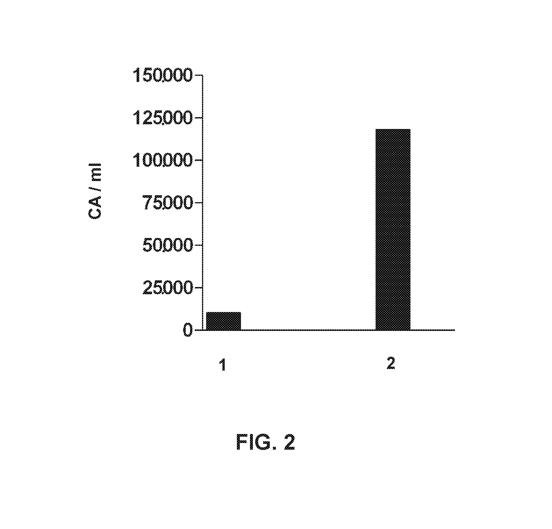
[0099]The trial includes 6 patients, two diagnosed with ischemic stroke (Patient 1: 85-year-old woman, Patient 2: 59-year-old man), two diagnosed with multiple sclerosis (Patient 3: 51-year-old woman, Patient 4: 32-year-old man), and two of Parkinson's disease (Patient 5: 61-year-old man, Patient 6: 68-year-old woman).

[0100]Blood (20 ml) from each patient was collected in the neurology service and deposited in tubes with sodium citrate. After less than 2 hours from extraction, the isolation of the apoptotic bodies was performed by means of serial centrifugations by the method of the first aspect of the invention, as indicated below:

[0101]Blood samples were subjected to centrifugation: 250 g for 10 min at 18° C. The plasma phase of the sample from patient 1 was diluted in a 1×TBS buffer in a ratio of 1:3 (step a1) and centrifuged at 850 g for 20 min at 18° C. (step b). The supernatant from the previous centrifugation was transferred to a clean pol...

example 2

Isolation of Apoptotic Bodies. Performance

[0104]The trial includes 2 patients, two diagnosed with ischemic stroke (patient 1: 76-year-old man, Patient 2: 58-year-old man).

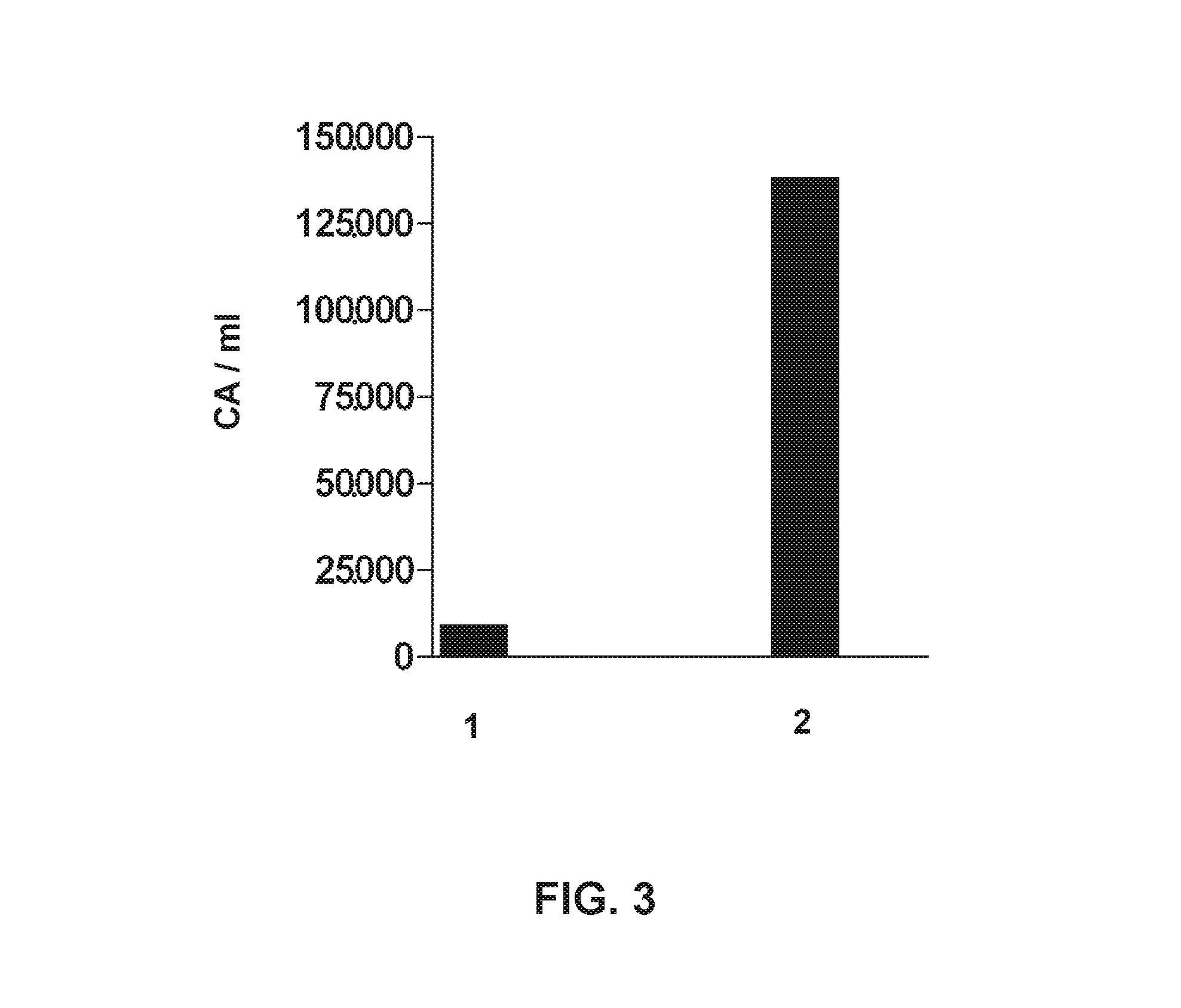
[0105]Blood (20 ml) from each patient was collected in the neurology service and deposited in tubes with sodium citrate. Within less than 2 hours after extraction, the isolation of apoptotic bodies was performed as follows:

[0106]Blood samples were subjected to centrifugation: 160 g for 20 min at 18° C. The plasma phase (containing the apoptotic bodies) of sample from patient 2 was transferred to a sterile polypropylene tube (step a) and centrifuged at 700 g for 30 min at 18° C. (step b). The plasma phase of the sample from patient 1 was diluted in a 1×TBS buffer in a ratio of 1:3 (step a1) and centrifuged at 700 g for 30 min at 18° C. (step b). The supernatant from the previous centrifugation was transferred to a clean polycarbonate tube and centrifuged at 14,000 g for 30 min at 9° C. (step c). The supernatant from...

example 3

Ischemic Stroke

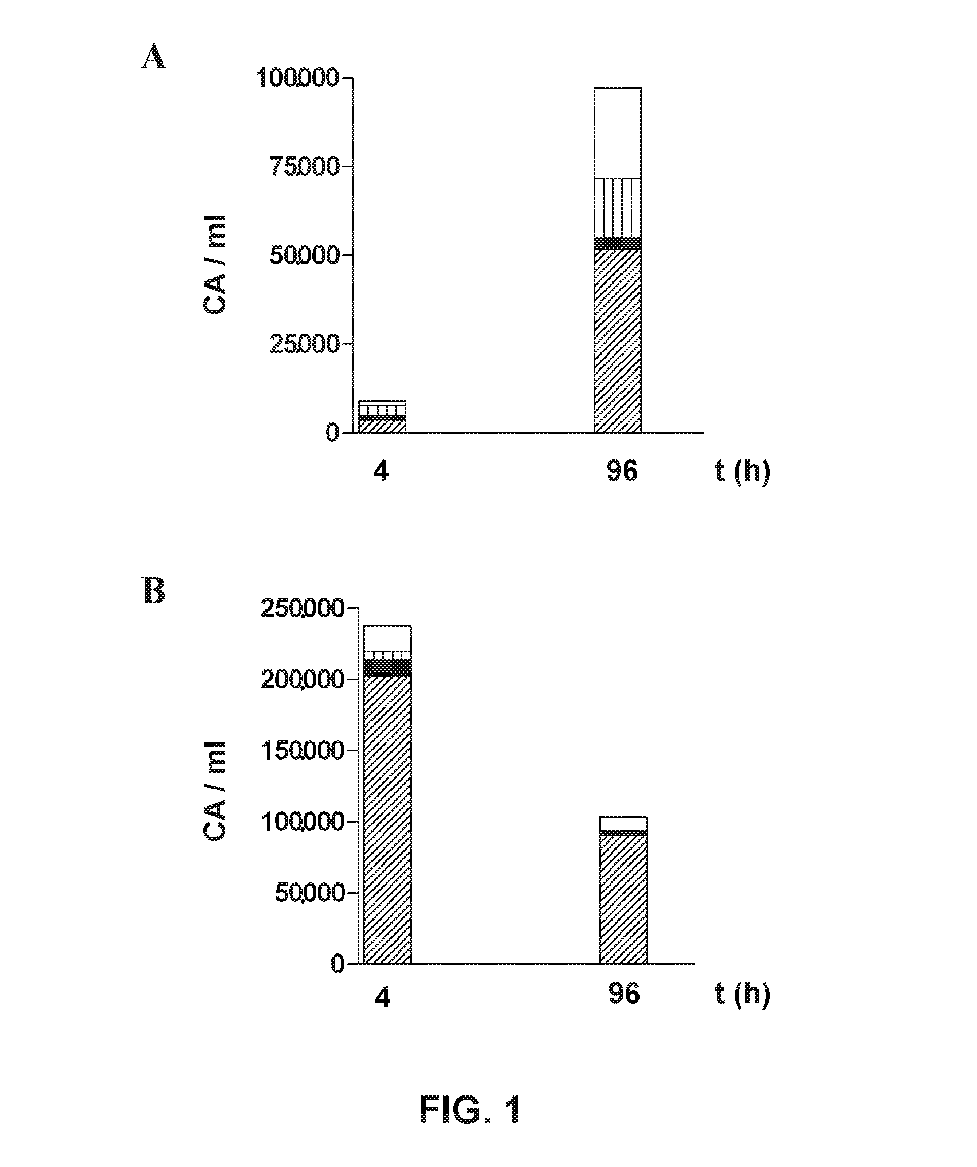
[0110]The trial includes two patients, a 78-year-old woman and a 70 year-old woman, who were attended by the Neurology service on suspicion of ischemic stroke. A simple CAT scan was performed under perfusion protocol, which showed ischemic injury in the area of the right middle cerebral artery in patient 1, and in the area of the left middle cerebral artery in patient 2. Below, are the reported clinical data for each patient;

[0111]aetiology as per TOAST classification:[0112]Patient 1: Cerebral infarction of undetermined origin.[0113]Patient 2: Cerebral infarction of cardioembolic origin.

[0114]radiological assessment: (i) initial (determination of the volume of initial infarction obtained from the CAT—perfusion test) and (ii) final, at 96 h from the onset of symptoms (a simple CAT scan was performed to determine final infarction volume).

[0115]Patient 1: initial infarction volume: 22,310 mm3, infarction volume at 96 h: 57,127 mm3. Ischemic lesion increased 2.5 folds dur...


PUM

PropertyMeasurementUnit
temperatureaaaaaaaaaa

Abstract

The invention pertains to the field of the study of apoptosis, especially the field of the prognosis and monitoring of diseases wherein apoptosis occurs. Concretely, the invention relates to a method for isolating apoptotic bodies from a sample of body fluid, and to methods for the prognosis and evaluation of the efficiency of a treatment for vascular, neurodegenerative and / or oncological diseases, based on the use of said method for isolating apoptotic bodies.

Description

TECHNICAL FIELD[0001]The present invention belongs to the field of apoptosis. Specifically, it relates to a method for the isolation of apoptotic bodies from body fluids and the use of said method and isolated apoptotic bodies for the evaluation of apoptotic processes, particularly in pathologies such as vascular, neurodegenerative and oncological diseases.BACKGROUND OF THE INVENTION[0002]Apoptosis is a kind of ordered cell death, which depends on the implementation of a cascade of molecular events that culminate in the total disintegration of the cell. In the late stage of apoptosis, and as a result of cell fragmentation, small membranous vesicles called apoptotic bodies are generated.[0003]Under physiological conditions, apoptosis is involved in morphogenesis and in tissue remodeling during the development, and the regulation of the immune system and homeostasis. However, this process also plays a key role in the cell and tissue death that occurs in various pathologies, such as va...

Claims


Application Information

Patent Timeline
Patent Type & Authority Applications(United States)
IPC IPC(8): G01N33/50
CPCG01N33/5091G01N2800/52G01N2800/32G01N2800/2835G01N2800/285G01N2800/324G01N2800/28G01N2333/705G01N2510/00G01N33/5005G01N2800/7028A61P25/00A61P35/00A61P9/00C12N5/0602G01N33/49
Inventor SEGURA MARTIN, TOMASAYO MARTIN, OSCARSERRANO DE LAS HERAS, GEMA
Owner SEGURA MARTIN TOMAS
Learn More