
Cardiac function monitor and/or intervention system attached outside or inside of heart

a technology of function monitor and intervention system, applied in the field of medical devices, can solve the problems of poor intuitive, microcosmic and accurate reflection, pulmonary congestion and peripheral edema, and the need to end invasive operations, so as to improve the condition of patients

Inactive Publication Date: 2017-04-20
ZHOU XIAOHUI
View PDF3 Cites 7 Cited by
  • Summary
  • Abstract
  • Description
  • Claims
  • Application Information

AI Technical Summary

Benefits of technology

The present invention is a device that combines monitoring and treatment for cardiac function. It can attach to the heart and accurately measure the electrical and biochemical properties of the heart's inner layer. This allows for specific treatment methods such as drug administration, pressure regulation, and electrical / magnetic stimulation. The device works to improve the condition of patients by monitoring and addressing the state of cardiac function.

Problems solved by technology

The basic manifestations of heart failure are dyspnea, fatigue, limited exercise tolerance and fluid retention, wherein the fluid retention may further lead to pulmonary congestion and peripheral edema.
However, these techniques indirectly and integrally reflect cardiac function and physiological condition, but are poor in intuitive, microcosmic and accurate reflection.
In addition, the invasive operation must be ended with the termination of the monitoring and a long-term and dynamic monitoring cannot be achieved.
This limits the clinical significance of these devices.
However, it is not feasible due to short supply of donors and limitation of social, economic and technical factors, therefore different techniques developed to treat end stage heart failure.
LVAD mainly regulates the left ventricular systolic dysfunction, but it can't restore shape and function of the heart.
This is an expensive procedure as surgery is required to implant this device.
However, there are some problems associated with LVAD due to its not being used extensively.
Surgical cost of LVAD implantation and a continuous medical cost for a postoperative protection are high.
LVAD requires a continuous external power, serving as a driving force in order to work normally, and the strong external electric and magnetic fields may cause damage to the electrical & mechanical malfunction of LVAD.
Moreover, external power supply or other circulating auxiliary equipment limits the activity of the patient to reduce his quality of life.
Advancement in basic clinical application and basic research revealed that these devices have certain limitations in application as below.
They are difficult to be regulated and controlled once they have been implanted in the body.
These equipments rely on the structure and natural properties of their materials, and a dynamic, quantitative, time-set, real-time, opportune and timely regulation is not possible.
In particular, it is difficult to use the LVAD, CSD, Heartnet and QVR directly and effectively with other medicine intervention, treatment or electromagnetic stimulation.
However, The LVAD, CSD, Heartnet and QVR can't be used effectively with fore-mentioned treatments in a convenient, fast and economic way.

Method used


Image

  • Cardiac function monitor and/or intervention system attached outside or inside of heart
  • Cardiac function monitor and/or intervention system attached outside or inside of heart
  • Cardiac function monitor and/or intervention system attached outside or inside of heart

Examples


embodiment 1

Preparation of a Cardiac Tube-Network

[0066]It comprises following steps:

(1) performing computer-aided design (CAD) modeling using a conventional method in the field, wherein designs could be derived from digitized image reconstruction on a heart of a patient, e.g., image data could be obtained from finely layered three-dimensional reconstruction or scanning techniques like MRI or CT;

(2) by utilizing liquid silicone, latex, conductive hydrogel, silicone, rubber or polymer plastic materials, printing the cardiac tube-network by a three-dimensional printing technology;

Or alternatively,

① manufacturing a solid structure of the device by a 3D printing device utilizing different materials like blue, green, red, black and white wax;

② soaking the solid structure of the blue wax into liquid silicone, latex, conductive hydrogel, silicone, rubber or polymer plastic material for 1 s to 24 hours;

coating with curing agent to form a membrane shaped structure; or it is exposed to a temperature ran...

embodiment 2

[0067]A surface attached cardiac function monitor and / or intervention system comprises of a cardiac support device and a cardiac function monitor device and / or a cardiac function intervention device;

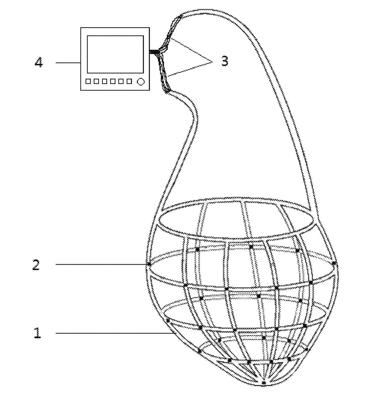
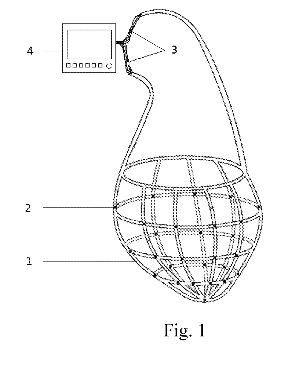
[0068]The cardiac support device is a cardiac tube-network. Structure is shown in FIG. 1, 1—cardiac tube-network; 2—physiological and biochemical sensor, 3—wire, 4—cardiac function monitor device.

[0069]The cardiac support device is attached on an external surface of a ventricle or an atrium or adhered on an internal surface of the cardiac chamber. The cardiac function monitor device is connected with the physiological and biochemical sensor which could be embedded in the tube walls, filled in the aperture of the tube walls or adhered on an internal or external surface of the cardiac support device.

embodiment 3

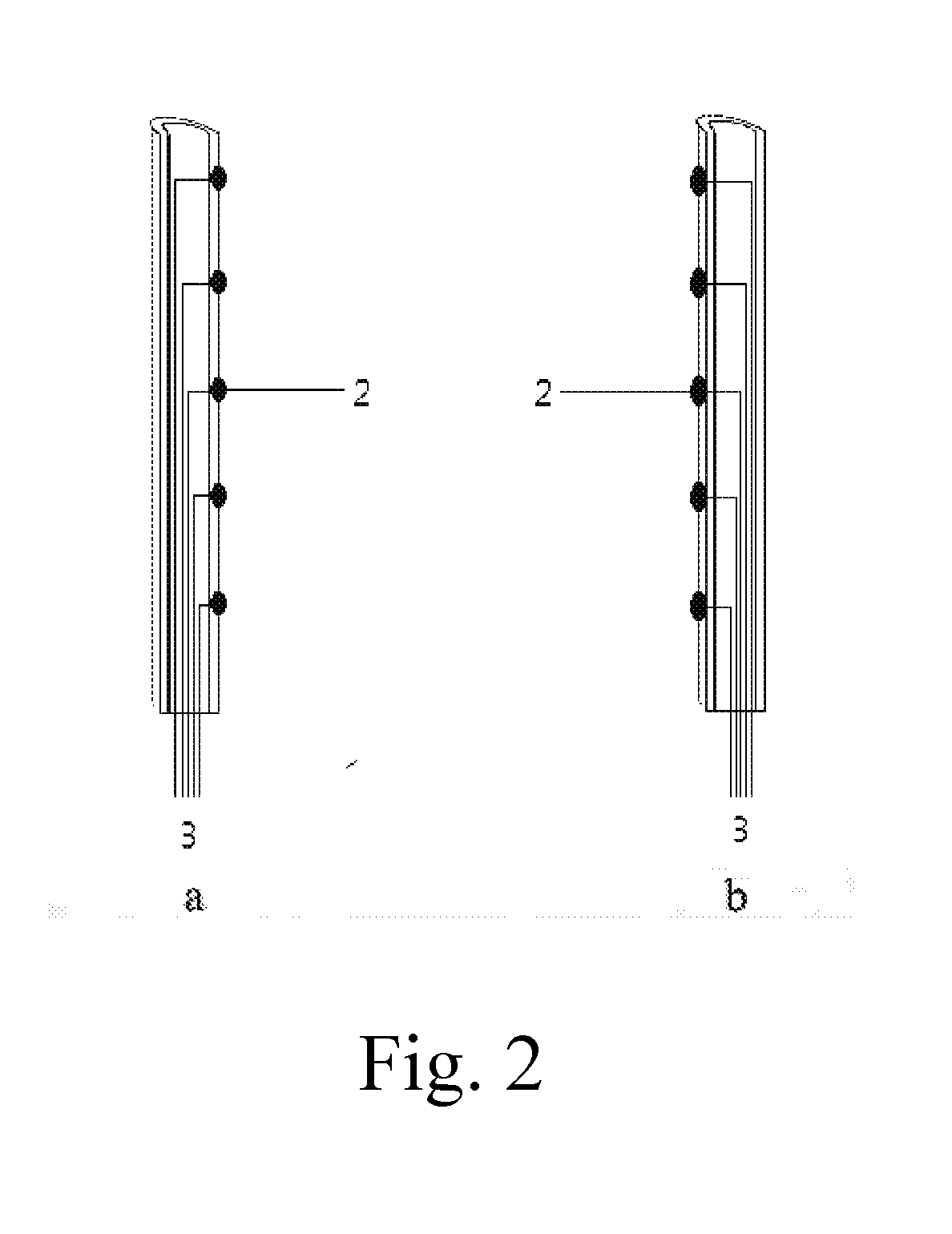
[0070]The basic structure is identical to the embodiment 2. Tube-network is composed of hollow tubes. All the hollow tubes are completely communicated or form a plurality of independent regions. It is intercommunicated within the region, and is not communicated between the regions. The wire of the physiological and biochemical sensor passes through the hollow tube of the tube-network and connects the function monitor device on one end of the tube-network. The structure is shown in FIG. 2, when the tube-network is attached on an external surface of the ventricle / atrium, the physiological and biochemical sensor is adhered on an internal side (see FIG. 2a) of the tube-network; and when the mesh is adhered on an internal surface of the internal cardiac chamber, the physiological and biochemical sensor is adhered on an external side of the tube-network (see FIG. 2b).


PUM


Abstract

A surface attached cardiac function monitor and / or intervention system comprises of a cardiac support device, a cardiac function monitor device and / or intervention device. Cardiac support device is attached on an external or internal surface of a cardiac chamber and supports it. The cardiac function monitor device is connected with a biochemical and physiological sensor. The physiological and biochemical sensor transmits variations of biochemical and physiological parameters that are received by the cardiac function monitor device. The intervention device has at least one member selected from pressure intervention device, an electrical / magnetic stimulation intervention device and a medicine intervention device. This medical system of the present invention could help in diagnosis as well as treatment of the heart failure and other myocardial diseases to improve the condition of patient. It could also be helpful for the monitoring, diagnosis and treatment of diseases of lungs, kidney, liver, spleen, stomach and bladder, etc.

Description

CROSS REFERENCE OF RELATED APPLICATION[0001]The present application claims priority under 35 U.S.C. 119(a-d) to CN 201610781862.1, filed Aug. 31, 2016.BACKGROUND OF THE INVENTIONArea of Invention[0002]The present invention relates to the technical field of medical devices, and more particularly to a medical device used for the monitoring, diagnosis and treatment of heart failure and other myocardial diseases. It could also be helpful for the diagnosis and treatment of diseases of lungs, kidney, liver, spleen, stomach and bladder.DESCRIPTION OF RELATED ARTS[0003]Heart failure, a clinical syndrome of ventricular filling or ejection abnormality caused by disorders in cardiac structure and function, is a common pathological state of the heart. The basic manifestations of heart failure are dyspnea, fatigue, limited exercise tolerance and fluid retention, wherein the fluid retention may further lead to pulmonary congestion and peripheral edema.[0004]Although progress has been made in rece...

Claims


Application Information

Patent Timeline
IPC IPC(8): A61B5/00A61B5/0402A61B5/0205A61B5/01A61N1/362A61B5/02A61N2/00A61H9/00A61M5/31A61M39/10A61N2/02A61B5/145A61M60/191A61M60/289A61M60/468
CPCA61B5/4836A61N2/02A61B5/0402A61B5/02055A61B5/01A61B5/14539A61B5/026A61B5/02028A61N2/004A61H9/00A61M5/31A61M39/10A61N1/362A61B5/14503A61B5/00A61B5/6869A61M31/00A61N1/3625A61N1/3627A61N2/002A61M60/191A61M60/468A61M60/289A61B5/0215A61N1/0597A61N1/3629A61B5/333A61M60/148
Inventor ZHOU, XIAOHUIHAN, LEIZHOU, YUYANZHOU, LILI, GUOHUALI, YONGZHEN
Owner ZHOU XIAOHUI
Learn More