Method for acquiring t2* and vascular images from magnetic resonance imaging system

a magnetic resonance imaging and vascular imaging technology, applied in the field of imaging techniques implemented by magnetic resonance imaging systems, can solve the problems of low signal-to-noise ratio (snr) and lower tissue contrast, and achieve the effect of shortening the total image acquisition time and reducing the burden on patients

Active Publication Date: 2017-05-18
GACHON UNIV OF IND ACADEMIC COOPERATION FOUND +1
View PDF1 Cites 2 Cited by
  • Summary
  • Abstract
  • Description
  • Claims
  • Application Information

AI Technical Summary

Benefits of technology

[0018]When both T2* and vascular images are concurrently acquired using the pulse sequence according to one embodiment of the present invention, accurate T2* image can be acquired by using the readout gradient without the flow compensation and also accurate T2* and vascular images can be concurrently acquired. Therefore, the clinical judgment for blood flow rate of the blood vessel and the clinical judgment for acute stroke can be concurrently made, and so the present invention can be widely utilized in clinical practice.
[0019]Furthermore, the method for concurrently acquiring T2* and vascular images according to one embodiment of the present invention can ensure a quality of images which is comparable with that of images obtained by established protocols, and can shorten the total image acquisition time by 40% as compared with the case of sequentially acquiring traditional T2* weighted and MRA images, As such, the present invention has the advantages reducing a burden on patients when clinically applied. Particularly, this is thought to be very useful in the case of acute ischemic stroke patients.

Problems solved by technology

FLASH techniques have advantages such as a rapid image acquisition speed, short TE (echo time) and TR (repetition time), and low flip angle, but they have disadvantages as compared with spin eco imaging techniques; for example, the contrast of the tissue is lower, many artifacts are generated, and the signal-to-noise ratio (SNR) is low.
Such imaging techniques are useful to diagnose cerebrovascular diseases, which are second only to cancer as the ranking cause of death of Korean people, but there is a problem in that is necessary to go through for the acquisition process for each individual image.

Method used


Image

  • Method for acquiring t2* and vascular images from magnetic resonance imaging system
  • Method for acquiring t2* and vascular images from magnetic resonance imaging system
  • Method for acquiring t2* and vascular images from magnetic resonance imaging system

Examples


Embodiment Construction

[0028]Hereinafter, preferred embodiments of the method for acquiring images through the magnetic resonance imaging system according to the present invention are described in detail with reference to the attached drawings. However, description concerning well-known functions and configurations which can make the subject matter of the present invention unnecessarily vague will be omitted.

[0029]The configurations of the MRI system as applied to the present invention are widely known in the art and so the description thereof is omitted.

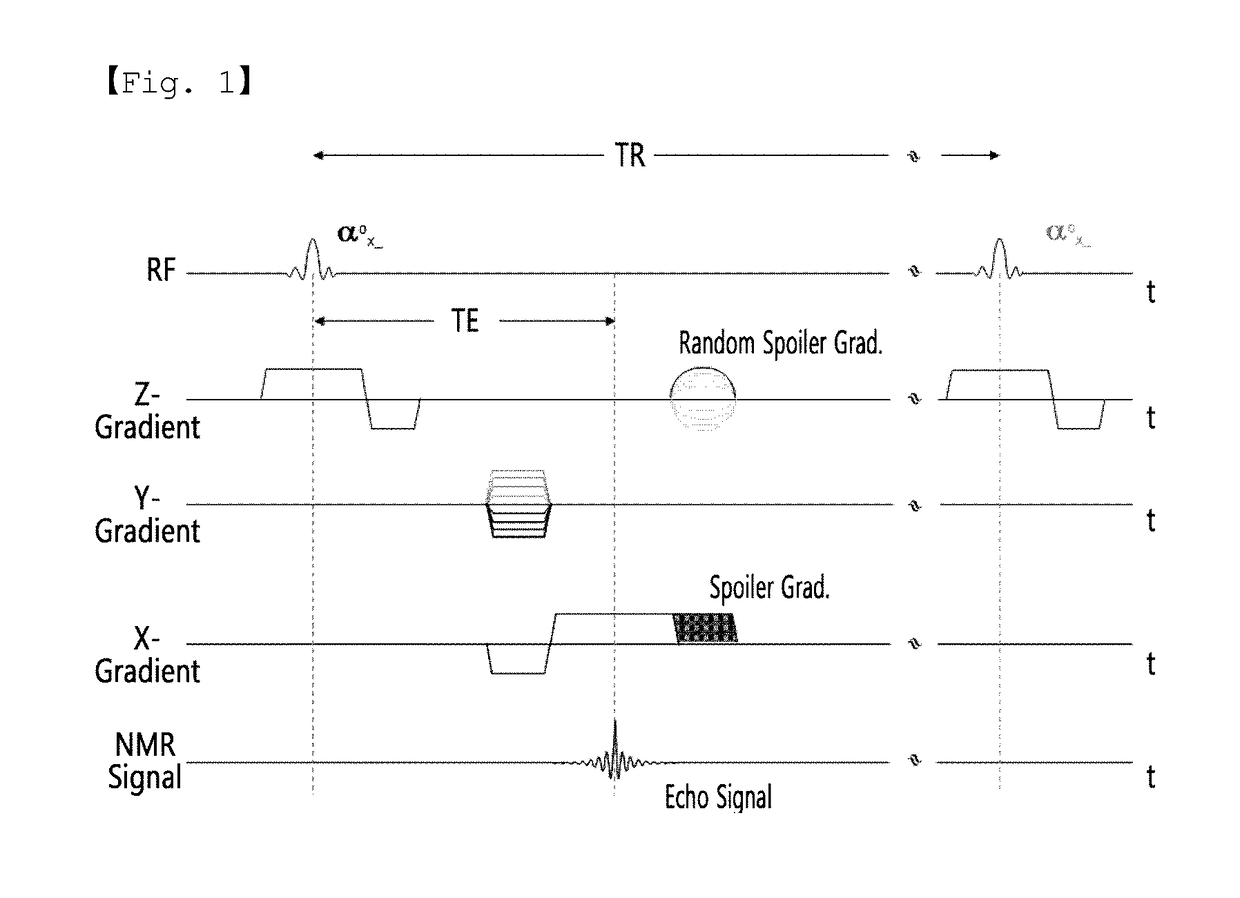
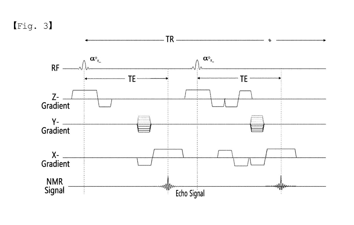
[0030]FIG. 3 represents the pulse sequences used in the method for concurrently acquiring T2* and vascular images according to an embodiment of the present invention.

[0031]In FIG. 3, Z-gradient, Y-gradient and X-gradient represent a slice selection, a phase decoding and a readout gradient, respectively.

[0032]As shown in FIG. 3, the readout gradient without a flow compensation is applied in a negative direction and then applied in a positive direction so a...


PUM


Abstract

According to the present invention, accurate T2* and vascular images are concurrently acquired by acquiring a T2* image without a flow compensation and a T2* image with a flow compensation and subtracting the two images to reconstitute an image showing the flow phenomenon. Furthermore, an accurate T2* image can be acquired by using the readout gradient without the flow compensation and also the accurate T2* and vascular images can be concurrently acquired. The clinical judgment for blood flow rate of the blood vessel and the clinical judgment for acute stroke can be concurrently made, and so the present invention can be widely utilized in clinical practice.

Description

TECHNICAL FIELD[0001]The present invention relates to an imaging technique implemented by an MRI (Magnetic Resonance imaging) system, and more specifically to an imaging technique capable concurrently acquiring T2* and vascular images from an MRI system.BACKGROUND ART[0002]Diagnostic imaging methods include devices utilizing a variety of techniques such as X-ray, CT, ultrasonic, RI imaging, or MRI. Among them, MRI is the least harmful to the human body as compared with the other diagnostic imaging methods and forms images of the constituents in the human body. Hence, MRI is a very important device in clinical practice.[0003]MRI devices can obtain tissue parameters such as spin density, T1, T2, chemical shift, magnetic transition, chemical exchange saturation transfer, blood flow, or spectroscopy, which are unique information of the living body. Various biometric information images can also be obtained through such parameters.[0004]Magnetic Resonance Angiography (MRA) is a method for...

Claims


Application Information

Patent Timeline
Patent Type & Authority Applications(United States)
IPC IPC(8): A61B5/026A61B5/055
CPCA61B5/055A61B5/0263G01R33/00G01R33/50G01R33/5635G01R33/56509G06T5/50G06T7/0012G06T2207/10088G06T2207/20224G06T2207/30101G06T2207/30104A61B5/0042
Inventor CHUNG, JUN YOUNGKIM, EUNG YEOP
Owner GACHON UNIV OF IND ACADEMIC COOPERATION FOUND
Why Eureka
Try Eureka