
Biopolymer-Nanoparticle Composite Implant for Tumor Cell Tracking

a biopolymer and nanoparticle technology, applied in the direction of powder delivery, drug compositions, medical preparations, etc., can solve the problems of most significant challenges in the management of cancer, accompanied by significant morbidities, and current lymph node staging via lymphadenectomy

Active Publication Date: 2017-07-27
NORTHEASTERN UNIV +2
View PDF2 Cites 5 Cited by
  • Summary
  • Abstract
  • Description
  • Claims
  • Application Information

AI Technical Summary

Benefits of technology

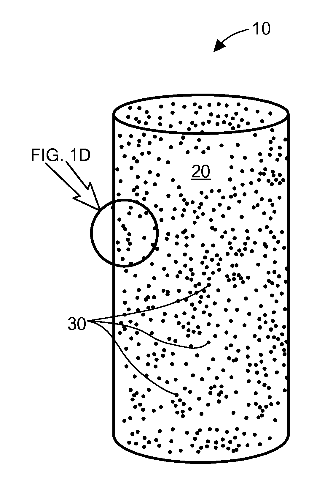
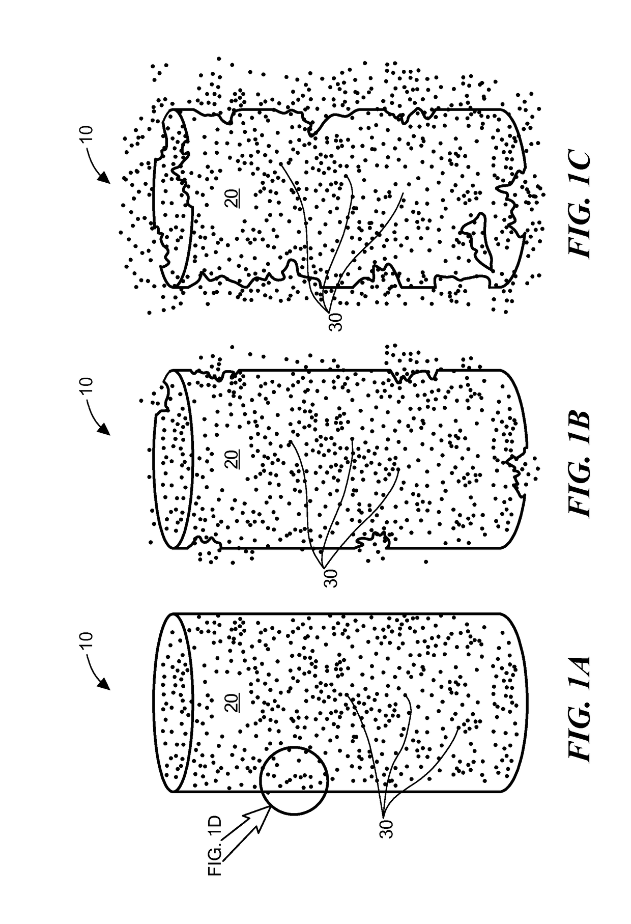
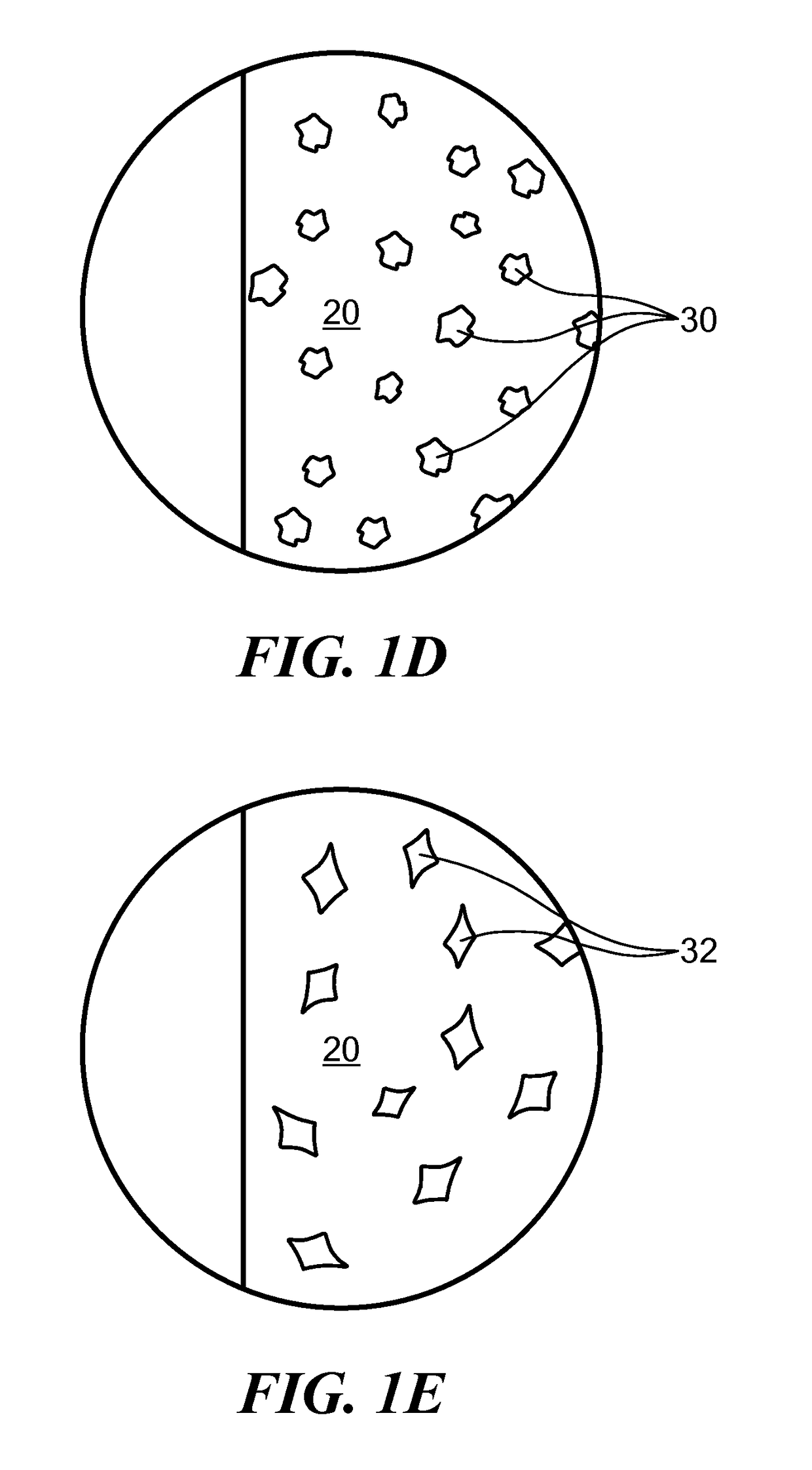
The patent describes a method for detecting and treating metastatic tumor cells in a patient. The method involves implanting a device in the region of tumor cells, which includes a matrix material and nanoparticles functionalized to bind to the tumor cells. The nanoparticles are dispersed throughout the matrix material and can be detected using various imaging techniques such as fluorescence imaging, X-ray computed tomography, magnetic resonance imaging, and ultrasound imaging. The device can also include a brachytherapy spacer or a radiotherapy fiducial marker for treatment purposes. The technical effects of this patent include improved detection and treatment of metastatic tumor cells in patients.

Problems solved by technology

Metastasis accounts for over 90% of all cancer-associated suffering and death, and it presents the most significant challenges in the management of cancer.
The ultimate goal, to efficiently detect and isolate the rare population of tumor cells in a viable state and with high purity from the vast number of surrounding blood cells, still presents a formidable technological challenge.
Current lymph node staging via lymphadenectomy is accompanied by significant morbidities.

Method used


Image

  • Biopolymer-Nanoparticle Composite Implant for Tumor Cell Tracking
  • Biopolymer-Nanoparticle Composite Implant for Tumor Cell Tracking
  • Biopolymer-Nanoparticle Composite Implant for Tumor Cell Tracking

Examples


example 1

[0282]In one example, 250-300 mg of PLGA was dissolved in a minimum amount of dimethyl sulfoxide (DMSO). Docetaxel was dissolved in DMSO, and the two solutions were mixed using sonication to obtain a viscous uniform slurry. The slurry was transferred to a 1 ml syringe using a Luer stub adapter (0.5 in; 18G) attached to a silicon tube (inner diameter 0.8 mm). The paste was infused at a predetermined flow rate into the silicon tubing using an infusion pump. The tube was dried overnight at 45-50° C. The crystallized implants were taken out of the tubing using brachytherapy stubs, cut into 5 mm lengths, and stored at room temperature in the dark.

example 2

[0283]A prototype implant with fluorescent gold nanoparticles embedded in PLGA was fabricated and implanted in a tissue mimic. The tissue mimic was provided by the material commercially available as SuperFlab. FIG. 4A illustrates an in vitro release of the fluorescent gold nanoparticles from the implant monitored using UV-visible spectroscopy over time, from 0 minutes to 4.5 hours. FIG. 4B shows the corresponding concentration profile over time. The prototype implants are shown in the top image of FIG. 5. Image B shows a CT image of the implant in the tissue mimic, and image C shows an optical fluorescence image of the implant in the tissue mimic.

example 3

[0284]Implants were fabricated with the drug docetaxel (DTX) dispersed within a PLGA matrix material as described in Example 1 above. The implants (also called spacers in the study) were implanted in tumors in PC3 tumored mice. Four groups were tested: a control group that was not treated; a control group with “blank” implants (implants that contain no drugs or functionalized nanoparticles); a group that received the drug DTX intravenously only; and a group that received implants loaded with the drug DTX.

[0285]The implants were found to inhibit tumor growth and shrink the tumor as the drug was released intratumorally with minimal visible adverse effects to the mice. See FIG. 7A, which illustrates the average change in tumor volumes over time. 89% of the mice with the implant loaded with DTX survived after 40 days, whereas 0% of the mice from the other treatment groups survived at 40 days. See FIG. 7B, which illustrates Kaplan-Meier survival curves for the mice at 40 days.

[0286]In a ...


PUM

PropertyMeasurementUnit
lengthaaaaaaaaaa
diameteraaaaaaaaaa
diameteraaaaaaaaaa

Abstract

A method of detecting migration of tumor cells is provided by implanting in a region of tumor cells one or more implants having a matrix material of a biocompatible and biodegradable polymer, and a plurality of nanoparticles dispersed within the matrix material and functionalized to bind tumor cells. Nanoparticles bound to the tumor cells that have migrated out of the region can be detected by various imaging modalities. The implant can be in the shape of a brachytherapy spacer or radiotherapy fiducial maker or can be a coating on a brachytherapy spacer or fiducial marker. A method of treating cancer is provided by implanting one or more brachytherapy spacers or fiducial markers including the matrix material and an anti-cancer therapeutic agent dispersed within the matrix material.

Description

CROSS REFERENCE TO RELATED APPLICATIONS[0001]This application claims priority under 35 §119(e) of U.S. Provisional Application No. 62 / 028,880, filed on Jul. 25, 2014, entitled “Biomaterial for Response Assessment and Nodal Detection (BRAND)”, the disclosure of which is hereby incorporated by reference.STATEMENT REGARDING FEDERALLY SPONSORED RESEARCH OR DEVELOPMENT[0002]N / ABACKGROUND[0003]Metastasis is the spread of cancer from the primary site (the source tumor) to one or more other parts of the body. Metastasis accounts for over 90% of all cancer-associated suffering and death, and it presents the most significant challenges in the management of cancer.[0004]Circulating tumor cells (CTCs) can serve as biological markers (biomarkers) for non-invasive early detection of metastasis, tumor aggressiveness, tumor response to treatment, or prognosis, and can enhance understanding of the biology of metastasis. The ultimate goal, to efficiently detect and isolate the rare population of tumo...

Claims


Application Information

Patent Timeline
Patent Type & Authority Applications(United States)
IPC IPC(8): A61K49/00A61L31/06A61L31/16A61B5/00A61L31/18A61K49/04A61B90/00A61N5/10A61K31/337A61L31/14
CPCA61K49/0065A61K31/337A61L31/06A61L31/16A61L31/14A61K49/0054A61N2005/1023A61K49/04A61B90/39A61N5/1027A61B5/4887A61L2300/416A61L31/18A61L31/042A61L31/048A61L31/148A61K9/0024A61K45/06A61K9/204A61K49/0423A61L2300/256A61L2400/12A61B2090/3966A61P35/04C08L67/04C08L71/02C08L33/26C08L5/08A61K2300/00
Inventor KUMAR, RAJIVSRIDHAR, SRINIVASNGWA, WILFREDCORMACK, ROBERTMAKRIGIORGOS, GERASSIMOS
Owner NORTHEASTERN UNIV
Learn More