Method for producing cerebellar progenitor tissue

a progenitor tissue and cerebellar technology, applied in the field of cerebellar progenitor tissue production, can solve the problems of largely elusive how the generated cellular components are assembled to form the intricate structure of the cerebellum, and achieve the effect of efficient induced

Active Publication Date: 2017-09-07
RIKEN +1
View PDF0 Cites 3 Cited by
  • Summary
  • Abstract
  • Description
  • Claims
  • Application Information

AI Technical Summary

Benefits of technology

[0033]According to the present invention, a cerebellar progenitor tissue can be efficiently induced in vitro from human pluripotent stem cells. A midbrain-hindbrain boundary progenitor tissue can be formed from human pluripotent stem cells for the first time in vitro. The dorsal-ventral polarity observed in vivo can be reproduced in this midbrain-hindbrain boundary progenitor tissue. According to the present invention, a cerebellar progenitor tissue (cerebellar plate tissue) having a continuous epithelial structure can be self-organized in an aggregat...

Problems solved by technology

However, it remains largely elusive how the generated cellular components are assembled to form the intricate structure of the cerebellum.
Although...

Method used


Image

  • Method for producing cerebellar progenitor tissue
  • Method for producing cerebellar progenitor tissue
  • Method for producing cerebellar progenitor tissue

Examples


example 1

[Example 1] Formation of Midbrain-Hindbrain Boundary Neural Progenitor Tissue from Human Pluripotent Stem Cell

(Method)

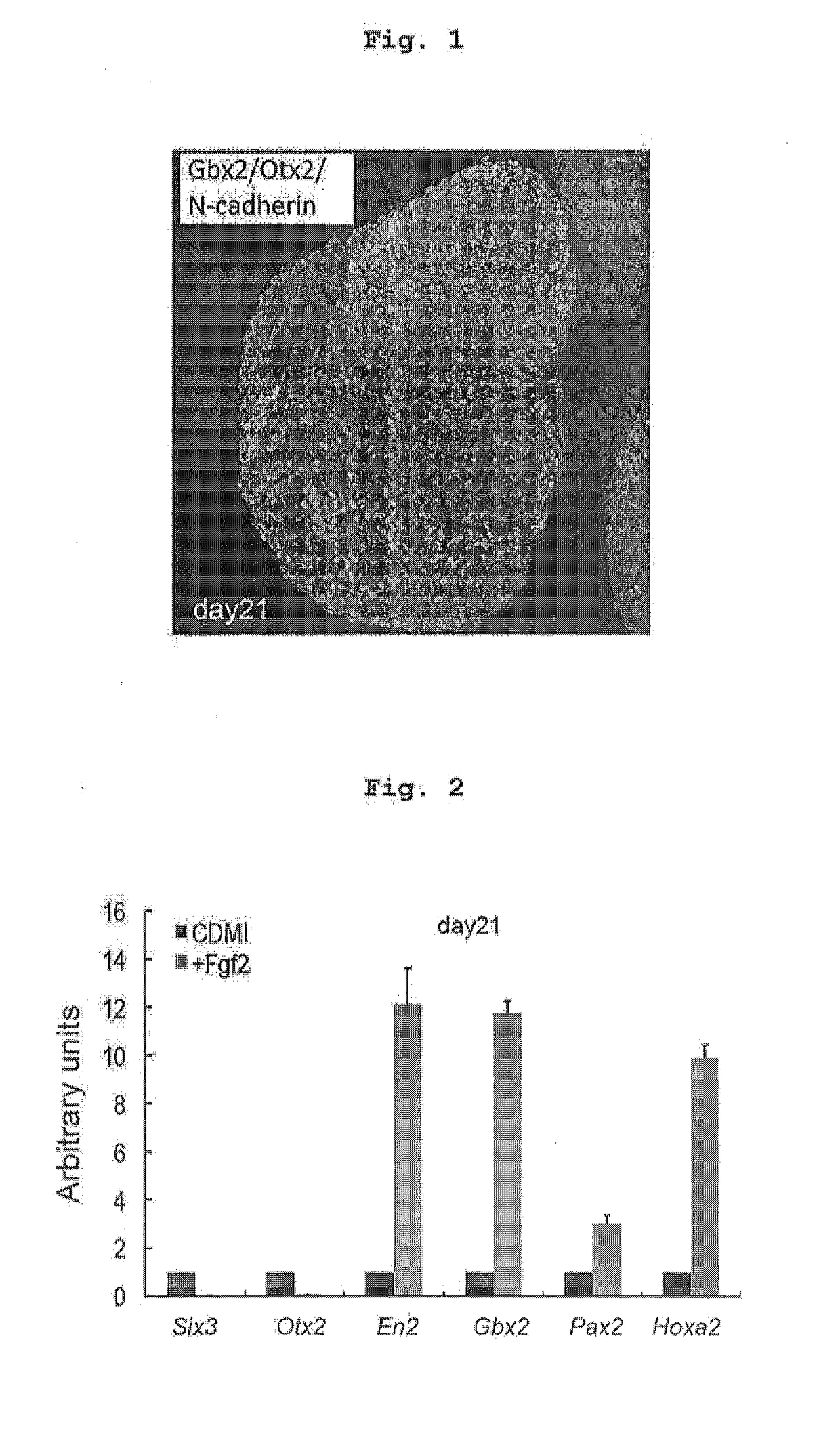
[0177]Human ES cells (KhES-1) subjected to maintenance culturing on MEF by a conventional method were used. To differentiate human ES cells by the serum-free floating culture of embryoid body-like aggregates with quick reaggregation (SFEBq method), human ES cells were enzymatically dispersed to single cells, according to the method of Nakano et al. (Cell Stem Cell, 10(6), 771-785, 2012), and reaggregated using a low-cell-adhesive V-bottom 96 well plate (SUMITOMO BAKELITE). 6,000 cells were seeded per well, and cultured in, as a differentiation medium, CDM medium [chemical synthesis medium without a growth factor (growth-factor-free Chemically Defined Medium; gfCDM; Wataya et al., Proc. Natl. Acad. Sci. USA, 105, 11796-11801, 2008) supplemented with insulin (7 μg / ml) and apo-transferrin (15 μg / ml)] under 5% CO2 at 37° C.

[0178]The seeding day is taken as day 0 of diffe...

example 2

[Example 2] Self-Formation of Cerebellar Plate Tissue Having Continuous Epithelial Structure from Human Pluripotent Stem Cells

(Method)

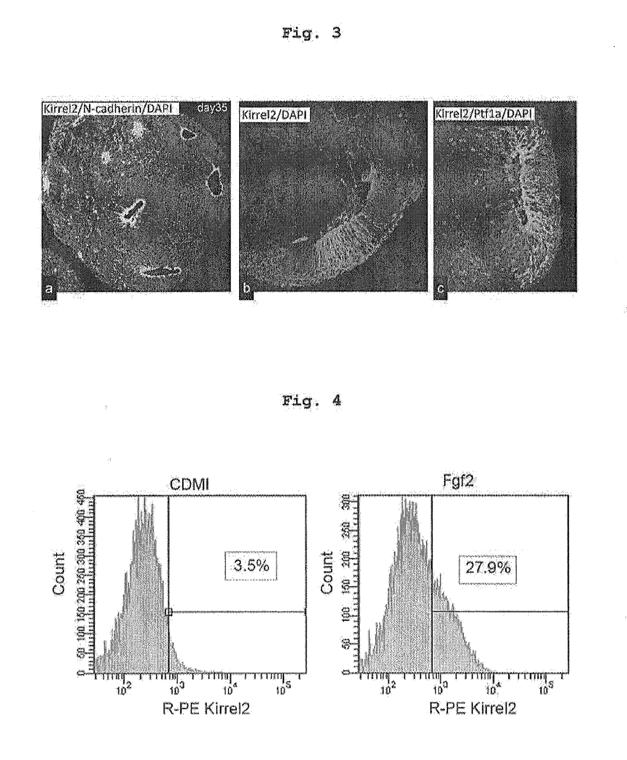
[0184]Human pluripotent stem cell aggregates were cultured according to the method of Example 1. The was aggregates were transferred to a low-cell-adhesion 10 cm dish on and after day 21, and cultured in a neural culture medium (Neurobasal / GlutaMaxI / N2, all Life Technologies). On day 28 of culturing, the total amount of the medium was exchanged with the same neural culture medium, and differentiation of the tissue was analyzed by the immunofluorescence method on day 35 of culturing.

(Results)

[0185]A plurality of N-cadherin positive neuroepithelial structures were formed in one aggregate, and not less than 70% of neuroepithelial cells were positive to cerebellar GABAergic neural progenitor-specific kirrel2 (FIG. 3 left, middle). Not less than 80% of the neuroepithelial structure expressing Kirrel2 also expressed GABAergic neural progenitor-specific Ptf1...

example 3

[Example 3] Differentiation of Purkinje Cell and Cerebellar Interneuron by Dispersion Culture of Human Pluripotent Stem Cell-Derived Cerebellar Plate Tissue

(Method)

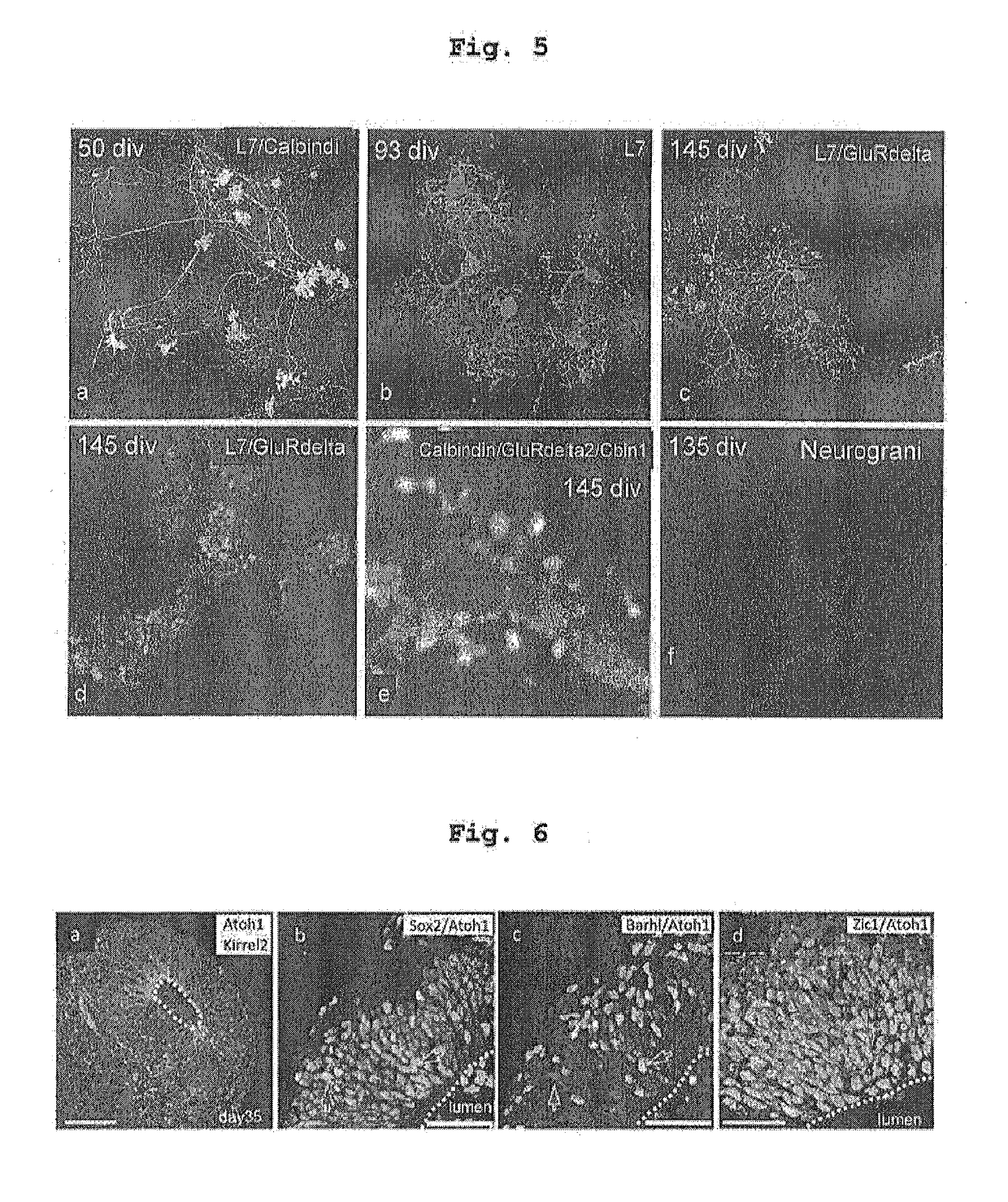
[0188]Human pluripotent stem cell aggregates were cultured according to the methods of Examples 1 and 2, and Kirrel2 positive cells were separated by FACS using an antibody to cerebellar GABAergic neural progenitor marker Kirrel2 on day 35 of culturing. Separated Kirrel2 positive cells, and cerebellar granule cell progenitors prepared from the upper rhombic lip of embryonic day 14 mouse were dispersed into single cells by cell dispersion enzyme (TrypLE, Life Technologies), and cocultured on a cover glass coated with poly D-lysine / laminin in DMEM / F12 / 10% FBS medium (5% CO2, 37° C.). In coculture, human pluripotent stem cell-derived Kirrel positive cells (1 vol) relative to mouse-derived cerebellar granule cell progenitors (10 vol) were mixed. After 6-12 hr, DMEM / F12 / N2 / BSA / Tri-iodothyronine (T3) was added to decrease the F...


PUM


Abstract

The present invention provides a method for producing a human cell aggregate containing a midbrain-hindbrain boundary neural progenitor tissue, including subjecting an aggregate of human pluripotent stem cells to suspension culturing in a serum-free medium containing insulin, and treating, in the suspension culturing, the aggregate of human pluripotent stem cells or a human cell aggregate derived therefrom with a ROCK inhibitor, a TGFβ signal inhibitor, and a first fibroblast growth factor. Furthermore, the human cell aggregate containing the midbrain-hindbrain boundary neural progenitor tissue is subjected to suspension culturing in a serum-free medium to induce formation of a neuroepithelial structure by neural progenitor in the neural progenitor tissue, whereby the human cell aggregate containing the cerebellar plate tissue can be obtained.

Description

TECHNICAL FIELD[0001]The present invention relates to a technique for inducing differentiation of human pluripotent stem cells into cerebellar progenitor tissue in vitro.BACKGROUND ART[0002]The cerebellum, a major component of the motor system, is a highly ordered brain structure with several well-defined types of cells. The early development of the cerebellum is conserved among amniotes including human. The initial phase of cerebellar development is the formation of the isthmic organizer, which lies at the midbrain-hindbrain boundary (MHB). Under its inductive influence, the cerebellar anlage arises in the dorsal region (alar plate) of rhombomere 1 (r1) (non-patent documents 1-3). Cerebellar cells are generated in two distinct germinal zones in r1. One is the ventricular zone (VZ) of the cerebellar plate, which expresses the basic helix-loop-helix (bHLH) transcription factor Ptf1a. The Ptf1a+ progenitors produce GABAergic neurons of the cerebellar cortex (Purkinje cells and interne...

Claims


Application Information

Patent Timeline
IPC IPC(8): C12N5/0793
CPCC12N5/0619C12N2500/90C12N2501/33C12N2506/08C12N2506/03C12N2501/21C12N2501/19C12N2501/113C12N2506/02C12N2500/25C12N2501/15C12N2501/727C12N2501/115C12N5/0618C12N2501/119C12N2513/00C12N5/10C12N5/0602C12N15/09
Inventor YOSHIKIMUGURUMA, KEIKO
Owner RIKEN
Why Eureka
Try Eureka