Tissue graft

a technology of tissue grafts and skin substitutes, which is applied in the field of tissue grafts, can solve the problems of limited data available on the engineering of human lymphatic capillaries, no data available on the bioengineering of prevascularized dermo-epidermal skin substitutes containing functional human lymphatic capillaries, and achieves rapid and efficient oxygen access, rapid and efficient regeneration, and differentiation of skin transplants.

Active Publication Date: 2017-09-14
UNIV ZURICH
View PDF0 Cites 3 Cited by
  • Summary
  • Abstract
  • Description
  • Claims
  • Application Information

AI Technical Summary

Benefits of technology

[0009]Additionally so far, limited data is available on the engineering of human lymphatic capillaries and no data is available on the bioengineering of prevascularized dermo-epidermal skin substitutes containing functional human lymphatic capillaries. In the human dermis, lymphatic vessels play a major role in tissue fluid homeostasis and immune cell trafficking. Dermal lymphatic capillaries exhibit a wide lumen, anchoring filaments, and no or an incomplete basement membrane, and lack mural cell coverage. These features enable lymphatic capillaries to respond to interstitial liquid pressure by taking up and removing excess tissue fluid. After wounding, also the lymphatic endothelium is ruptured; thus, the draining capacity of the lymphatic vessels is compromised. As a consequence, accumulation of tissue fluid arises. Persistent local interstitial fluid, as well as delayed removal of local debris and inflammatory cells, impedes wound healing. In contrast, induction of lymphangiogenesis and immune cell recruitment were shown to accelerate skin regeneration.
[0054]With the help of the present invention both human blood and lymphatic vessels can be engineered in one tissue or organ graft. The in vitro generated network of capillaries significantly supports perfusion of the dermal component, hence, providing rapid and efficient access to oxygen and nutrients, which assures rapid take, proliferation, and differentiation of the skin transplant (Böttcher-Haberzeth et al., Tissue engineering of skin, Burns 36, 450-460 (2010)). Bioengineering a preformed network of lymphatic capillaries into dermo-epidermal skin grafts should help circumvent seroma formation by improving lymphatic drainage and accelerating the establishment of tissue fluid homeostasis.

Problems solved by technology

No data is available on the bioengineering of prevacularized dermo-epidermal skin grafts for intended clinical use for the treatment of skin defects.
Additionally so far, limited data is available on the engineering of human lymphatic capillaries and no data is available on the bioengineering of prevascularized dermo-epidermal skin substitutes containing functional human lymphatic capillaries.
After wounding, also the lymphatic endothelium is ruptured; thus, the draining capacity of the lymphatic vessels is compromised.
Persistent local interstitial fluid, as well as delayed removal of local debris and inflammatory cells, impedes wound healing.

Method used


Image

  • Tissue graft
  • Tissue graft
  • Tissue graft

Examples


example 1

[0069]Production of Uncompressed Prevascularized Dermo-Epidermal Skin Grafts Containing Blood and Lymphatic Capillaries

[0070]The transplantation of human dermo-epidermal skin grafts containing vascular (blood and lymph) plexus onto rats was monitored. First, skin grafts were created in vitro using CD31 positive (CD31+) HDMECs, human CD90 positive (CD90+) fibroblasts, and human keratin5 positive (K5+) keratinocytes in fibrin hydrogels.

[0071]Both cell types constituting the dermal compartment of the graft were arranged underneath several layers of keratinocytes, the epidermal compartment (made visible and verified by confocal micrograph). These skin grafts were then transplanted onto wounded backs of immunoincompetent nu / nu rats using a Fusenig chamber to avoid competitive, lateral ingrowth / overgrowth of rat keratinocytes. Two weeks after transplantation, the human skin substitute was surgically removed from the rat underlying tissue and analysed for dermal structure and neovasculariz...

example 2

[0087]Production of Uncompressed Prevascularized Dermo-Epidermal Skin Grafts Containing Blood Capillaries Generated by SVF Cells

[0088]The transplantation of human dermo-epidermal skin grafts containing a blood vascular plexus onto rats was monitored. First, skin grafts were created in vitro using SVF cells and keratinocytes in fibrin hydrogels.

[0089]SVF cells were arranged underneath several layers of keratinocytes, the epidermal compartment (made visible and verified by confocal micrograph). These skin grafts were then transplanted onto wounded backs of immunoincompetent nu / nu rats using a Fusenig chamber to avoid competitive, lateral ingrowth / overgrowth of rat keratinocytes. Two weeks after transplantation, the human skin substitute was surgically removed from the rat underlying tissue and analysed for dermal structure and neovascularization. The vascularized neodermis supported stratification of the overlaying epidermis. Immunofluorescence analysis after 2 weeks revealed the pres...

example 3

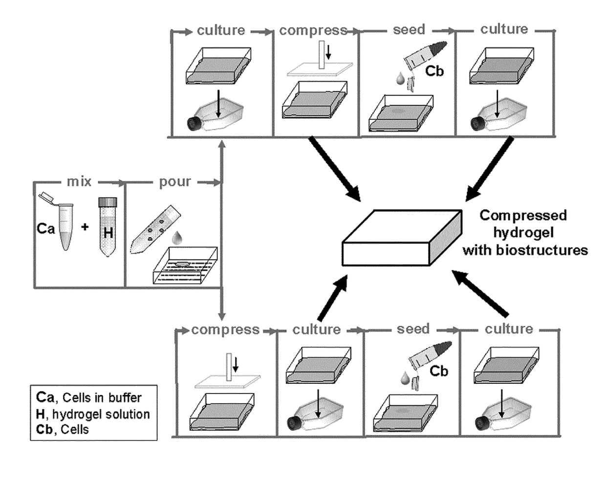
[0098]Production of Compressed Prevascularized Dermo-Epidermal Skin Grafts

[0099]Materials and Methods for Example 3:

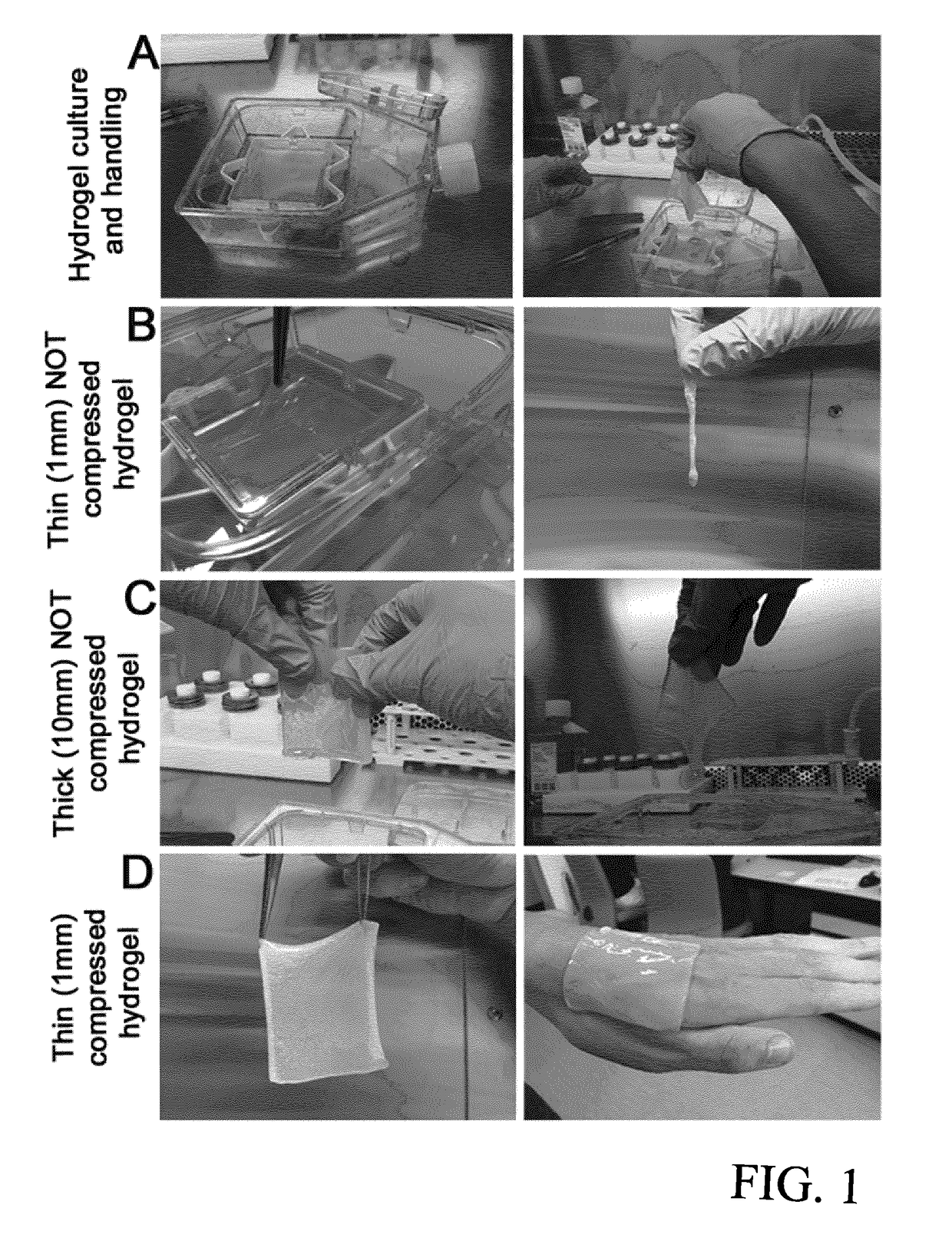
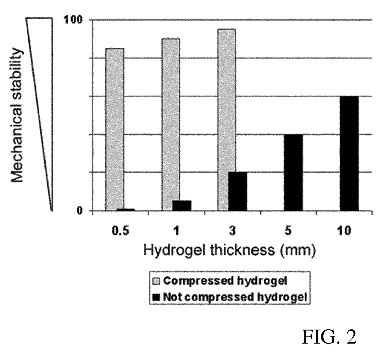
[0100]Human cells (keratinocytes, fibroblasts and endothelial cells) were isolated as described above. Tissue grafts were prepared from hydrogels of 7×8 cm size as described below with reference to FIGS. 7-10. To obtain mechanical stability, modified plastic compression was performed with the compression device according to EP 13 174 441.

[0101]Preparation of the Hydrogel (as shown in FIG. 7):

[0102]As shown in FIG. 7, an insert frame (A) and an insert (B) are placed into a tissue culture flask of 115 cm2. 1 million fibroblasts / endothelial cells (ratio 1:1) are resuspended in 4 ml endothelial cell medium (“cells”). 18±1 ml of collagen hydrogel is poured in a tube (“hydrogel”). 850 μl of Acetic acid filtered are added to the “hydrogel” (“mixture”). The “mixture” is mixed by gently pivoting the tube.[0103]Addition of 7.5±0.2 ml of Reconstitution buffer to the cells (“cells...


PUM

PropertyMeasurementUnit
thicknessaaaaaaaaaa
physiological sizeaaaaaaaaaa
thicknessaaaaaaaaaa

Abstract

The present invention is directed to a method of producing a tissue graft, comprising at least steps of providing a gel, seeding the gel with cells of at least a first and / or cells of a second type, and culturing of the cells of the first and / or cells of the second type in said gel until the formation of at least one first biostructure in the gel by the cells of the first type and / or the cells of the second type.

Description

TECHNICAL FIELD[0001]The present invention relates to a device containing at least one biostructure, and a method of making the device, for use in therapeutic treatment and / or for in vitro testing of human or other mammalian tissues. For example, the device may be used on skin wounds for burned, injured, or diseased skin, and provides structures and functions as in normal uninjured skinPRIOR ART[0002]Human skin is composed of three main layers: the epidermis (outermost), the dermis (middle) and the hypodermis (deeper most). When a deep wound is created (full-thickness skin wound), after e.g. a burn accident, the epidermis and the full dermis (in some cases also the hypodermis) are damaged and surgical intervention is required. Small full-thickness skin wounds are optimally treated by means of full-thickness skin autografts: epidermis and whole dermis are harvested from a healthy part of the body and transplanted on the wound. In contrast, large full-thickness skin defects, treated b...

Claims


Application Information

Patent Timeline
Patent Type & Authority Applications(United States)
IPC IPC(8): A61L27/60A61L27/36G01N33/50A61L27/24A61L27/38A61L27/52A61L27/22
CPCA61L27/60A61L27/52A61L27/3691A61L27/225A61L27/24G01N33/5005A61L27/3834A61L27/3813A61L27/3808A61L27/3891A61L27/3804A61L27/50
Inventor REICHMANN, ERNSTMARINO, DANIELAKLAR, AGNIESZKA SYLWIA
Owner UNIV ZURICH
Why Eureka
Try Eureka