Methods to generate gastrointestinal epithelial tissue constructs

a technology of gastrointestinal epithelium and constructs, which is applied in the field of methods to generate gastrointestinal epithelium constructs, can solve the problems of not improving crypt survival, not precisely mimicking the in vivo biochemical and biophysical microenvironments of crypts, and being extremely difficult to generate a long-term proliferative intestinal epithelium in vitro. achieve the effect of maintaining the viability of stem cells

Active Publication Date: 2018-01-04
THE UNIV OF NORTH CAROLINA AT CHAPEL HILL
View PDF0 Cites 8 Cited by
  • Summary
  • Abstract
  • Description
  • Claims
  • Application Information

AI Technical Summary

Benefits of technology

[0007]To overcome the problems noted above in mimicking the lamina propria, biomimetic scaffolds were fabricated from w

Problems solved by technology

Culture of live crypts in vitro has been attempted since the 1970s, but it has proven to be extremely difficult to generate a long-term proliferative intestinal epithelium in vitro.6, 7 This is thought to be a result of the complexity of in vivo microenvironments that constitute the intestinal epithelial stem-cell niche, which has been impossible to recapitulate in vitro.
Standard 2D culture of crypts in dishes yields short-term growth of a monolayer of cells.11 Performing 3D culture by embedding crypts or stem cells in collagen gel alone (i.e. without a feeder layer of supporting cells) does not improve crypt survival.9 The loss of proliferative ability of crypt cells under these culture methods suggests the loss of intestinal stem cells in vitro.
While this

Method used


Image

  • Methods to generate gastrointestinal epithelial tissue constructs
  • Methods to generate gastrointestinal epithelial tissue constructs
  • Methods to generate gastrointestinal epithelial tissue constructs

Examples


example 1

[0095]Demonstration of Long-Term Colonic Epithelial Monolayers on both 2d and 3d Scaffolds

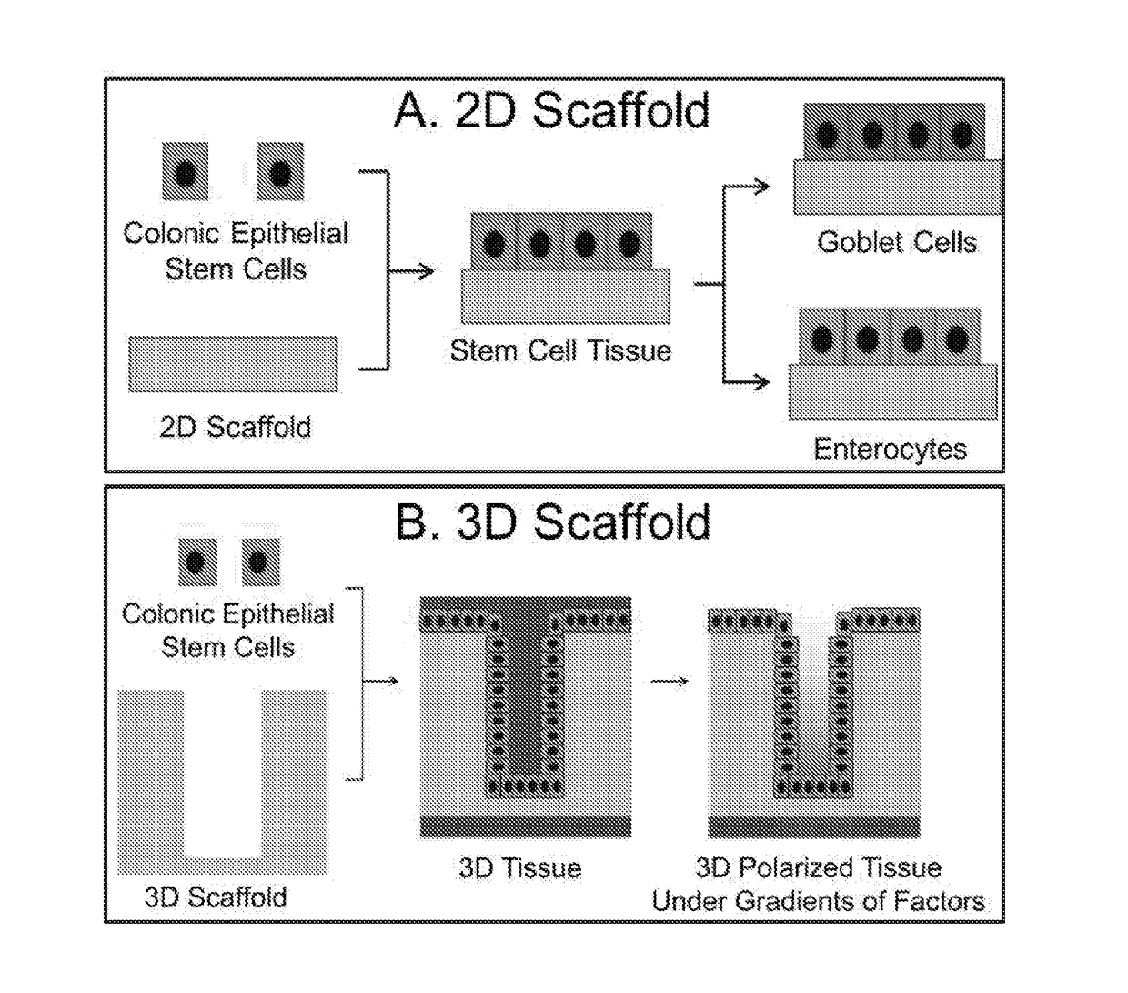
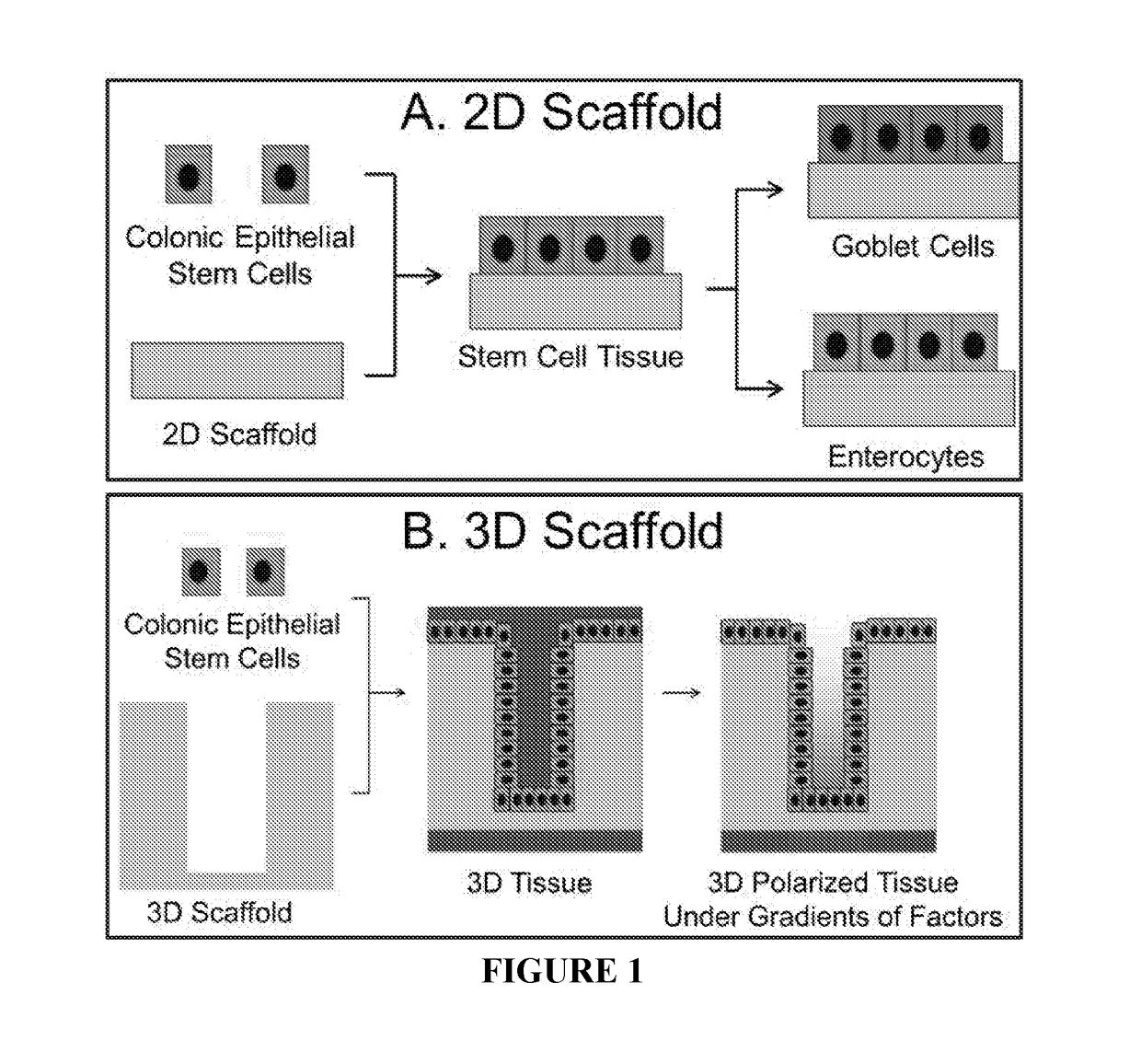
[0096]As a demonstration of the current invention, two types of scaffolds, 2D and 3D scaffolds, were fabricated from collagen and demonstrated to support the long-term culture of cells obtained from primary colonic crypt tissue including the stem cells (FIG. 1). A 2D colonic epithelial monolayer was generated by culturing isolated crypts or stem cells on the surface of the planar, 2D scaffolds (FIG. 1A). Under this condition, the biochemical microenvironment was provided by the soluble growth factors (Wnt-3A, R-spondin, noggin and EGF), while the biophysical microenvironment was provided by the collagen scaffold which mimics the lamina propria in terms of permeability, stiffness, and ECM components. The monolayer contained proliferative stem cells. By changing the growth factors in the medium, the monolayer could be selectively differentiated at will into specialized cells, such as goblet cells...

example 2

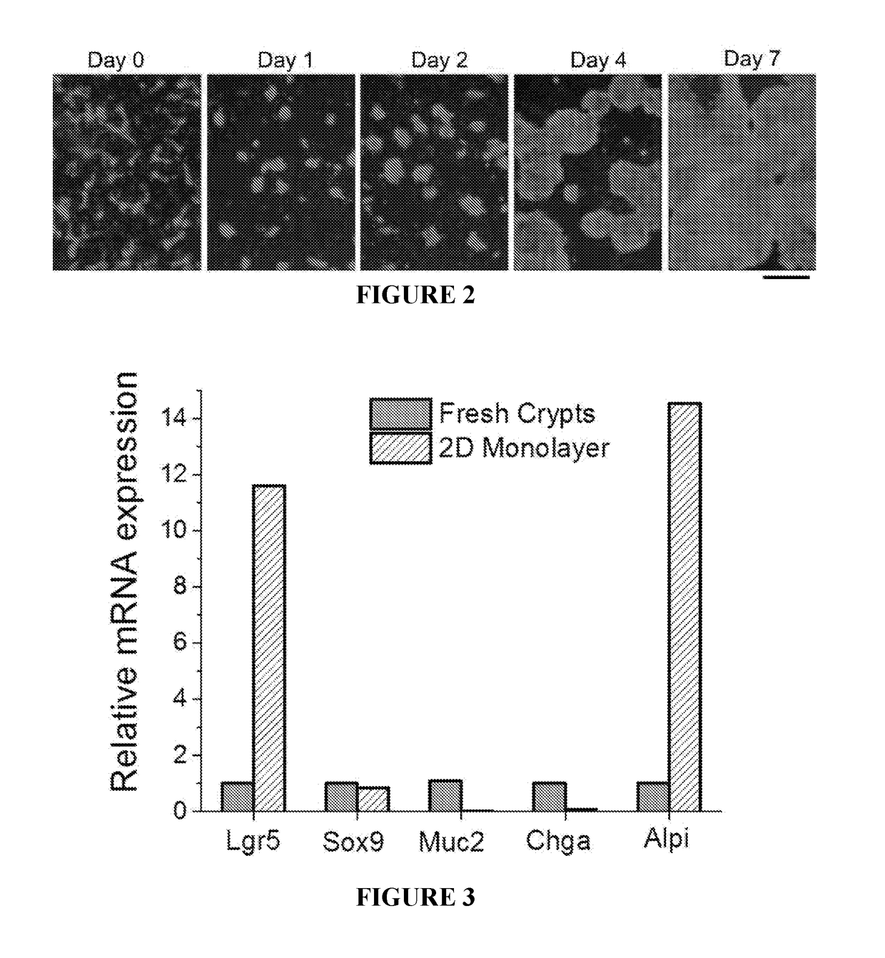
[0116]The 2d Monolayer Possesses In Vivo-Like Proliferating Capability, Morphology and Function

[0117]In vivo, the intestinal epithelium is one of the most rapidly proliferating tissues in the mammalian body. Consistent with this, the in vitro 2D monolayer of colonic epithelial cells cultured on the collagen scaffold was highly proliferative. To demonstrate this proliferative capability, fragments of 2D monolayer were plated on the collagen scaffold in a Transwell insert (FIG. 7A, top panel). The perimeter of the insert is indicated by the white dotted line). By day 5, the cells generated a continuous 2D monolayer that occupied the entire surface of the insert with a surface area of 137 mm2 (FIG. 7B, bottom panel). This result demonstrates that the 2D monolayer culture technique can generate a large piece of colonic epithelial tissue in vitro, which is impossible to accomplish with the 3D organoid culture technique.

[0118]To investigate if the in vitro tissue possesses in vivo-like mo...

example 3

[0120]Scaffold Composition and Properties Determine the Morphology of Colonic Epithelium

[0121]We have shown that the colonic epithelial cells form a 2D monolayer of proliferative cells on the surface of a collagen gel. To explore other types of ECMs that can be used to build biomimetic scaffolds, we mixed Matrigel (protein concentration 8.7 mg / mL) and collagen (2 mg / mL) at different volumetric ratios and polymerized the resulting hydrogel at 37° C. for 1 h. The colonic epithelial cells were plated on the scaffolds and cultured under ENR-WB (see below for definition) for 5 days. The cells formed different morphologies (FIG. 8). The cells spread to form a 2D monolayer when the Matrigel composition was less than 25%, while the cells formed 3D spheroids when the Matrigel composition was higher than 50%. Our result show for the first time that 3D organoids can be formed on the top surface of Matrigel, without requiring the encapsulation of cells. One advantage is that all organoids are l...


PUM


Abstract

A method of making a live cell construct is carried out by: (a) providing a non-cellular support having a top surface and a bottom surface, (b) contacting live undifferentiated cells to the non-cellular support, and then (c) propagating a gastrointestinal epithelial cell monolayer on said top surface. In some embodiments, the live cells in the monolayer include: (i) undifferentiated cells (e.g., stem or progenitor cells); and (ii) optionally, but in some embodiments preferably, differentiated cells (e.g., enterocytes, Paneth cells, enteroendocrine cells, tuft cells, microcells, intra-epithelial lymphocytes, and/or goblet cells). Constructs formed by such methods and methods of using the same (e.g., in high through-put screening) are also described.

Description

RELATED APPLICATIONS[0001]This application claims the benefit of U.S. Provisional Patent Application Ser. No. 62 / 110,147, filed Jan. 30, 2015, the disclosure of which is incorporated by reference herein in its entirety.GOVERNMENT SUPPORT[0002]This invention was made with Government support under Grant number RO1 EY)24556 awarded by the National Institutes of Health. The United States Government has certain rights to this invention.BACKGROUND OF THE INVENTION[0003]Most organs in the body possess an epithelial layer composed of multiple cell types. For example, the mammalian intestine is lined with a single layer of epithelial cells which invaginate into the underlying mesenchyme to form tubular glands (e.g., in the small intestine called “crypts of Lieberkühn”). The proliferative compartments of intestinal epithelium are located at the base of these crypts where the stem cells and transit-amplifying cells reside. These cells fuel the rapid renewal (˜5 days in mice1) of intestinal epi...

Claims


Application Information

Patent Timeline
IPC IPC(8): C12N5/071C12Q1/08
CPCC12N5/0697G01N2500/10C12N2533/54C12Q1/08C12N5/0679C12N2501/11C12N2501/15C12N2501/415C12N2501/727C12N2501/998C12N2501/999C12N2513/00C12N2533/90C12N2535/00C12N2537/10
Inventor ALLBRITTON, NANCYWANG, YULISIMS, CHRISTOPHERMAGNESS, SCOTTBULTMAN, SCOTT
Owner THE UNIV OF NORTH CAROLINA AT CHAPEL HILL
Why Eureka
Try Eureka