[0012]The present invention provides an
extracorporeal therapeutic device for delivery of a pre-selected molecule or cell products into a
mammal, for example, into the
circulatory system or body fluids of a
mammal. An embodiment of the invention enables molecules to be introduced into the
circulatory system or a
body cavity without invasive
surgical procedures. Once the device is deployed, it delivers the molecule directly into the
blood stream or
body fluid. In addition, the device of the invention is adapted to produce and thereafter secrete the pre-selected molecule or cell product into the
blood stream or
body fluid over a determined period of time. The extracorporeal device and method provide an easy and reproducible
system for delivering therapeutically effective amounts of a
gene product, for example, a
hormone,
growth factor, anti-coagulant, immunomodulator, or the like, directly into the
blood stream or
body fluid of the recipient without the disadvantages of an invasive
implantation procedure.
[0013]An extracorporeal device that administers a preselected molecule(s) into the mammal over a predetermined period presents advantages over the prior art. An extracorporeal device has the
advantage of being easily taken out of the circulation
system compared to the efforts required to remove an
implanted device. Accordingly, the present invention provides an extracorporeal device for delivering, over a determined period of time, a preselected molecule or cell products into the
systemic circulation of a mammal. In another aspect, the present invention provides a method for non-surgically introducing the device into
blood circulation of a mammal that is capable of delivering the preselected molecule or cell products into
systemic circulation.
[0017]The term “
capsule” as used in this specification embraces any hollow structure dimensioned to fit within the lumen of a tube or conduit used in an extracorporeal circuit and does not occlude or prevent blood or fluid flow. In one embodiment, the
capsule is held in place within the extracorporeal blood circuit by anchoring element(s). For example, the
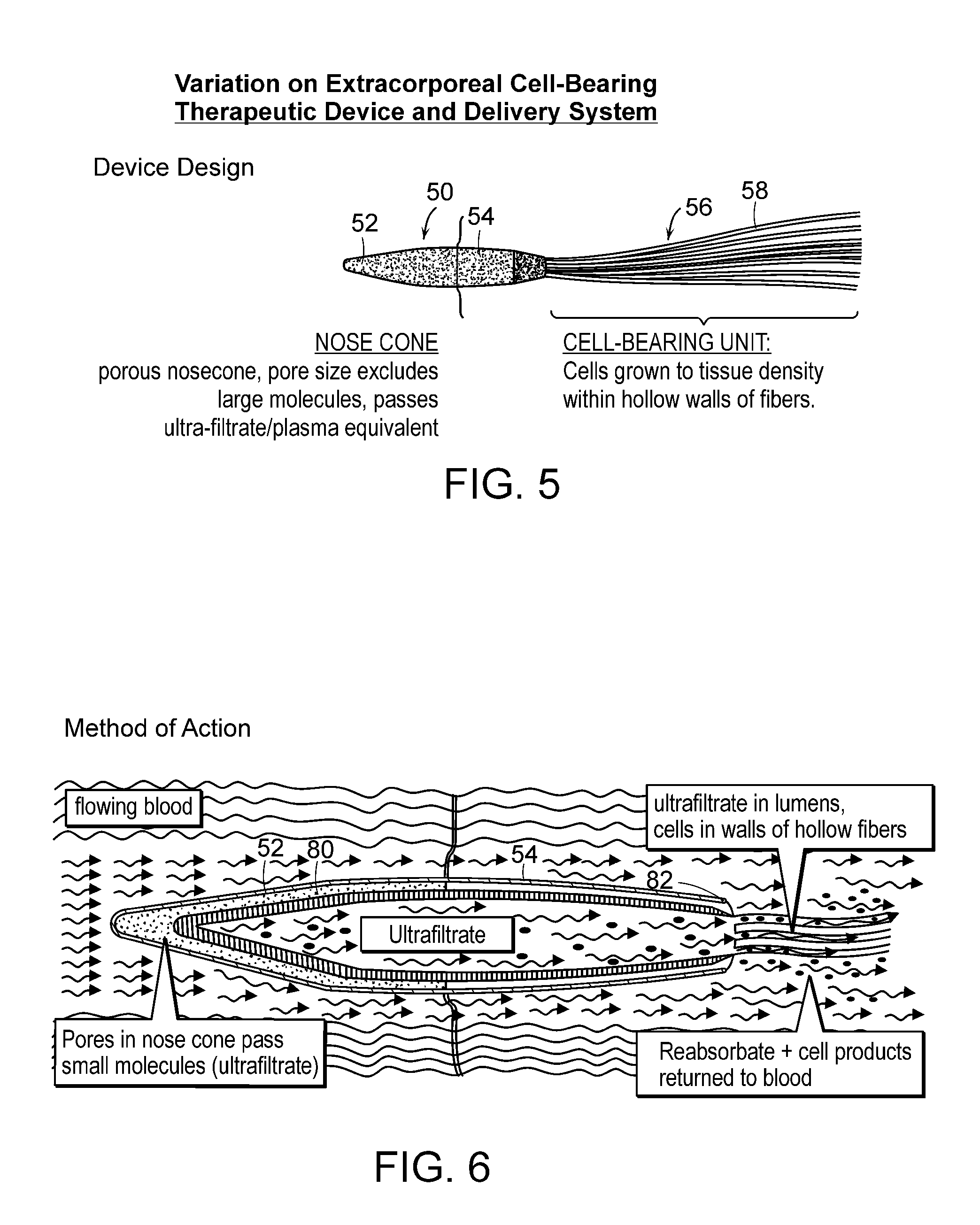
capsule may be retained upstream of the anchoring element, alternatively, the anchoring element may be located downstream of the anchoring element and retained in place by an attachment, for example, a hook or tether, extending from the anchoring element to the capsule. In addition, the capsule may be conical or wedge-like in shape to decrease the turbulence of blood flowing past the capsule. In a preferred embodiment, the capsule is formed from a material that can filter particles such that particles (including cells) below a certain size can pass through and particles above a certain size are prevented from passing through. The filter forms an ultrafiltrate from the blood to minimize the entry of proteins greater than 100,000 molecular weight so that immunoglobulins can be excluded from the
bathing media around the cells, especially for nonautologous cells not to activate an immunologic response.
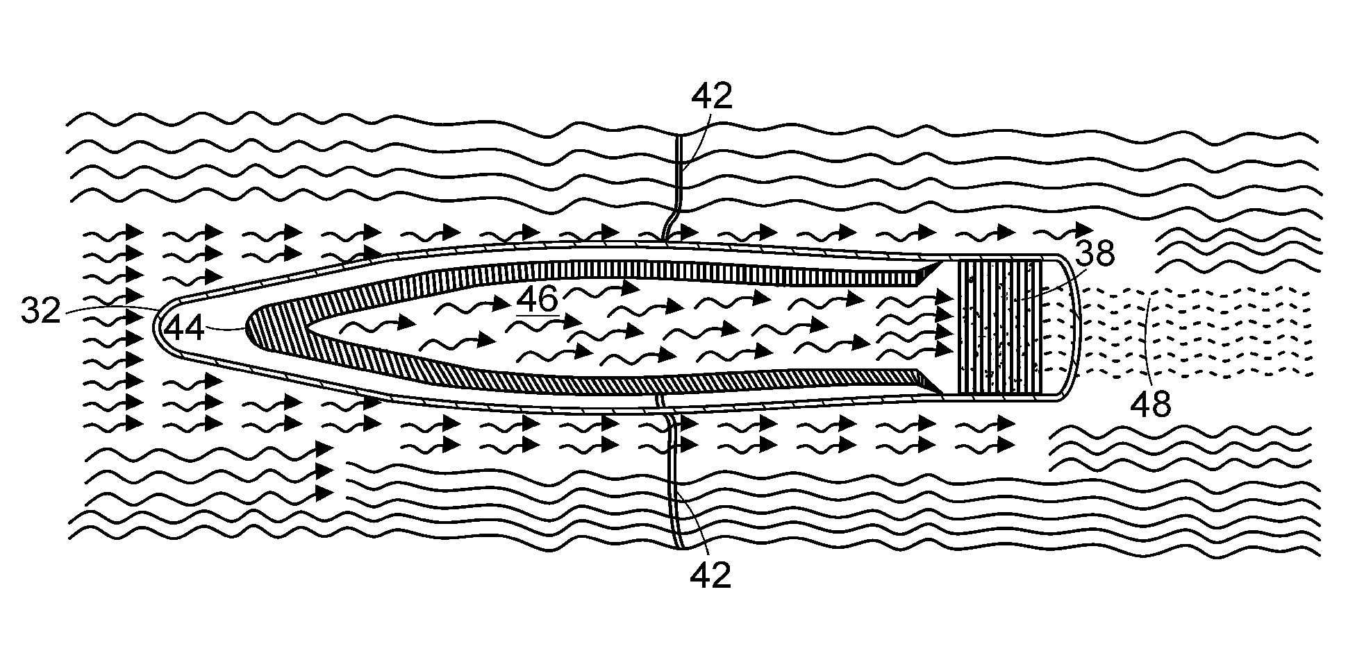
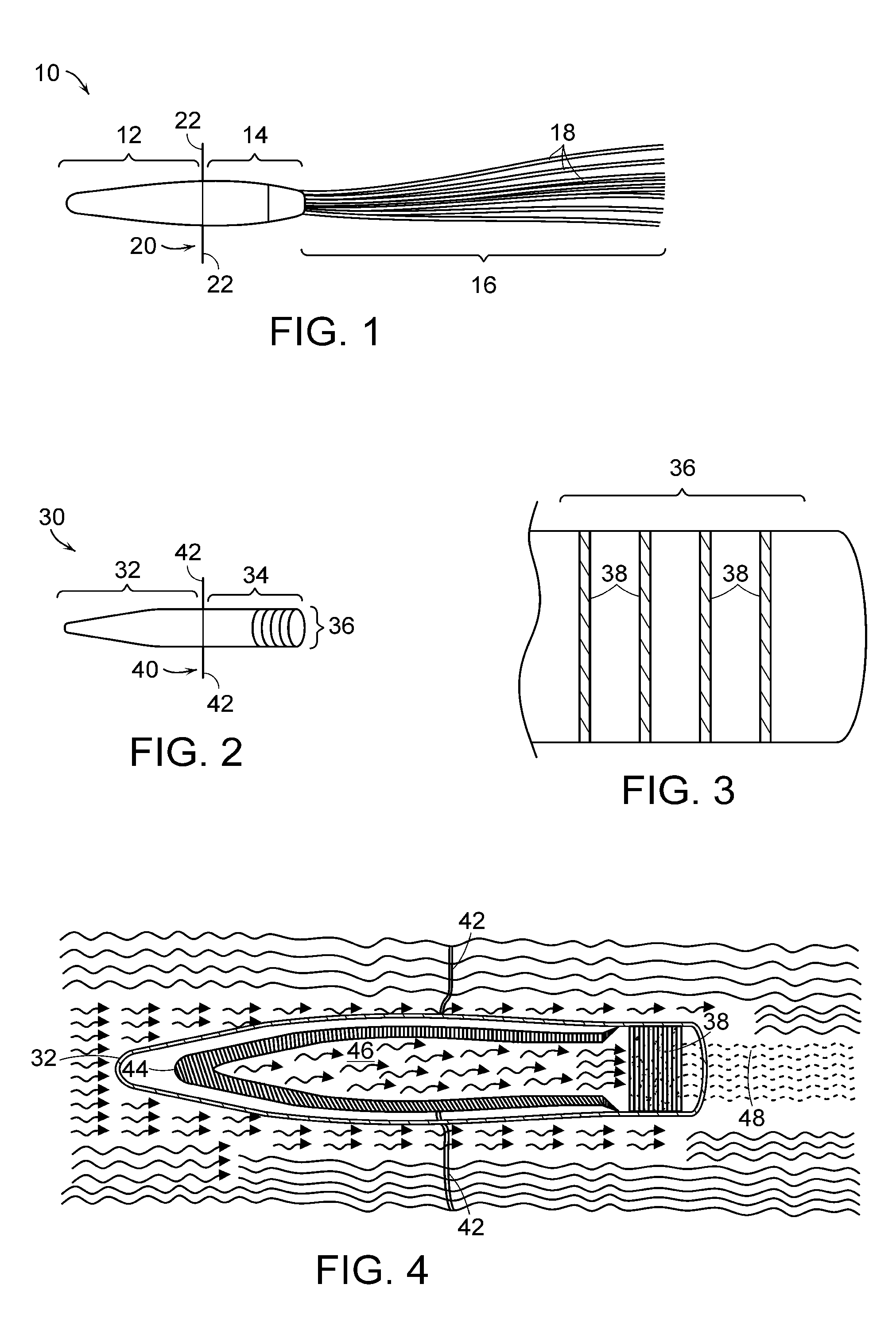
[0022]Preferred embodiments of the device include three configurations. Each preferred configuration isolates the therapeutic cells to minimize the immune response. In a first configuration, a device consists of a
cartridge, a cell bearing unit which may be in the form of tubes attached to the
cartridge and an anchoring
system. The therapeutic cells are disposed within the tubes and the cells are isolated by the size of the pores in the tube. In a second configuration, the cell bearing unit is in the form of disks that are disposed in the
cartridge. The therapeutic cells on the disks are protected from immunologic rejection by isolating the disks in the cartridge and providing pores in the cartridge that prevents the cells from being exposed to undesirable elements while allowing free physiologic exchange for the cells within the extracorporeal blood or fluid
stream. A third configuration is a combination of the configurations described above. Other configurations are possible.
[0025]In another aspect of the invention, an extracorporeal therapeutic system includes a housing defining an
interior space, a substrate including a carbon material coated with
niobium disposed within the housing, and at least one cell disposed on the substrate. This aspect or any of the following aspects can have any of the following features. The substrate can have a trabecular structure, and the substrate can be initially separate from the housing. The substrate can be coated with collagen IV. The substrate can also be coated with any three-dimensional biomatrix material that protects the integrity and durability of the carbon material, and the biomatrix material can promote or increase
cell expansion, attachment, and / or viability. The housing can include an inlet for receiving a fluid and an outlet for releasing a processed fluid. At least one
scaffold to retain the substrate can be disposed within the housing, and at least one flow separator can be disposed between the inlet and the substrate. At least one baffle can also be disposed between the inlet and the substrate. The substrate can be frozen.
[0030]In another aspect of the invention, an extracorporeal
cell based therapeutic device includes (a) an anchor system which can be capable of attaching the device to an extracorporeal tube, which when attached to the inner wall of the tube permits blood in the tube to pass therethrough; and (b) a capsule including a plurality of pores and having viable cells disposed therein, so that the capsule, when introduced into the tube, can be retained within the tube by the anchor system and the pores permit nutrients to enter the capsule to maintain viability of the cells disposed therein. This aspect or any of the following aspects can have any of the following features. The capsule can be defined by a semi-permeable membrane, and the semi-permeable membrane can include a material selected from
polyvinylidene fluoride, polyvinylchloride,
polyurethane, polyalginate,
polystyrene,
polyurethane,
polyvinyl alcohol,
polyacrylonitrile,
polyamide, polymethylmethacrylate,
polyethylene oxide, polytetrafluorethylene,
isocyanate,
cellulose acetate,
cellulose diacetate,
cellulose triacetate, cellulose
nitrate,
polysulfone, and mixtures thereof. The viable cells can be disposed on a plurality of filaments, and the filaments can be metallic. The pores can be dimensioned to prevent passage of antibodies therethrough, and the cells can be disposed on at least one disk. The cells can be eukaryotic cells, such as mammalian cells. The capsule can be adapted to be separated from the tube by attaching and detaching the anchor system from the tube. The capsule can include at least one hollow
fiber. The pores can permit solutes less than 150 kD to pass therethrough, and the device can be configured to provide a therapeutically significant amount of a molecule with or without using an artificial
blood pump.

