Low-energy defibrillation with nanosecond pulsed electric fields

a nanosecond pulsed electric field and low-energy technology, applied in the field of defibrillation, can solve the problems of reducing survival chances, nspefs not allowing pore expansion, and the first shock of the automated external defibrillator does not always terminate the vf or restore the organized rhythm, so as to reduce the risk of inhomogeneity, uniform tissue activation, and uniform tissue activation

Active Publication Date: 2018-02-22
OLD DOMINION UNIVERSITY RESEARCH FOUNDATION
View PDF11 Cites 0 Cited by
  • Summary
  • Abstract
  • Description
  • Claims
  • Application Information

AI Technical Summary

Benefits of technology

[0013]In some embodiments, nsPEFs interact with living matter using dielectric displacement, thereby allowing deeper penetration of the electric fields (e-fields) and more uniform activation of tissue. In these embodiments, deeper penetration of the e-fields and more uniform activation of tissue are critical advantages for defibrillation. Further to these embodiments, as a result of the aforementioned critical advantages, nsPEF defibrillation is less susceptible to inhomogeneities and provides uniform and simultaneous activation of cardiomyocytes.
[0014]In some embodiments, nsPEF defibrillation causes cell membrane nanoelectroporation whereas conventional methods employ long-pulses that cause electroporation. In these embodiments, pores within the cell membrane opened by nsPEFs are smaller in diameter than those opened by long pulses. Further to these embodiments, the pores opened by nsPEFs within cell membranes grow in diameter while the externally applied voltage is still on, and the briefness of nsPEFs does not allow pore expansion. In these embodiments, the smaller pore size resulting from nsPEFs greatly limits the transmembrane traffic through the pores and reduces the loss of critical solutes by the cell, thereby making nanoelectroporation less injurious than long-pulse electroporation.
[0015]In some embodiments, nsPEFs relies on displacement currents rather than on the conduction curr...

Problems solved by technology

Several studies have tested improved biphasic waveforms methodologies that further reduce the required defibrillation energy, but these results have indicated only marginal improvements.
Additionally, the first shock from the automated external defibrillator does not always terminate VF or restore the organized rhythm.
In clinical practice, patients often receive several shocks which delay the defibrillation and decrease the chances for survival even if cardiopulmonary resuscitation is performed.
Further to these embodiments, the pores opened by nsPEFs within cell membranes grow in diameter while the externally applied voltage is still on, and the briefness of nsPEFs does not allow p...

Method used


Image

  • Low-energy defibrillation with nanosecond pulsed electric fields
  • Low-energy defibrillation with nanosecond pulsed electric fields
  • Low-energy defibrillation with nanosecond pulsed electric fields

Examples


example 1

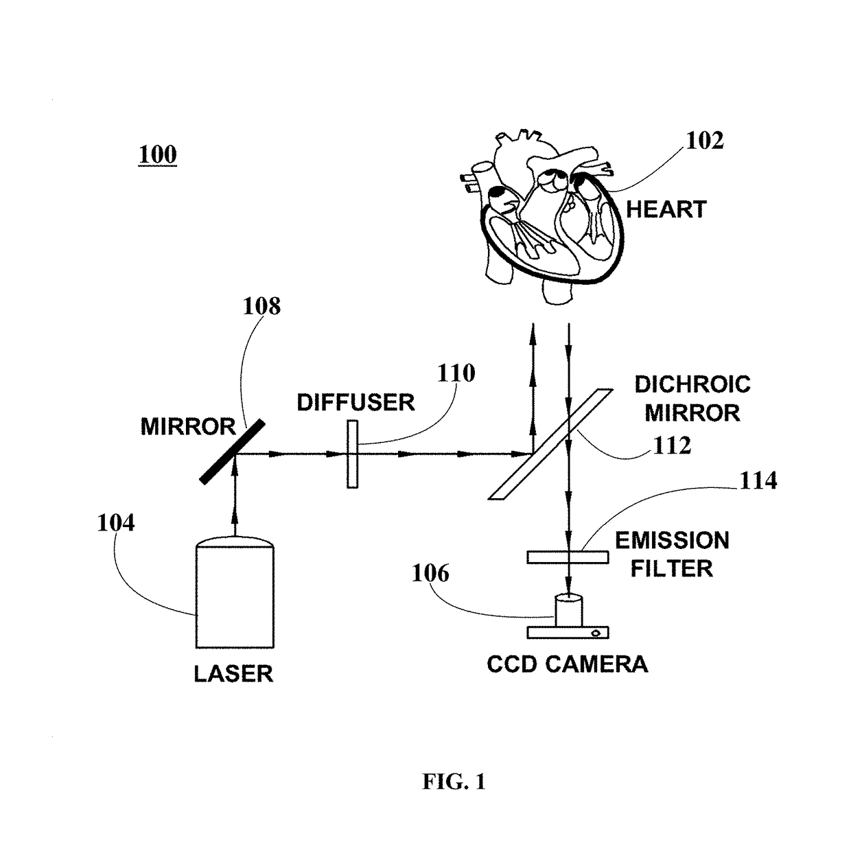
[0061]Three (3) New Zealand white rabbits were handled and euthanized according to the approved animal protocol. The three rabbits were heparinized (500 IU / kg) and brought to a surgical plane of anesthesia with 2.5-4% isoflurane. The hearts were surgically removed, the aorta immediately cannulated, and the hearts flushed with and immersed in cold cardioplegic solution (in mM: glucose 280, KCl 13.4, NaHCO3 12.6, mannitol 34). The hearts were placed within optical mapping system 100 and retrogradely perfused with Tyrode solution (in mM: NaCl: 130, KCl: 4.0, CaCl2: 1.8, MgCl2: 1.0, NaHCO3: 24, NaH2PO4: 1.2, glucose: 5.6) bubbled with 95% O2 / 5% CO2, at a pressure of 60-80 mmHg, with pH kept between 7.35 and 7.45 and temperature of about 37.5±0.5° C.

[0062]In an example, Langendorff-perfused heart 102 is injected with 5 mL bolus of the voltage-sensitive fluorescent probe Di-4-ANBDQBS (about 10 μM). In this example, an electromechanical uncoupler blebbistatin inhibitor (about 10 μmol / L) is...

example 2

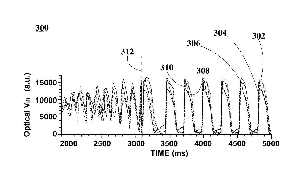
[0071]Twelve (12) New Zealand white rabbits of either sex (3-4 kg) were handled and euthanized according to the approved animal protocol. The twelve rabbits were heparinized (500 IU / kg) and brought to a surgical plane of anesthesia with 2.5-4% isoflurane. The hearts were rapidly removed, the aorta cannulated and flushed with ice cold Tyrode solution (in mM: NaCl: 128.2, NaCO3: 20, NaH2PO4: 1.2, MgCl2: 1.1, KCl: 4.7, CaCl2: 1.3, glucose: 11.1). The hearts were placed within optical mapping system 100, where they were perfused and superfused with warm oxygenated Tyrode solution (37±0.5° C.) at a constant pressure of 60-80 mmHg. After 30 min equilibration, 10-15 mM of 2, 3-butanedione monoxime was added to eliminate contractions.

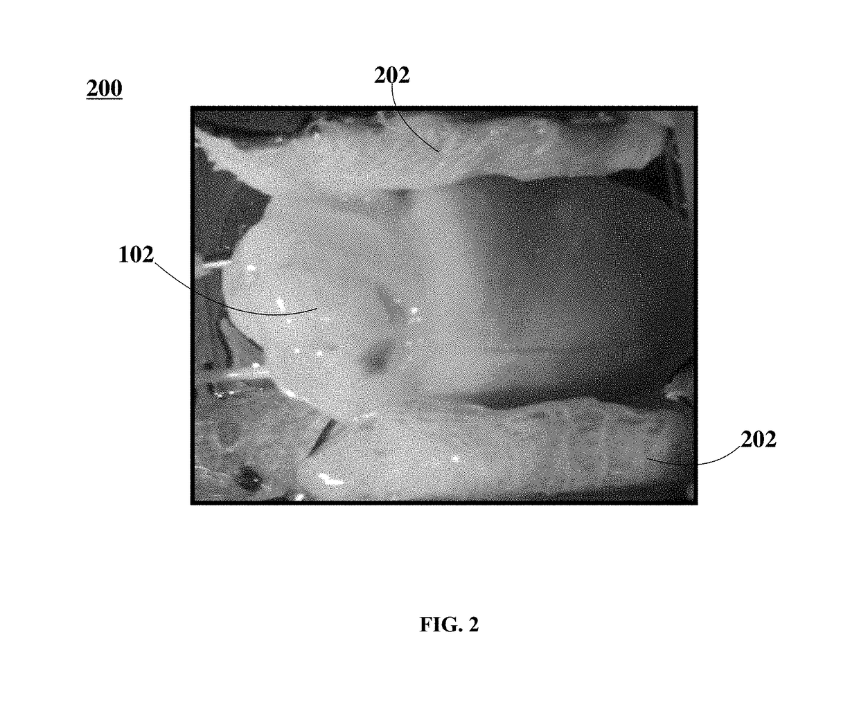
[0072]FIG. 5 is a photographic representation depicting a Langendorff-perfused rabbit heart in an optical mapping system, according to an embodiment. In FIG. 5, optical mapping system 500 includes Langendorff-perfused heart 102, electrodes 504, and inset image ...


PUM


Abstract

Methods for terminating fibrillation in a fibrillating heart employing nanosecond pulsed electric fields (nsPEFs) are disclosed. nsPEF defibrillation demonstrates its effectiveness as a new defibrillation modality, achieving reliable defibrillation with energies that are an order of magnitude lower than those needed for conventional defibrillation (millisecond shocks with mono- and bi-phasic waveforms). Tests did not reveal any negative effect of nsPEF defibrillation on cardiac tissue, in particular, cardiac tissue treated with nsPEFs does not exhibit a baseline shift in the optical transmembrane potential signal (distinctive feature that indicates electroporation), or changes in action potential duration or shape. The mechanism of nsPEF defibrillation is likely different from conventional defibrillation since it does not rely on membrane charging but on the basis of displacement currents that flow within nanoseconds after the shock is applied. nsPEFs provide the technology for the next generation of defibrillators that help emergency medical services to treat patients effectively.

Description

CROSS-REFERENCE TO RELATED APPLICATIONS[0001]This application claims priority to U.S. Provisional Application Ser. No. 62 / 127,462, filed Mar. 3, 2015, which is hereby incorporated by reference.BACKGROUNDField of the Disclosure[0002]The present disclosure relates generally to defibrillation, and more specifically, to systems and methods for heart defibrillation using nanosecond pulsed electric fields.Background Information[0003]The American Heart Association (AHA) estimates that each year about 420,000 people in the United States experience Emergency Medical Service (EMS)-assessed out-of-hospital cardiac arrests [1]. Among them 23% have an initial rhythm of ventricular fibrillation (VF) or ventricular tachycardia (VT) or are shockable by an automated external defibrillator [1-2]. For decades, the most common lifesaving procedure for terminating VF and VT is by applying an intense electric shock, conventionally known as defibrillation.[0004]Defibrillation is applied employing an elect...

Claims


Application Information

Patent Timeline
IPC IPC(8): A61N1/39
CPCA61N1/3906A61N1/327
Inventor PAKHOMOV, ANDREI G.ZEMLIN, CHRISTIAN W.
Owner OLD DOMINION UNIVERSITY RESEARCH FOUNDATION
Why Eureka
Try Eureka