Systems And Methods For Phase-Sensitive Inversion Recovery MR Imaging With Reduced Sensitivity To Cardiac Motion

a phase-sensitive inversion recovery and mr imaging technology, applied in the field of systems and methods for phase-sensitive inversion recovery mr imaging with reduced sensitivity to cardiac motion, can solve the problems of image contrast, uninterpretable or misinterpreted clinical images, image data corruption, etc., and achieve the effect of reducing spatial misregistration and reducing scan times

Active Publication Date: 2018-07-19
DUKE UNIV +1
View PDF0 Cites 3 Cited by
  • Summary
  • Abstract
  • Description
  • Claims
  • Application Information

AI Technical Summary

Benefits of technology

[0033]Exemplary embodiments of the present disclosure can provide a system and method for magnetic resonance (MR) imaging that can overcome limitations associated with standard phase-sensitive inversion recovery (PSIR) imaging techniques and similar MR imaging techniques by collecting paired reference and inversion recovery datasets within a shortened time frame. This can reduce spatial misregistration between the paired datasets for both segmented and single shot dataset acquisitions, and for acquisitions using or not using respiratory navigators. Further, scan times may be reduced for acquisitions using respiratory navigators.

Problems solved by technology

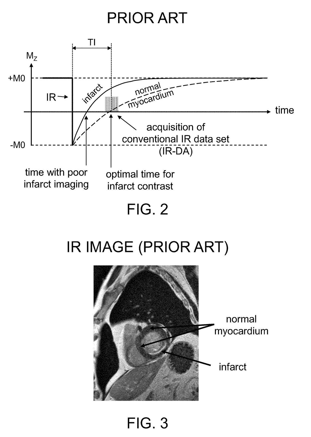
Such inverted or ‘wrong’ image contrast can occur if the time delay between IR and data readout is set too short.
Accordingly, use of an incorrect TI value can result in uninterpretable or misinterpreted clinical images.
Without such timing, different IR-DA sequences that are used to reconstruct a single image may obtain image data when the heart has different shapes within the cyclical heartbeat sequences, leading to corrupted image data.
Without such timing and triggered data acquisition, image data may be corrupted due to cardiac-induced motion of the imaged tissues.
Normal breathing by the subject or other bodily movement while MR image data is being collected can also lead to image corruption, because the region being imaged can move relative to the MRI apparatus.
However, many subjects do not hold their breath perfectly and a small non-negligible diaphragmatic drift may occur, leading to motion-based image corruption.
Additionally, involuntary swallowing during a breath-hold can corrupt the images.
Conventional PSIR techniques, e.g., as illustrated in FIG. 5, are susceptible to imaging artifacts arising from spatial misregistration of the conventional data and reference phase data.
Nonetheless, it is commonly assumed that by acquiring both datasets (IR-DA and REF) in an identical manner—e.g., in the same time point window within the cardiac cycle (centered over the same cardiac phase of two consecutive heart beats) and with the same spatial and temporal resolution—image artifacts are reduced to their lowest level, and that image artifacts would be worse if both datasets were not acquired in an identical manner.
These common assumptions can lead to suboptimal clinical results in patients because of the extended time periods needed to obtain the PSIR paired datasets (especially when obtaining a plurality of such paired datasets in segmented acquisitions), which may introduce various motion artifacts during the extended imaging procedure.
Another limitation of conventional PSIR techniques is that they are not well-suited for single-shot imaging during free-breathing acquisitions.
However, the requirement of acquiring two paired image datasets (IR-DA and REF) during PSIR imaging increases the likelihood of substantial breathing motion between the two datasets.
As a consequence, the final PSIR image can have motion artifacts due to spatial misregistration between IR-DA and REF datasets even though each of the two paired datasets was acquired in a single shot.
Although moving the IR-DA and REF data acquisitions closer together in time could result in fewer motion artifacts, it is commonly thought that such a shortened interval is not possible in conventional PSIR imaging procedures because the two datasets need to be acquired at the same cardiac phase in separate heartbeats.
Imperfect breath holds or free breathing imaging procedures, however, can lead to substantial shifts in cardiac position due to respiration-induced motion, thereby degrading spatial registration between the IR dataset and the corresponding reference dataset, leading to artifacts in the final PSIR image.
In clinical practice, imperfect breath holds (or an inability to hold the breath for an extended period) are common, resulting in significant artifacts for the resulting PSIR images.
Even when a subject can perform a perfect sustained breath hold, there may be ectopic heartbeats (such as a premature ventricular contraction) during the PSIR acquisition, which can also lead to spatial misregistration errors in the reconstructed PSIR image.
An important limitation of conventional PSIR techniques is that they are not well-suited for 3D (or 2D) respiratory-navigated procedures.
This approach can produce significant artifacts, because the reference dataset is usually acquired more than one second after the navigator data is acquired.
If any motion of the subject occurs (e.g. breathing) between acquisitions of the IR-DA dataset and the reference dataset, spatial misregistration artifacts will occur even if the single navigator indicates that the image data is “good” data.
However, the need for a second navigator results in a significant lengthening of the overall scan time needed for data acquisition.
Scan times can become so excessive that this version of respiratory navigated PSIR is rarely attempted in clinical practice.
To overcome this limitation, some navigated PSIR techniques do not navigate the reference data, assuming that the errors in misregistration will not greatly affect image quality.
However, as discussed above, this assumption is usually invalid, and because of poor resulting image quality, this type of respiratory-navigated PSIR (with non-navigated reference datasets) is also rarely attempted in clinical practice.
This technique works only with 2D datasets, and cannot correct for through-plane cardiac position shifts that typically occur with free breathing or poor breath-holding.

Method used


Image

  • Systems And Methods For Phase-Sensitive Inversion Recovery MR Imaging With Reduced Sensitivity To Cardiac Motion
  • Systems And Methods For Phase-Sensitive Inversion Recovery MR Imaging With Reduced Sensitivity To Cardiac Motion
  • Systems And Methods For Phase-Sensitive Inversion Recovery MR Imaging With Reduced Sensitivity To Cardiac Motion

Examples


Embodiment Construction

[0033]Exemplary embodiments of the present disclosure can provide a system and method for magnetic resonance (MR) imaging that can overcome limitations associated with standard phase-sensitive inversion recovery (PSIR) imaging techniques and similar MR imaging techniques by collecting paired reference and inversion recovery datasets within a shortened time frame. This can reduce spatial misregistration between the paired datasets for both segmented and single shot dataset acquisitions, and for acquisitions using or not using respiratory navigators. Further, scan times may be reduced for acquisitions using respiratory navigators.

[0034]In certain embodiments of the disclosure, a method for triggered cardiac PSIR imaging is provided in which the paired reference and inversion recovery (IR) datasets are acquired within the duration of a single cardiac cycle (e.g. the time between consecutive R-waves or heartbeats). In one embodiment, these two paired datasets are acquired within the sam...


PUM


Abstract

A magnetic resonance imaging system and method are provided for improved phase-sensitive magnetic resonance imaging of tissues affected by cardiovascular pulsatile motion. A magnetically-prepared image dataset and corresponding reference image dataset (for phase sensitivity) are obtained within the duration of a single cardiac cycle. The paired datasets can be single-shot or segmented datasets and a navigator sequence can optionally be provided with each paired dataset. The system and method take advantage of the shape symmetry of the cardiac cycle to acquire the paired dataset in a shorter time interval, thereby reducing misregistration artifacts. The magnetic preparation can include inversion recovery pulses, FIDDLE sequences, other magnetic preparation sequences, or combinations thereof. The reference dataset can be acquired at a lower resolution than the corresponding magnetically-prepared dataset without compromising image quality.

Description

CROSS-REFERENCE TO RELATED APPLICATION(S)[0001]The present application relates to and claims priority from U.S. Provisional Patent Application Ser. No. 62 / 446,027 filed on Jan. 13, 2017, the disclosure of which is incorporated herein by reference in its entirety.FIELD OF THE DISCLOSURE[0002]The present disclosure relates to a method and a system for providing improved phase-sensitive inversion recovery (PSIR) magnetic resonance imaging including, more specifically, improved PSIR imaging of tissue affected by cardiovascular pulsatile motion.BACKGROUND INFORMATION[0003]Magnetic resonance (MR) imaging is a known technology that can produce images of the inside of an examination subject without radiation exposure. In a typical MR imaging procedure, the subject is positioned in a strong, static, homogeneous base magnetic field BO (having a field strength that is typically between about 0.5 Tesla and 3 Tesla) in an MR apparatus, so that the subject's nuclear spins become oriented along th...

Claims


Application Information

Patent Timeline
Patent Type & Authority Applications(United States)
IPC IPC(8): G01R33/563G01R33/56
CPCG01R33/5602G01R33/5635G01R33/4818G01R33/56325G01R33/56509G01R33/5673G01R33/5676
Inventor REHWALD, WOLFGANG G.WENDELL, DAVID C.JENISTA, ELIZABETH R.CHEN, ENN-LINGKIM, RAYMOND J.
Owner DUKE UNIV
Why Eureka
Try Eureka