Three dimensional microtissue bioprinter

a bioprinter and microtissue technology, applied in the field of three-dimensional bioprinting, can solve the problems of inability to print contact live cells, slow throughput of bioprinters, and inability to allow microtissue printing, etc., to achieve the effect of improving the position control of dispensed materials, high cell viability, and not being able to use conventional cell aggregation techniques

Active Publication Date: 2018-08-30
THE GENERAL HOSPITAL CORP
View PDF4 Cites 16 Cited by
  • Summary
  • Abstract
  • Description
  • Claims
  • Application Information

AI Technical Summary

Benefits of technology

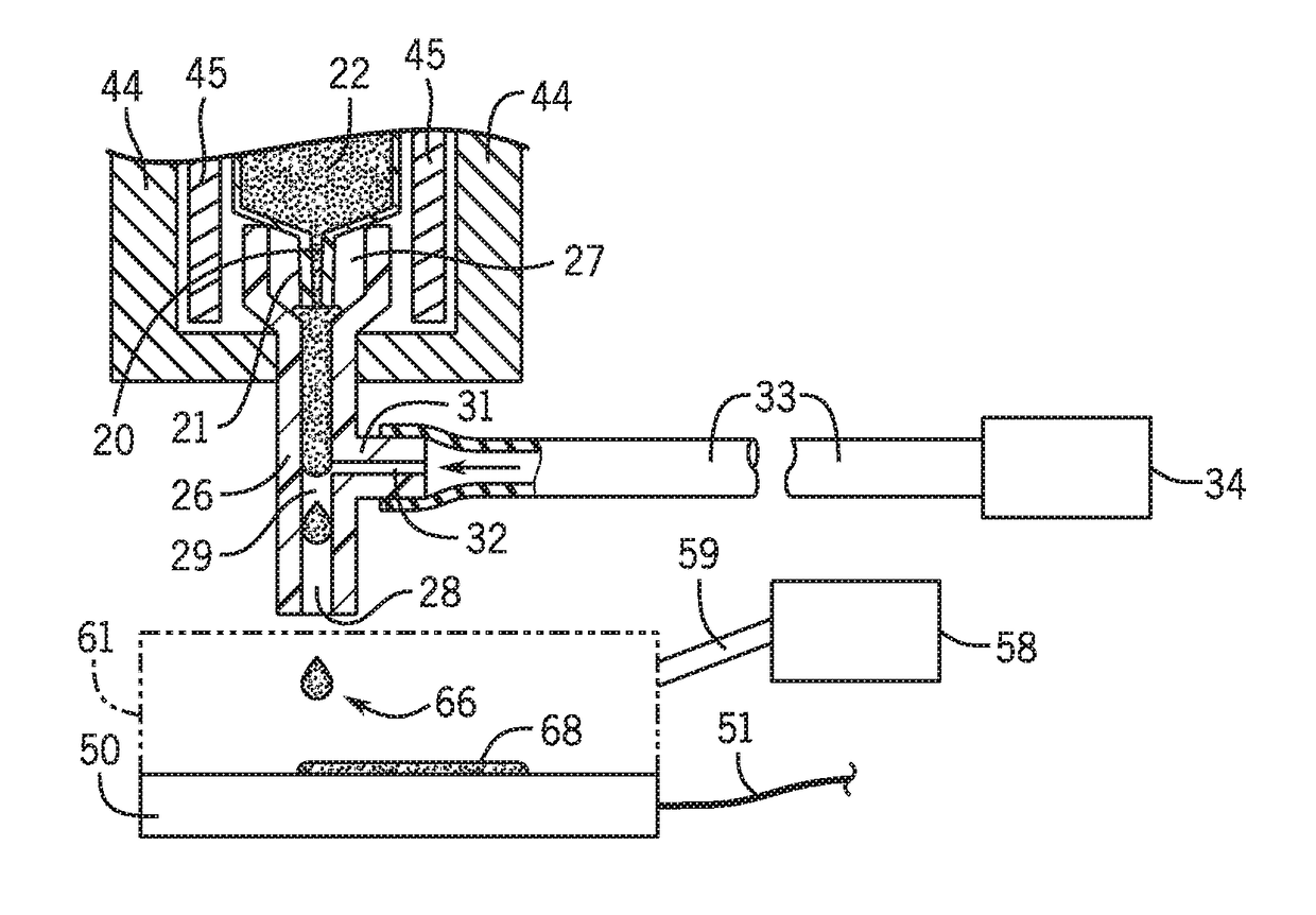
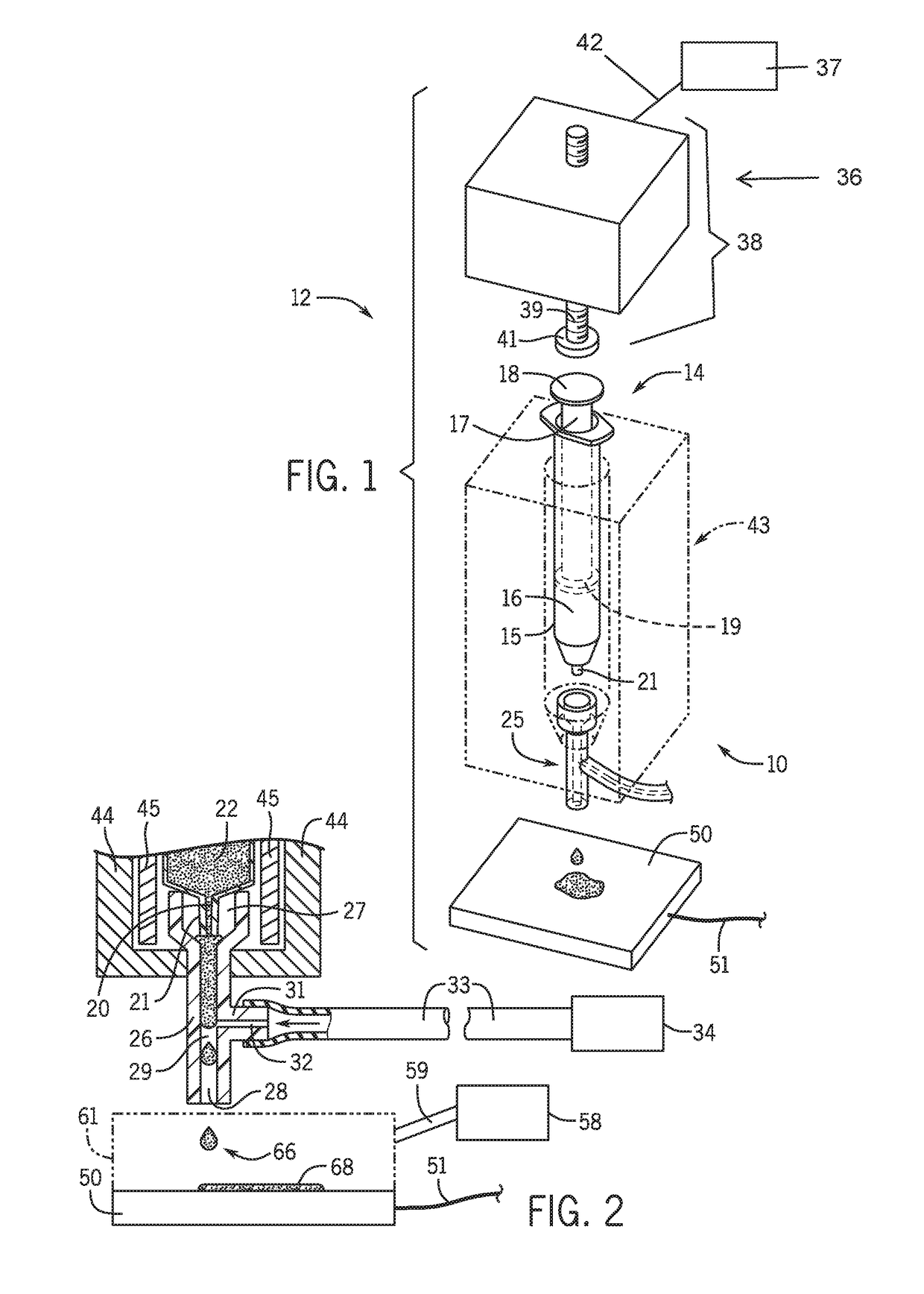
[0021]We are able to print micro tissues with morphology similar to the native tissue. In order to accomplish this, we print the droplets of bioink onto a temperature controlled surface to partially solidify the printed bio-ink during printing and control humidity to keep the printed bioink from drying and to maintain high cell viability. Our precise technology allows us to print tissue systems morphologically comparable to native tissues.
[0022]Our system can use a coaxial setup or a non-coaxial setup for delivery of pulsed air. A non-coaxial setup has better position control of the dispensed material.
[0023]Our results show that our printed tumor micro tissues, with the same components as native tumor tissue, are much more drug resistant to chemotherapy drugs than the cell aggregates. Additionally, there are many types of cells that do not aggregate and are not amenable to conventional cell aggregating techniques. We can still print these non-aggregating tissues using our bioprinter / technique. This represents a much more realistic testing scenario.
[0024]Our bioprinted microtissue is predeveloped and mechanically enhanced in vitro. Once injected in vivo, the tissues self-assemble into large tissue amalgamations which can repair the defects.

Problems solved by technology

Bioprinters are limited by slow throughput inherent in the small size of their building materials as well as the vast number of building units that must be deposited.
This technique does not allow microtissue printing as the resultant tissues are a couple of centimeters in size.
Contact live cell bioprinting can only perform low throughput printing.
Other commercial printers are unable to print tissue with architecture and morphology similar to native tissues.
This technique uses continuous air flow and continuous dispensing material flow in a coaxial setup, which cannot control the volume of each droplet and cannot dispense a single droplet.
This technique ordinarily dispenses thousands of droplets at a time, and in commercial systems, such as those available from Nisco Engineering AG, the material is limited to alginate.
Animal models do not fully resemble human diseases because of species differences.
However, weak mechanical properties, volume shrinkage and long time in vivo growth make it inapplicable to joint cartilage or bone repair.

Method used


Image

  • Three dimensional microtissue bioprinter
  • Three dimensional microtissue bioprinter
  • Three dimensional microtissue bioprinter

Examples


example 1

[0061]Example 1 describes the development of injectable mesenchymal stem cells (MSCs) microtissues for repairing cartilage defects using 3D bioprinting. This is a representative project for our high speed bioprinting in which we bioprint micro-cartilage and bone tissues using human bone marrow mesenchymal stem cells (MSCs). We have successfully bioprinted micro bone tissue and MSC-incorporated-tissue for cartilage and bone regeneration. Our bioprinted micro-tissue is predeveloped and mechanically enhanced in vitro. Once injected in vivo, the tissues self-assemble into large tissue amalgamations which can repair the defects.

Introduction

[0062]Articular cartilage exhibits poor intrinsic capacity for repair, and tissue engineering is a new approach for articular cartilage repair. Bone marrow mesenchymal stem cells (MSCs) and bulk hydrogel have been used to repair the cartilage defects in open surgery. As a minimally invasive approach is always preferred for joint surgery, injectable tis...

example 2

[0082]Example 2 describes high throughput 3D bioprinting of tumor micro tissues for drug development and personalized cancer therapy.

Introduction

[0083]Cancer caused about 25% of all deaths in the United States in 2014. In vitro anti-cancer drug screening is widely used in the pharmaceutical industry and chemosensitivity test for patients. However, only 2D assays and simple 3D culture models were used previously. But these models do not resemble the native tumor microenvironment, or yield inaccurate prediction of drug sensitivity. In this example, we report a novel 3D bioprinted model of tumor microtissues (TMTs), which resemble the native tumor characteristics, for high throughput screening. We characterized the 3D bioprinted tumors in various of aspects.

Methods

[0084]3D bioprinting: The bioink composed of tumor cells, stromal cells and ECM is loaded into our custom developed 3D bioprinter of the present invention. Micro tumor tissues were directly bioprinted into each well of a 96 o...

example 3

[0100]We 3D bioprinted liver tissues. Specifically, we have successfully bioprinted vascularized human micro liver tissues using HepG2 human liver cells and human endothelial cells. As shown in FIG. 21, the vascularized micro liver tissues show significant higher cell density (A) and viability (B) comparing with non-vascularized micro liver tissues (C and D).

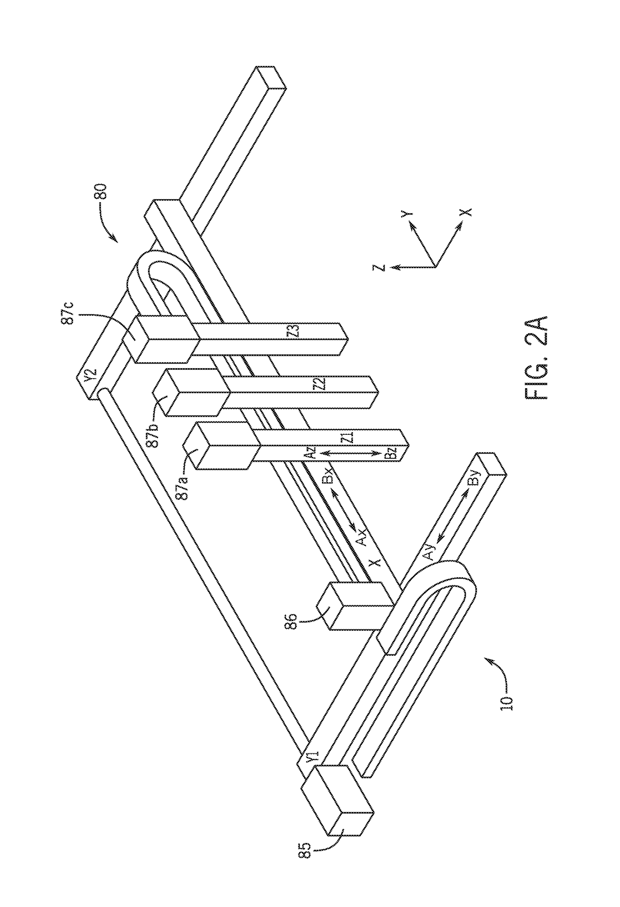
[0101]Thus, the invention provides a three dimensional microtissue bioprinter and methods for using the three dimensional microtissue bioprinter. We have disclosed a computer controlled programmable 3D bioprinter for generating live micro tissues with precisely controlled micro-scale accurate XYZ motion and volumetric nanoliter dispensing capability. We have printed many types of tissue with many different types of cells including pluripotent stem cells, bone marrow mesechymal stem cells, osteoblasts, fibroblast, human endothelial cells, liver cells, and many different tumor cells.

[0102]We have bioprinted micro tumor tissues for...


PUM

PropertyMeasurementUnit
volumeaaaaaaaaaa
inner diameteraaaaaaaaaa
diameteraaaaaaaaaa

Abstract

A bioprinter comprises one or more dispensing units. Each dispensing unit may include (i) a syringe including a hollow body and a plunger dimensioned to translate in the body wherein the body has an exit orifice; (ii) an actuator in contact with a proximal end of the plunger; (iii) a controller for moving the actuator; and (iv) a nozzle having a wall defining a fluid path extending from an inlet of the nozzle to an outlet of the nozzle. The inlet of the nozzle is in fluid communication with the exit orifice of the syringe body. The nozzle includes a fluid passageway in fluid communication with a source of fluid and the fluid path. The bioprinter can be used in a method of preparing microtissue comprising dispensing a bioink from one or more dispensing units of the bioprinter on a plate. The microtissue may comprise cartilage cells or tumor cells or liver cells.

Description

CROSS-REFERENCES TO RELATED APPLICATIONS[0001]This application claims priority from U.S. Patent Application No. 62 / 214,551 filed Sep. 4, 2015.STATEMENT REGARDING FEDERALLY SPONSORED RESEARCH[0002]Not Applicable.BACKGROUND OF THE INVENTION1. Field of the Invention[0003]The invention relates to three-dimensional bioprinting, and more particularly to a computer controlled programmable three dimensional (3D) bioprinter generating live microtissues with precisely controlled microscale accurate XYZ motion and volumetric nanoliter dispensing capability.2. Description of the Related Art[0004]Bioprinters were developed to try and meet the challenge of printing three dimensional tissues. Bioprinters fabricate structures via a dropwise printing of cells with a material which serves as the “bio-glue”. Bioprinters are limited by slow throughput inherent in the small size of their building materials as well as the vast number of building units that must be deposited. Various techniques and techno...

Claims


Application Information

Patent Timeline
Patent Type & Authority Applications(United States)
IPC IPC(8): A61L27/38B33Y30/00B33Y50/02G01N33/50
CPCA61L27/3834B33Y30/00B33Y50/02G01N33/5011A61L27/3847A61L27/3852B33Y10/00A61L2430/02A61L2430/06A61L2400/06A61L27/3804A61L27/50B41J2/04B29C64/112B29C64/209C12M33/00B01L3/0268C12N5/0671C12N5/0697B01L2200/0673B01L2300/1822B01L2400/0478B01L2400/0487C12M21/08
Inventor PANG, YONGGANGGROTTKAU, BRIAN
Owner THE GENERAL HOSPITAL CORP
Why Eureka
Try Eureka