

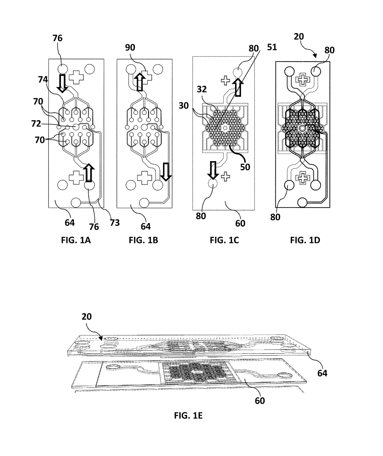
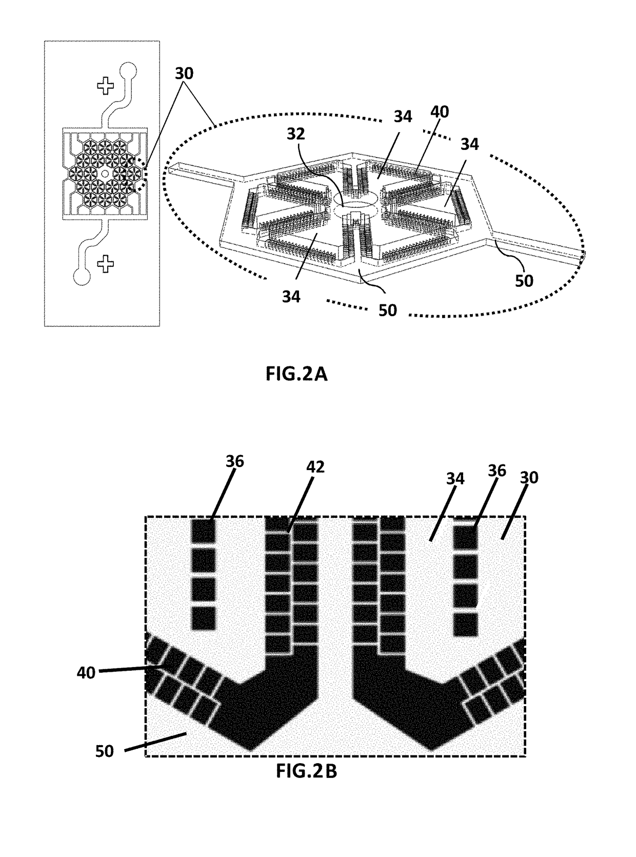
Microfluidic device for culturing cells
a microfluidic device and hepatocyte technology, applied in microstructural technology, biochemistry apparatus and processes, biochemistry apparatus, etc., can solve the problems of high failure rate, inability to adequately evaluate and predict the mechanisms of liver injury and drug toxicity in humans, and the efficacy and toxicity trials on animal models fail to reveal the specific human metabolic pathways of the substance being tested. , to achieve the effect of improving performan
- Summary
- Abstract
- Description
- Claims
- Application Information
AI Technical Summary
Benefits of technology
Problems solved by technology
Method used
Image

Examples
experiment 1
[0062]Cell Seeding / Culturing and Maintenance of the Microfluidic Devices
[0063]Both ipsc and primary hepatocytes were cryopreserved and directly thawed prior to seeding.
[0064]Seeding / Culturing of Cellartis hiPSC
[0065]Enhanced ips derived hepatocytes were purchased from Cellartis (Takarabio, Gothenburg, Sweden) and were handled according to the company's protocol. Briefly, cells were thawed in a 37° C. water bath and immediately transferred to 15 ml of thawing medium (InvitroGro HT from BioreklamationIVT)+0.1% PEST and Y-23627. Each vial contained approximately 12 M viable cells. Cells were incubated in the thawing medium at room temperature for 15-20 minutes and centrifuged at 100×g for 2 minutes. The thawing medium was aspirated and cells were gently re-suspended in plating medium (InvitroGro CP Bioreklamation IVT)+0.1% PEST. 96 well plates were immediately seeded by 150 pi of the cell suspension and placed in the incubator. To seed the devices, cell suspension was centrifuged again...
experiment 2
[0072]Cell Morphology and Long-Term Maintenance, Comparison Between Ipsc, Primary Hepatocytes and HepG2 Cell Line
[0073]FIG. 7 shows the tissue morphology of HepG2 cell line in the tissue chambers 30. Images are taken in day 5 after cell seeding. The cluster formation and tissue-like structure generation was observed starting from day 2 after seeding. The duration of experiments was 6 days for HepG2 cells. For ips-derived hepatocytes it was observed that during the 3 weeks after cell seeding day the cells form the 3D tissue-like structures (Data not shown). The tissue formation process started at day 2 after seeding after cells were attached to the bottom glass slide. This process was monitored on a daily basis and bright-filed microscope images were taken every second day. Primary cells did not attach to the bottom of the glass slide without an extra cellular matrix (ECM) coating and remained as cell clusters during the 7 days of experiment period (Data not shown). To monitor the ce...
experiment 3
[0085]Albumin secretion as a liver-specific biomarker was measured by means of enzyme-linked immunosorbent assay (ELISA) from Bethyl Inc. The assay was performed based on the manufacturer protocol. Collected supernatants were stored in −20° C. prior to the assay day. ELISA assays were run in clear flat bottom 96 well plates (Nunc™) and measured in a microtiter plate reader (FLUOstar Omega, BMG LABTECH, Germany) in absorbance mode at 450 nm wavelength. As seen in FIG. 11(a) the amount of secreted albumin per day for a period of 6 days was recorded for HepG2 cells under both pump-driven and gravity-driven conditions. The results show that the amount of secreted albumin for the devices under a steady flow condition was higher compared to the gravity-driven flow devices. However, by elaborating the top feed network and adjusting the hydraulic resistance of the feed channels a steady gravity-driven flow with desired flow rates under the whole 24-hr period may...
PUM
| Property | Measurement | Unit |
|---|---|---|
| width | aaaaa | aaaaa |
| width | aaaaa | aaaaa |
| diameter | aaaaa | aaaaa |
Abstract
Description
Claims
Application Information