
RNA Fixation and Detection in CLARITY-based Hydrogel Tissue

a technology of clarity-based hydrogel tissue and fixation, which is applied in the direction of instruments, optical elements, biochemistry apparatus and processes, etc., can solve the problems of limited to small volumes, difficult tract tracing and circuit mapping, and many such approaches may not be compatible with accessing the wealth of biological information contained in the rna of large intact volumes

Active Publication Date: 2019-04-25
THE BOARD OF TRUSTEES OF THE LELAND STANFORD JUNIOR UNIV
View PDF4 Cites 27 Cited by
  • Summary
  • Abstract
  • Description
  • Claims
  • Application Information

AI Technical Summary

Benefits of technology

This patent describes methods and systems for detecting RNA in intact tissue using carbodiimide-based chemistry. The method involves fixing the tissue with hydrogel subunits, polymerizing them to form a hydrogel-embedded specimen, and then fixing RNA in the specimen using carbodiimide-mediated crosslinking. The specimen is then cleared of cellular components and lipids, and a nucleic acid probe is used to detect the RNA. The method allows for the visualization of RNA in tissue using confocal microscopy, two-photon microscopy, light-field microscopy, and tissue expansion microscopy, as well as CLARITY™-optimized light sheet microscopy (COLM). Overall, the patent provides technical means for studying RNA expression in various types of tissue.

Problems solved by technology

Historically these two goals of content and context have been in opposition, since higher-resolution analyses have tended to require disassembling the system or accepting a limited field of view.
For example, one of the current challenges in neuroscience is to query molecular identity, activity level, and circuit wiring of individual cells within intact brain networks, which would require linkage of information spanning several orders of magnitude in spatial scale.
Such reconstructions have been valuable, but are often laborious, limited to small volumes, and susceptible to loss of information at section boundaries, making tract-tracing and circuit-mapping particularly difficult (Wanner et al., 2015).
To date these technologies have chiefly focused on interrogating proteins, whether transgenically-expressed or immunohistochemically-detected (with the exception of single probes tested in CLARITY-based hydrogel experiments in sectioned tissue; Chung et al., 2013; Yang et al., 2014), and many such approaches may not be compatible with accessing the wealth of biological information contained in the RNA of large intact volumes.

Method used


Image

  • RNA Fixation and Detection in CLARITY-based Hydrogel Tissue
  • RNA Fixation and Detection in CLARITY-based Hydrogel Tissue
  • RNA Fixation and Detection in CLARITY-based Hydrogel Tissue

Examples


example 1

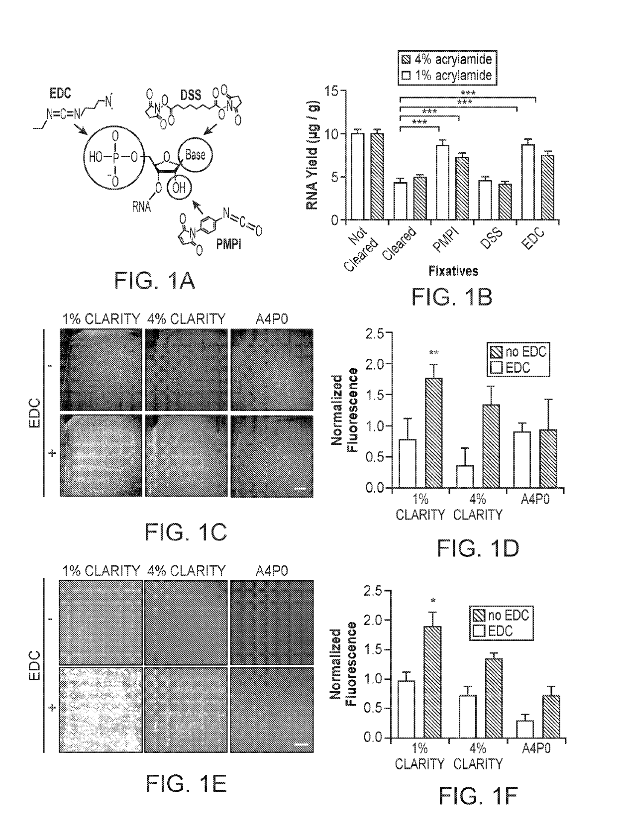
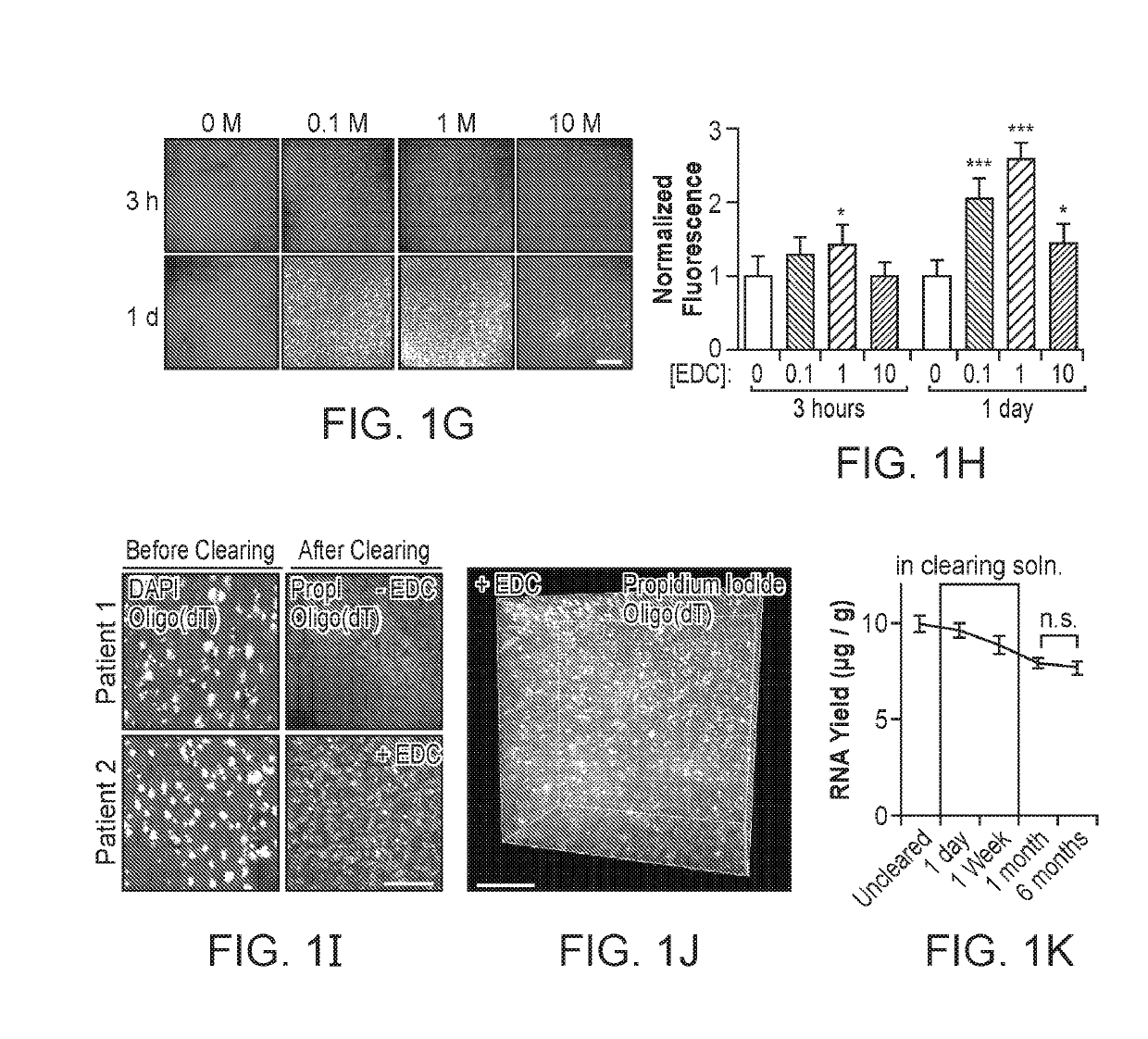
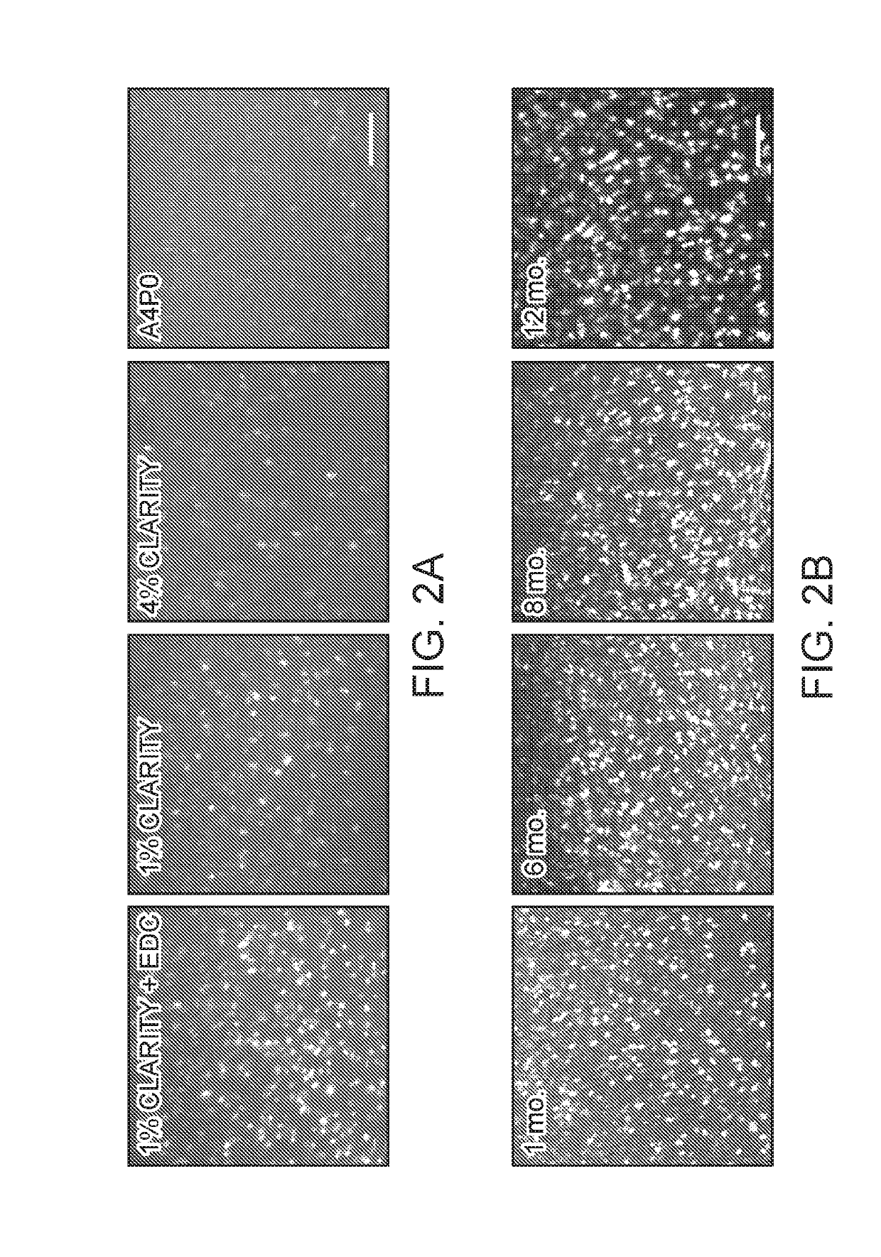
[0115]Advancing Clarified Tissue Chemistry with Carbodiimide-Based RNA Retention

[0116]Many existing clearing methods rely on incubation of tissue for prolonged periods of time at temperatures of 37° C. or greater (Chung et al., 2013; Tomer et al., 2014; Yang et al., 2014; Renier et al., 2014; Susaki et al., 2014; Tainaka et al., 2014); however, formalin is known to revert its crosslinks at elevated temperatures, and the bonds made to nucleic acids are particularly vulnerable (Masuda et al., 1999; Srinivasan et al., 2002). Therefore, to improve retention of RNA during high-temperature tissue clearing, we sought to introduce temperature-resistant covalent linkages to RNA molecules prior to clearing, by targeting functional groups on the RNA molecule for fixation to surrounding proteins or the hydrogel matrix.

[0117]We explored three tissue-chemistry strategies: EDC (1-Ethyl-3-3-dimethyl-aminopropyl carbodiimide) for linkage of the 5′-phosphate group to surrounding amine-containing prot...

example 2

[0122]Quantifying Diffusion of In Situ Hybridization Components into Clarified Tissue

[0123]After ensuring stable retention of RNAs, we next focused on access to target RNAs for specific labeling in transparent tissue volumes. Traditional in situ hybridization (ISH) uses labeled DNA or RNA probes, which are detected by enzyme-conjugated antibodies that catalyze the deposition of chromophores or fluorophores at the target location. Interrogation of RNA by these methods requires the penetration of each component to the target location. Since prior work had only shown detection of RNA in small volumes (100-500 μm thick; Chung et al., 2013; Yang et al., 2014), we sought to test the ability of ISH components to diffuse into intact EDC-CLARITY tissue.

[0124]We began by characterizing the diffusion of nucleic acid probes into EDC-CLARITY tissue. We incubated tissue blocks with 50-base DIG-labeled DNA or RNA probes, and visualized the diffusion profile of these probes by cutting cross-section...

example 3

In Situ Hybridization in EDC-CLARITY

[0127]Based on these findings that demonstrate stable retention of RNA with EDC-CLARITY and rapid penetration with short DNA probes, we next sought to develop a panel of oligonucleotide-based ISH techniques for application to large transparent tissue volumes. We began with digoxigenin (DIG)-labeled DNA oligonucleotide probes targeting somatostatin mRNA (3 probes) and amplified with anti-DIG HRP-conjugated antibody and TSA (FIG. 4A). In initial tests, we were readily able to resolve individual cells expressing somatostatin mRNA, demonstrating that specific mRNA species within the EDC-CLARITY hydrogel can be retained and are accessible to ISH probes (FIG. 4C).

[0128]However, using this technique in larger volumes revealed two major limitations: (1) the surface of the tissue sections showed non-specific staining that could result in false positives during cell detection, and (2) the signal was visible only to a depth of <300 μm (FIG. 4C). A similar pa...


PUM

PropertyMeasurementUnit
timeaaaaaaaaaa
timeaaaaaaaaaa
temperatureaaaaaaaaaa

Abstract

Methods, kits, and systems for fixation of RNA permitting its detection in intact tissue, such as, large volume of mammalian tissue are disclosed. The methods, kits, and systems utilize carbodiimide-based chemistry to stably retain RNAs in tissue clarified using CLARITY. Also provided herein are methods, kits, and systems for detection of RNAs in clarified tissue.

Description

CROSS-REFERENCE[0001]This application claims the benefit of U.S. Provisional Patent Application No. 62 / 293,490, filed Feb. 10, 2016, which application is incorporated herein by reference in its entirety.INTRODUCTION[0002]An exciting theme in modern biology is moving toward joint maximization of the content and context of molecular-level observations—that is, obtaining high-resolution and content-rich information about the biological system, while also maintaining this system largely or fully intact to preserve crucial contextual information. Historically these two goals of content and context have been in opposition, since higher-resolution analyses have tended to require disassembling the system or accepting a limited field of view. But the value of obtaining and integrating information about the identity, function and connectivity of cells in intact 3D volumes has been increasingly appreciated.[0003]For example, one of the current challenges in neuroscience is to query molecular i...

Claims


Application Information

Patent Timeline
Patent Type & Authority Applications(United States)
IPC IPC(8): C12Q1/6841C12Q1/6806G02B21/06G01N1/30C12Q1/6837
CPCC12Q1/6841C12Q1/6806G02B21/06G01N1/30C12Q1/6837G01N2001/305G02B21/0024C12Q1/68C12Q2523/101C12Q2543/10C12Q2565/601
Inventor DEISSEROTH, KARL A.SYWLESTRAK, EMILY L.RAJASETHUPATHY, PRIYAWRIGHT, MATTHEW
Owner THE BOARD OF TRUSTEES OF THE LELAND STANFORD JUNIOR UNIV
Learn More