
Method for the preselection of drugs for protein misfolding diseases

a protein misfolding and drug technology, applied in the field of alzheimer's disease treatment, can solve the problems of no spectral or structural resolution, brain damage already irreversible, and difficulty in drug development against alzheimer's disease, and achieve the effect of increasing -helix and reducing -sh

Active Publication Date: 2019-09-19
BETASENSE GMBH
View PDF0 Cites 2 Cited by
  • Summary
  • Abstract
  • Description
  • Claims
  • Application Information

AI Technical Summary

Benefits of technology

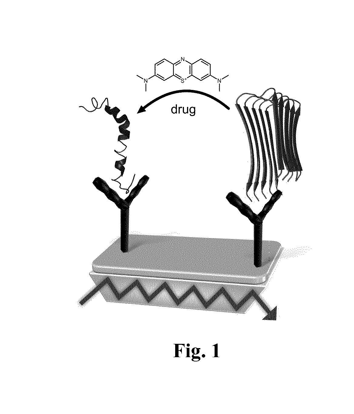
The present invention provides a screening assay that uses an infrared sensor element to analyze potential drugs that can help treat protein misfolding diseases. The assay involves modifying a germanium surface with antibodies that can bind to a specific target protein. The drug is then tested by adding a solution containing a potential drug to see if it affects the structure of the target protein. The analysis is done in real-time and without the need for labeling. The invention allows for the direct screening of drugs and the evaluation of their efficacy on a target protein. The use of infrared sensors in this assay provides a reliable and accurate method for analyzing the interaction between drugs and target proteins.

Problems solved by technology

The drug development against the Alzheimer's disease is challenging.
In these stages at which clinical symptoms appear the brain is already irreversible damaged.
Since, these techniques only provide kinetical information, but no spectral or structural resolution; they are not able to monitor secondary structure distributions of proteins.
This approach was successfully employed to identify small molecules that bind to monomeric Tau (Pickhardt et al., Current Alzheimer Res. 12(9):814-28 (2015)), but an effect on the secondary structure cannot be detected due to the lack of spectral and structural resolution.
Further techniques like surface enhanced Infrared absorption (SEIRA) spectroscopy provide spectral resolution, but the reproducibility of the measurements is very challenging due to the preparation of the rough gold surfaces and thus does not provide a robust platform for a protein-drug analysis.
Nevertheless, PET and MRT are very expensive and time-consuming techniques, which cannot be employed as mass screening method nor for preselection of drug candidates.
A further disadvantage is in the case of PET the usage of contrast agents, which also stress the patients.
Most importantly, ELISA and sFIDA do not provide direct information about the secondary structure distribution or changes of the secondary structure distribution by the drug.
Due to the preparation process of the Western blot the native secondary structure of the protein is lost.
Thus ELISA, sFIDA, or Western blots are not able to measure directly the intervention of a potential drug on the secondary structure distribution of a target protein.

Method used


Image

  • Method for the preselection of drugs for protein misfolding diseases
  • Method for the preselection of drugs for protein misfolding diseases
  • Method for the preselection of drugs for protein misfolding diseases

Examples


example 1

Blue “Cures” Alzheimer's Disease In Vitro

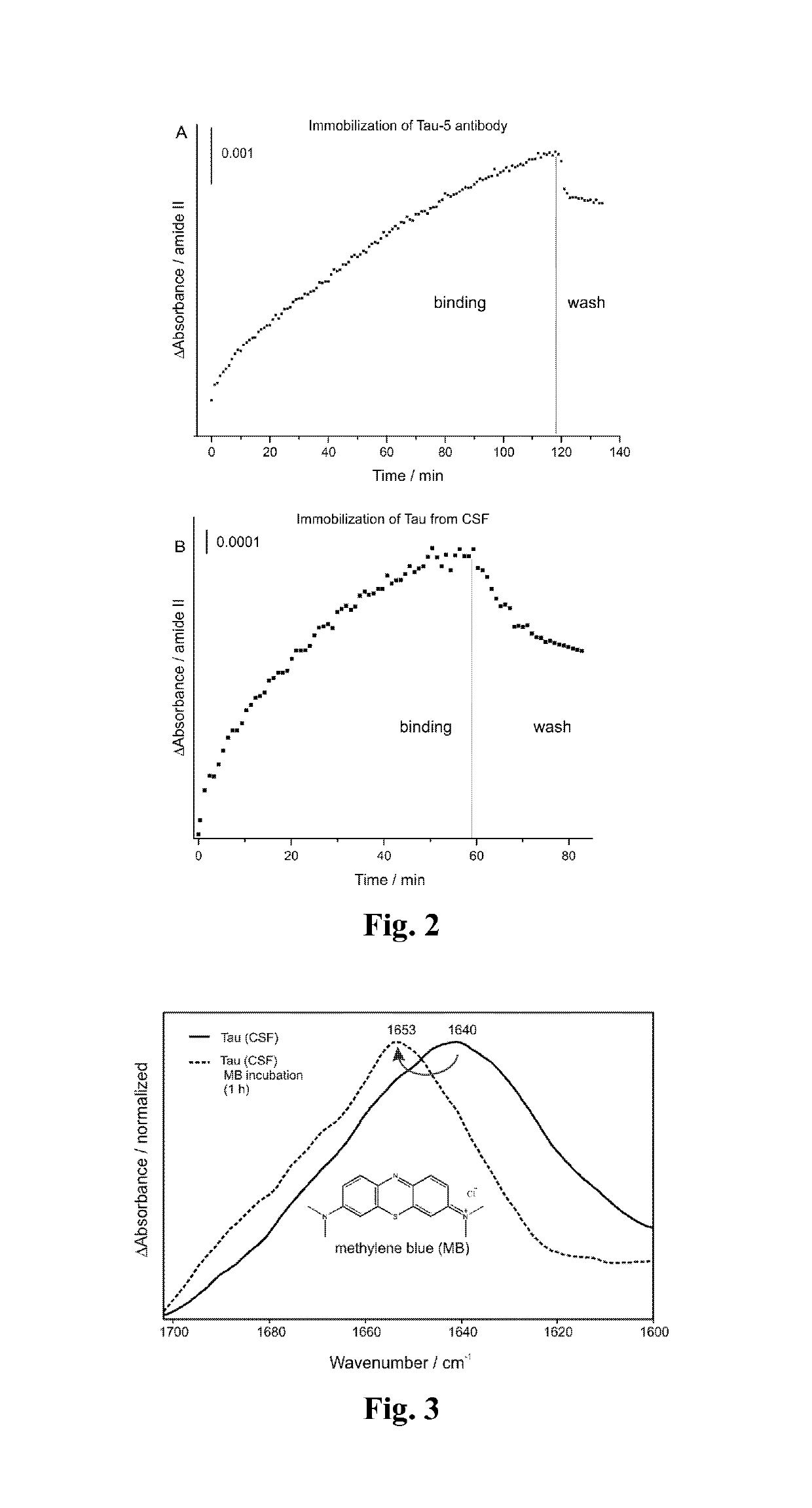
[0042]To monitor the drug effect of methylene blue the invented method was employed. We previously invented an immuno-ATR sensor, which differentiates AD with an accuracy of 90% based on CSF and 84% based on blood plasma analyzes (Nabers et al., Anal. Chem. 88(5):2755-62 doi:10.1021 / acs.analchem.5b04286 (2016)). First, we employed silane chemistry to modify the germanium surface (Schartner et al., JACS 135(10):4079-87 doi:10.1021 / ja400253p (2013)). Second, the monoclonal IgG1 antibody Tau-5 was covalently immobilized on the germanium surface. The immobilization is completed after 2 hours as presented in FIG. 2A by reaching an absorbance of 5 mOD. After washing the surface with binding buffer 1 the antibody remains stable (FIG. 2A). To obtain a highly specific surface the saturation with casein is crucial (Nabers et al., J. Biophotonics 9(3):224-34 doi:10.1002 / jbio.201400145 (2016)). Finally, a complex sample such as cerebrospinal fluid (CSF) ...

example 4

does not Affect Tau

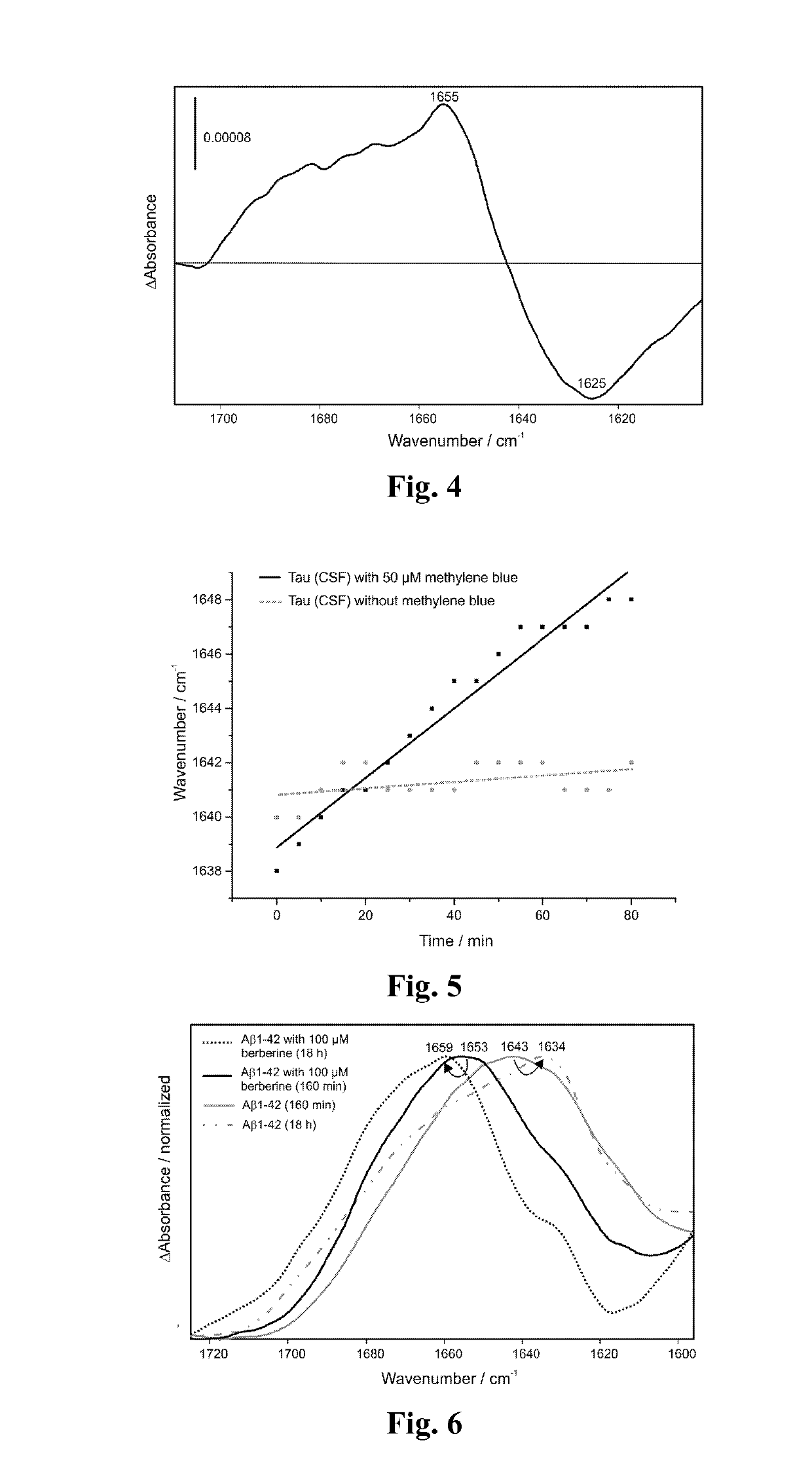
[0045]Furthermore, we studied the effect of berberine on the Tau protein (from CSF) under the same conditions as for methylene blue (FIG. 8). The amide band and its maximum are not affected by the berberine treatment, which indicates that berberine has no significant effect on the Tau protein in comparison to methylene blue (FIG. 3 and FIG. 8).


PUM


Abstract

The invention provides a method that gives direct information about the intervention of a potential drug on the secondary structure distribution of a targetbiomolecule, i.e., for a disease with misfolded protein, such as neurodegenerative diseases in a complex body fluid. The secondary structural change is monitored by vibrational spectroscopy. The method can be applied for prescreening of drug candidates for targeting of specific biomolecules. The effect of the drug on the secondary structure distribution is monitored label-free in real time and provides thereby direct information about the efficacy of the potential drug.

Description

[0001]The invention provides a method that gives direct information about the intervention of a potential drug on the secondary structure distribution of a target biomolecule, i.e., for a disease with misfolded proteins, such as many neurodegenerative diseases, by monitoring a secondary structural change by vibrational spectroscopy. The method can be applied for prescreening of drug candidates targeting specific biomolecules. The intervention of the drug is monitored label-free in real time and provides direct information about the efficacy of the potential drug to prevent pathological folding species.BACKGROUND OF THE INVENTION[0002]For example, treatment of Alzheimer's disease (AD) is an emerging field, which affects millions of human beings worldwide. The disease progression is characterized by the formation of plaques and tangles in the brain, which are based on aggregation processes of the Aβ peptide and Tau protein. The protein aggregation is driven by structural transition in...

Claims


Application Information

Patent Timeline
IPC IPC(8): G01N33/68G01N21/552
CPCG01N2333/4709G01N21/552G01N2800/52G01N2800/2821G01N33/6896G01N2021/3595G01N21/35G01N2500/04G01N2800/2835G01N2800/2828
Inventor GERWERT, KLAUSNABERS, ANDREASSCHARTNER, JONAS
Owner BETASENSE GMBH
Learn More