

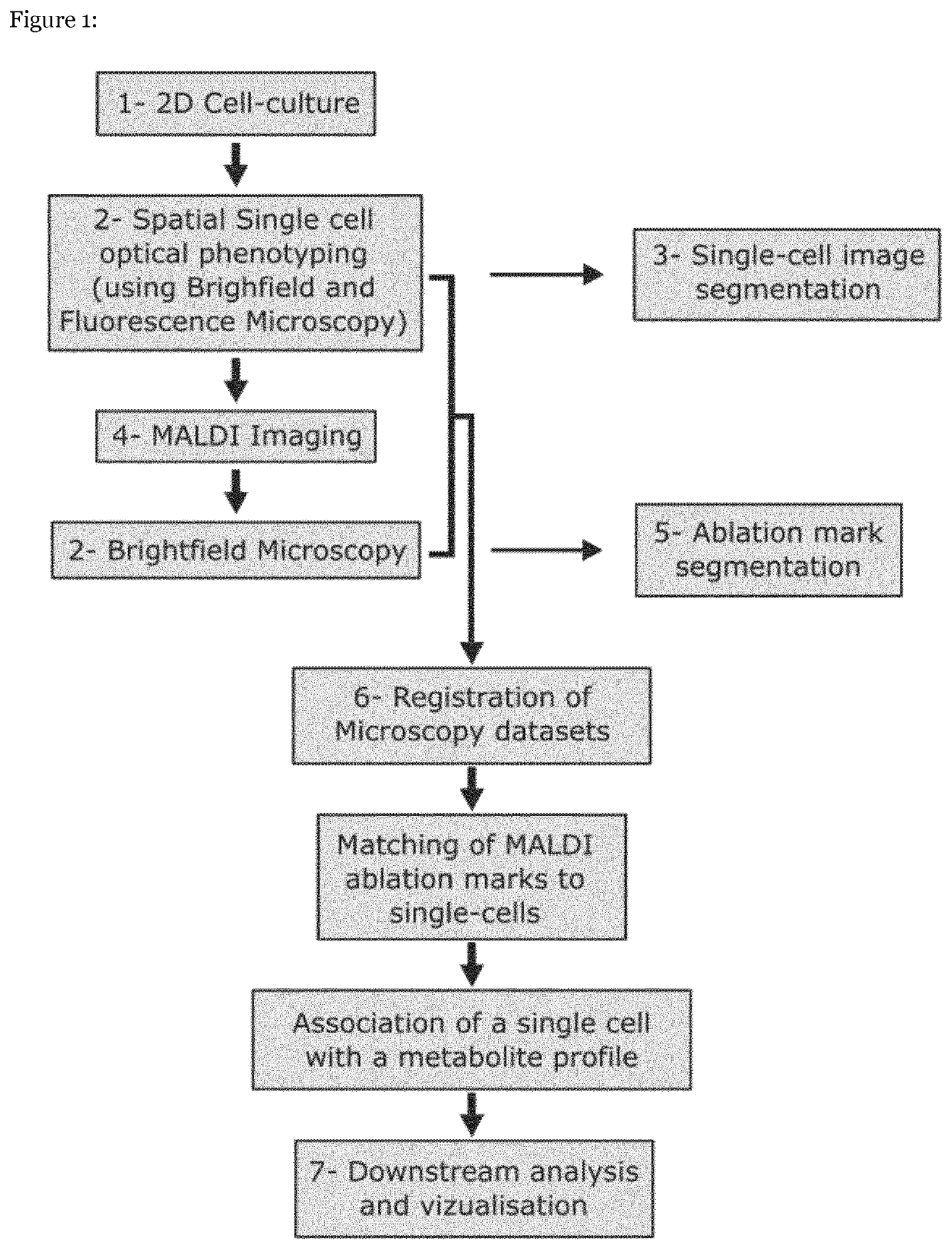
Single-cell imaging mass spectrometry
- Summary
- Abstract
- Description
- Claims
- Application Information
AI Technical Summary
Benefits of technology
Problems solved by technology
Method used
Image


Examples
example 2
of Intracellular Metabolome Changes in Stimultated Hepatocytes
[0074]Using the method of the invention, the inventors investigated the molecular content of the lipid droplets (LDs) in hepatocytes stimulated with several pro-inflammatory factors. Human hepatocytes exposed to inflammatory cytokines, such as TNF-α, accumulate neutral lipids such as triglycerides and diglycerides as well as cholesteryl ester in lipid droplets, resulting in macrovesicular steatosis (U. J. Jung, M.-S. Choi, Int. J. Mol. Sci. 15, 6184-6223 (2014)). As an orthogonal measure of LD accumulation, the inventors used the fluorescent dye LD54o which stains the LD core (J. Spandl, D. J. White, J. Peychl, C. Thiele, Li, Traffic. 10, 1579-1584 (2009)). Monolayers of differentiated human hepatocytes were cultured (the HepaRG cell line) on glass microscopy coverslips and stimulated with TNF-α as described in the Methods. Fluorescence of the LD540 staining for individual cells from the pre-MALDI fluorescent microscopy i...
example 3
ll Trends in Induced Hepatocytes
[0076]The inventors exploited the method of the invention to investigate changes in intracellular metabolome caused by different pro-inflammatory factors, considering the following conditions: (i) CTRL, untreated cells, (ii) FA, cells stimulated with an excess of fatty acids (oleic acid and palmitic acid) similar to the inventor's previous in vivo work where the diet enriched with these FAs recapitulated key features of human metabolic syndrome nonalcoholic steatohepatitis (NASH) and hepatocellular carcinoma (HCC) in mouse (M. J. Wolf et al., Cancer Cell. 26, 549-564 (2014)), (iii) LPS, cells stimulated with both FAs and a lipopolysaccharide, a pathogen-associated molecular pattern and (iv) TNF-α, cells stimulated with both FAs and the cytokine Tumor Necrosis Factor alpha, the central pro-inflammatory cytokine of the tumor necrosis factor superfamily (TNFSF). For each of the four conditions, three culture wells were considered as technical replicates ...
example 4
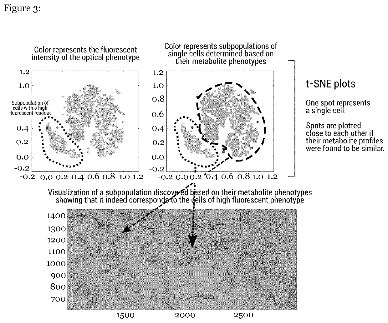
lecular Organization of Hepatocyte on Single-Cell Level
[0079]The inventors investigated the spatio-molecular organization of hepatocytes (FIG. 6). Among all metabolites detected, correlation analysis revealed the phosphatidylinositol phosphate PIP(38:4) to be the most associated with the cell-cell contact with the Spearman rs=0.36, p-value=2.5e-57. PIP(38:4) is a precursor of PIPS, a signaling phospholipid in the plasma membrane known to have transporter functions that, in the absence of gap junctions in the considered hepatocytes, can explain how physical contact between cells can induce the locally-concerted molecular response. Not all detected metabolites were found to be positively correlated with the cell-cell contact. For example, adenosine monophosphate (AMP) showed no correlation and oleic acid showed slightly negative correlation (FIG. 6C-D).
Materials and Methods
Cell Culturing and Stimulation
[0080]HepaRG cell culture (done in kind collaboration with the lab of Matthias Heik...
PUM
 Abstract
Description
Claims
Application Information