Biological Scaffolds, Products Containing Biological Scaffolds and Methods of Using the Same

a technology of biological scaffolds and biological scaffolds, applied in the field of new materials, can solve the problems of not being able to create the classic unilocular or “signal ring” and the limitations of the standard 2d culture model, and achieve the effect of maintaining the macroscopic and cellular structure and function of the scaffold

Pending Publication Date: 2020-03-12
OBATALA SCI INC
View PDF1 Cites 2 Cited by
  • Summary
  • Abstract
  • Description
  • Claims
  • Application Information

AI Technical Summary

Benefits of technology

[0009]In some embodiments, an illustrative device incorporating the scaffolds disclosed herein may comprise a combination of a) one or more cellular components derived from an adipose tissue derived cellular fraction (ATDCF), a mammalian platelet lysate and optionally, a cell population, and b) a human-derived and / or silk-based, static, three-dimensional (3-D) culture with microfluidic form with inlets and outlets. The combination of cells and the biological scaffold enables short-term and extended 3-D in vitro culture of various combinations of one or multiple cell subtypes present within the ATDCF, or in vivo implantation into both small and large animal models. In certain embodiments, the present disclosure provides a tissue-engineered, humanized, adipose depot that includes the combination of an adipose tissue-resident stromal vascular fraction (SVF) cell population with a mammalian platelet lysate, wherein the platelet lysate may be a human platelet lysate. The cell / biological scaffold combination enables short-term and extended three-dimensional in vitro culture of various combinations of one or multiple cell subtypes present within the SVF, or in vivo implantation into both small and large animal models. The silk, human platelet lysate, and ATDCF (SVF cells) allow for robust cell attachment, adipocyte differentiation, and the retention of the cellular diversity associated with both healthy and metabolically diseased human adipose depots. The constructs can be cultured for weeks or longer while maintaining their macroscopic and cellular structure and function. In addition, the biological scaffolds proposed herein are bioactive. biocompatible, free of donor DNA, human in origin, suitable for both autologous and allogeneic transplantation, and supportive of stromal / stem cell growth.

Problems solved by technology

While isolated human pre-adipocytes and adipocytes have been vastly utilized in two-dimensional (2D) cultures to identify signal transduction pathways relevant to healthy and diseased adipose tissue, the standard 2D culture model has substantial limitations.
Due to biomechanical constraints imposed by the 2D model, it is not possible to create the classic unilocular or “signet ring” adipocytes characteristic of human obesity.

Method used


Image

  • Biological Scaffolds, Products Containing Biological Scaffolds and Methods of Using the Same
  • Biological Scaffolds, Products Containing Biological Scaffolds and Methods of Using the Same
  • Biological Scaffolds, Products Containing Biological Scaffolds and Methods of Using the Same

Examples


example 1

on of Components of a Biological Scaffold

[0089]Isolation of Bone Marrow Mesenchymal Stem Cells (BM-MSC)

[0090]To isolate BM-MSC, first obtain bone marrow aspirate that has been anti-coagulated with heparin or citrate to prevent clotting. Maintain the bone marrow aspirate on wet ice at 4° C. prior to use and perform all steps with open containers within a BSL2 biological safety cabinet. Thoroughly mix Ficoll Paque solution (GE Healthcare™ Ficoll-Paque™ PREMIUM, 1.078 g / mL). Aliquot 20 ml of Ficoll Paque solution to a sterile 50 ml conical tube. Gently layer 10 ml of bone marrow aspirate onto the top of the Ficoll Paque layer and avoid any mixing. After sealing the tube, centrifuge for 30 minutes at 500×g at room temperature without any braking using a benchtop Sorvall™ Legend™ centrifuge. After returning the conical tube to the BSL2 biological safety cabinet, aspirate off the upper clear plasma layer and aliquot to a fresh sterile 50 ml conical tube. Next, aspirate the buffy coat laye...

example 2

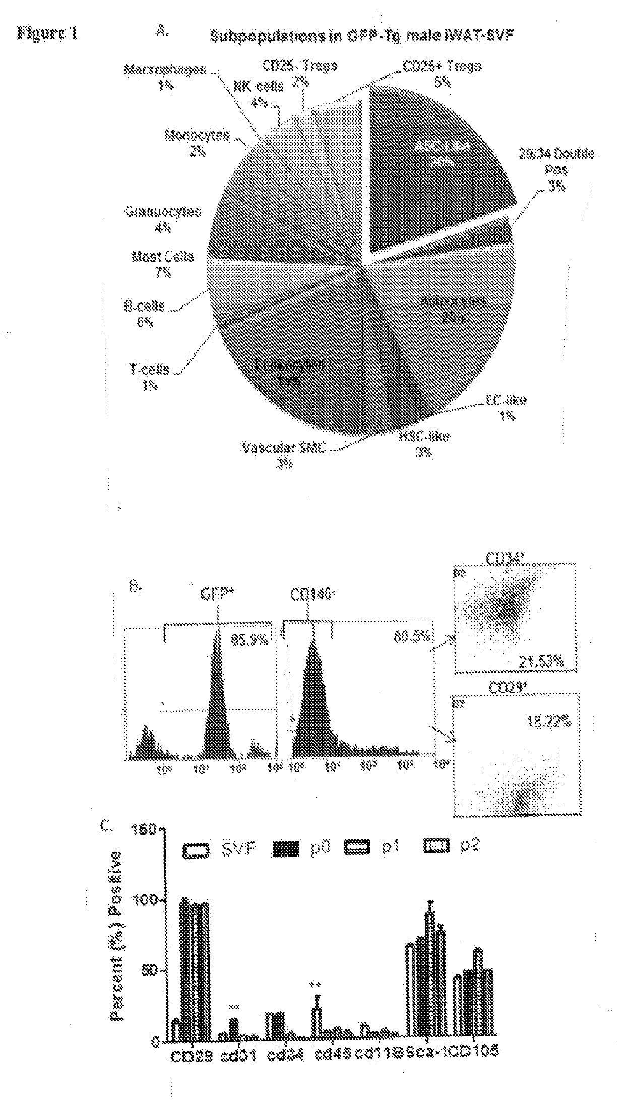
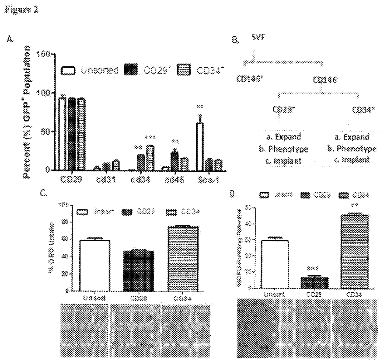
of Stromal Vascular Fraction (SVF) Cells and Adipose-Derived Stromal / Stem Cells (ASC)

[0095]To isolate SVF cells, first obtain a volume of at least 100 ml of lipoaspirate or 50 grams of subcutaneous adipose tissue from patients undergoing elective liposuction or abdominoplasty. The tissue can be stored at room temperature for up to 24 hours after the surgical procedure to allow for shipping or transit to the laboratory. Transfer the tissue upon receipt to the laboratory into a sterile BSL2 biological safety cabinet. If the tissue is received intact (after abdominoplasty), mince the tissue thoroughly into fragments of 2 to 3 mm3 using either two scalpels or two pairs of fine scissors that have been sterilized or autoclaved prior to use. After mincing the tissue or aliquoting the lipoaspirate, transfer the 100 ml volume of tissue to a sterile 250 ml capped Nalgene™ plastic centrifuge tube. Add an equal volume of sterile PBS and allow the tissue and liquid phases to separate over a 2 to...

example 3

tal for Human Platelet Lysates (hPL) Gel Preparation

[0166]When hPL and DMEM F12 combine, and are warmed to 37° C., the product is a thermos-responsive bioscaffold that is conducive to extended (1-2 month) 3-D culture and / or in vivo implantation via subcutaneous injections.

[0167]Methods

[0168]Preparation of hPL

[0169]Perform all steps within a biological safety cabinet unless otherwise indicated. Obtain desired amount concentrated expired human platelets in 15 mL or 50 mL conical tube. If not using immediately for 3-D cell culture, store at −20° C. before lysate preparation by three freeze / thawing cycles. Perform 3 freeze / thaw cycles to lyse platelets by osmosis. This will release maximum yield of growth factors for cell cultures. Centrifuge 8,000×g for 20 mins. Remove the solid (white) top layer of human platelets by aspiration.

[0170]hPL Supplemented Media Preparation

[0171]Prepare hPL supplemented media by adding 7.5% (v / v) hPL to 1% antibiotic / antimycotic and 91.5% Dulbecco's Modifie...


PUM

PropertyMeasurementUnit
temperatureaaaaaaaaaa
v/vaaaaaaaaaa
v/vaaaaaaaaaa

Abstract

The present disclosure provides biological scaffolds, methods for their synthesis and methods for their use. The biological scaffolds contain at least to components, the first, a mammalian cell platelet lysate and an adipose tissue-derived cell fraction (ATDCF) cell. Products that comprise the biological scaffold(s) may be a gel, a semi-solid or a solid substrate upon which at least one surface of the substrate is contacted with the biological scaffold to form a biocompatible material. The biological scaffold and biological scaffold containing products, can find utility in a vast range of medical device technologies and may be used for therapeutic purposes or for performing experimentation to test various pharmacological agents in vitro and in vivo.

Description

CROSS REFERENCE TO RELATED APPLICATIONS[0001]This International PCT Application claims the priority to and the benefit of U.S. Provisional Patent Application Ser. No. 62 / 457,366 filed on Feb. 10, 2017, the disclosure of which is incorporated herein by reference in its entirety.STATEMENT REGARDING FEDERALLY SPONSORED RESEARCH[0002]This invention was made with United States Government support under Federal Grant No. R21DK094254 awarded by the National Institutes of Health. The United States Government has certain rights in this invention. This invention was made in part using the facilities of the Cell Biology and Bioimaging Core that are supported in part by COBRE (NIH 8 P20-GM103528) and NORC (NIH 2P30-DK072476) center grants from the National Institutes of Health.BACKGROUND[0003]The present disclosure relates to novel biological scaffolds which are derived from adipose tissue, products utilizing such scaffolds, and methods of use thereof.[0004]While isolated human pre-adipocytes an...

Claims


Application Information

Patent Timeline
Patent Type & Authority Applications(United States)
IPC IPC(8): A61K35/35A61K35/19A61K35/28C12N5/077C12N5/0775
CPCA61K35/19A61K35/35A61K35/28C12N5/0653C12N5/0667A61P17/02A61P19/00A61P37/04A61P7/00G01N33/5014G01N33/5088A61L27/52A61L27/3633A61L27/3886A61L27/3895A61L27/3604C12M25/14C12N2533/90C12N2533/54C12N2513/00C12N2500/38C12N2501/33C12N2501/39C12N2501/999C12N5/0068
Inventor GIMBLE, JEFFREY MARTINWU, XIYINGFRAZIER, TRIVIA PENNS
Owner OBATALA SCI INC
Why Eureka
Try Eureka