

Cryopreservation
a cryopreservation and scaffold technology, applied in the field of cryopreservation, can solve the problems of inability to provide sufficient numbers of suitable cellularised scaffolds promptly, methods which may be applicable to these cell-free scaffolds or native tissues cannot be reasonably expected to apply to cellularised scaffolds, etc., to achieve the effect of maintaining cellular function
- Summary
- Abstract
- Description
- Claims
- Application Information
AI Technical Summary
Benefits of technology
Problems solved by technology
Method used
Image

Examples
example 1
and Methods
[0157]Confirming Scaffold Integrity
[0158]Histology
[0159]Samples are fixed for 24 hours in 10% neutral buffered formalin solution in PBS (pH 7.4; Sigma, UK) at RT, washed in dH2O, dehydrated in graded alcohol, embedded in paraffin and sectioned at 5 μm. Tissue slides are stained with haematoxylin and eosin (H&E; Leica, Germany).
[0160]DNA Quantification
[0161]DNA is isolated using a tissue DNA isolation kit following the manufacturer's instructions (PureLink Genomic DNA MiniKit, Invitrogen, UK).
[0162]ECM Component Quantification
[0163]Collagen, elastin and glycosaminoglycan (GAG) content can be quantified using the total collagen assay kit (Biocolor, UK), the FASTIN elastin assay and the GAG assay kit (Biocolor, UK) respectively—see [14]
[0164]Biomechanical Testing
[0165]To evaluate the biomechanical properties of oesophagi, specimens can be tested and subjected to uniaxial longitudinal tension until failure [12]. Uniaxial tension may be applied using an Instron 5565, with spec...
example 2
s
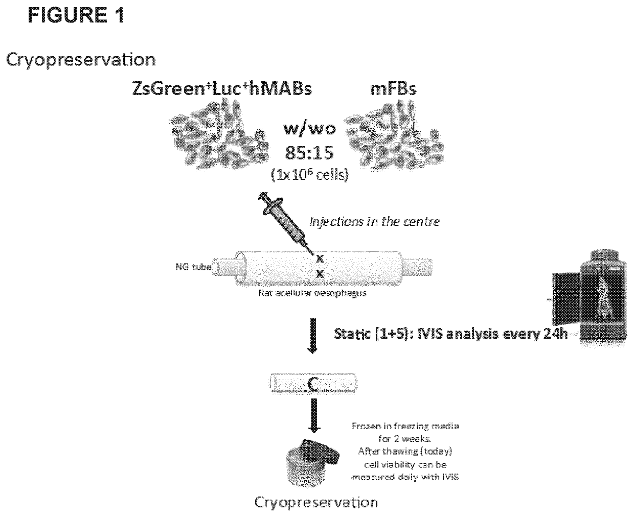
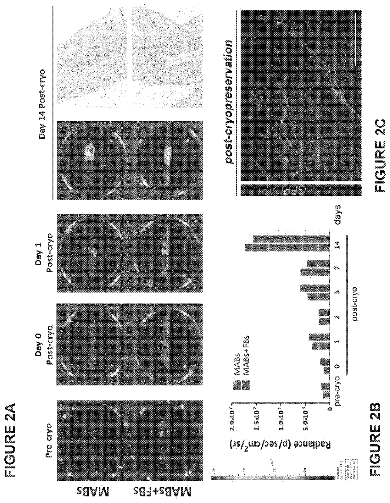
[0188]Rat decellularized oesophagi seeded with human mesoangioblasts (MABs), mouse fibroblasts (FBs) and mouse neural crest cells, were cultured in a bioreactor for up to 11 days and then frozen with the following protocol:[0189]The seeded scaffold (size 7+20 mm length) was placed in a cryovial (size: 2 mL) with 500 μL FBS.[0190]The vial was kept in ice throughout the process.[0191]Another 500 μL were added of a solution of Megacell medium containing 20% DMSO.[0192]The vial was transferred in a Nalgene freezing container and kept at −80° C. overnight.[0193]Samples were then placed and stored in the vapour phase of liquid nitrogen at approximately −160° C.[0194]After 2 to 4 weeks in the liquid nitrogen container, vials were rapidly thawed at 37° C. and samples transferred in 10-20 mL culture medium (Megacell supplemented with FBS and antibiotic) at 37° C. under mild agitation for 20 minutes.[0195]Samples were then transferred to a culture petri dish with fresh culture medium and lef...
example 3
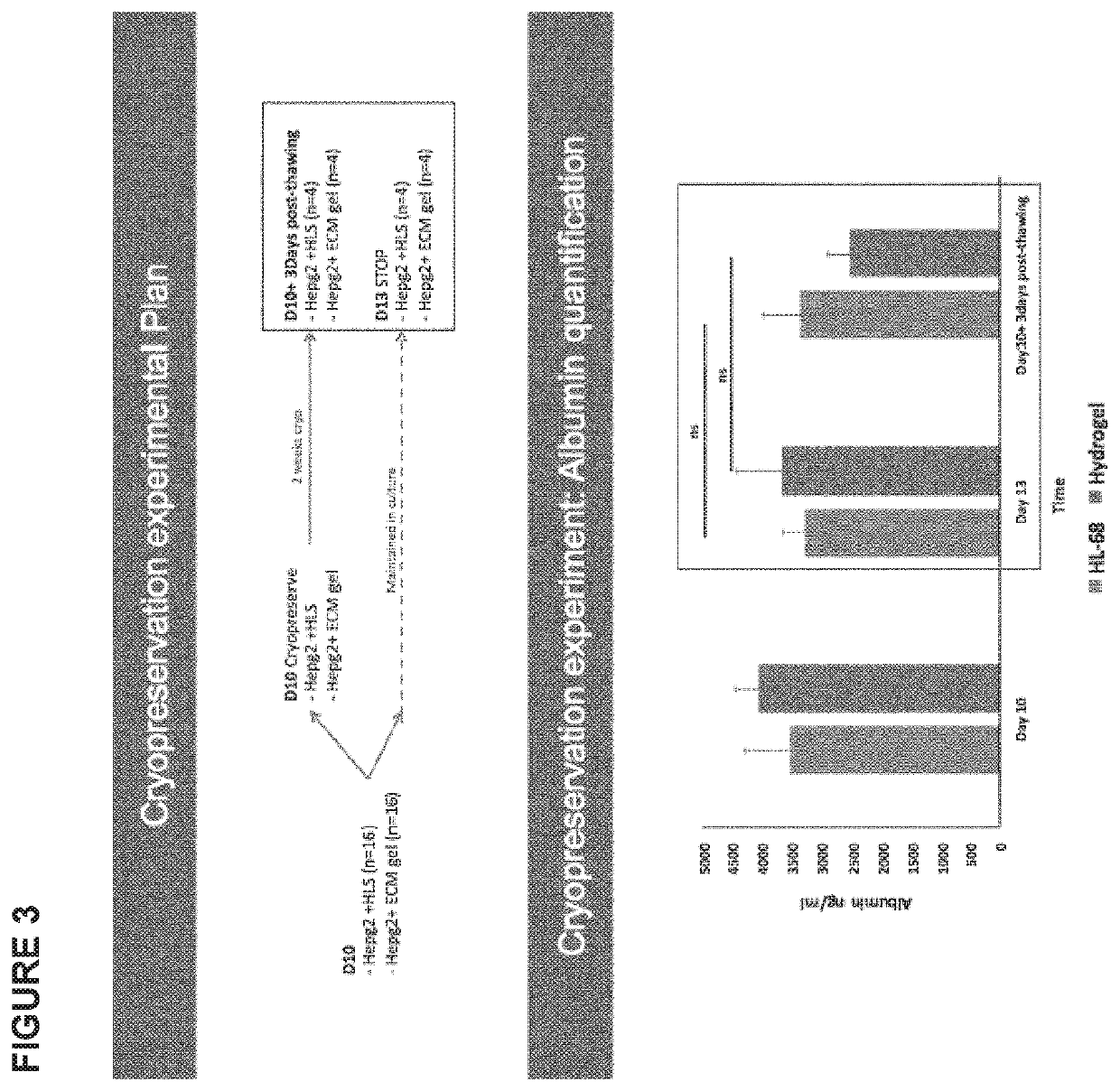
[0198]Human decellularized liver cubes (5×5×5 mm) or human decellularized liver-derived Hydrogel cubes (5×5×5 mm) seeded with HepG2 cell line, cultured in static conditions for up to 10 days were frozen with the following protocol:[0199]The seeded scaffold was placed in a cryovial (size: 2 mL) with 500 μL FBS.[0200]The vial was kept in ice throughout the process.[0201]Another 500 μL were added of a solution of alpha MEM containing 20% DMSO.[0202]The vial was transferred in a Nalgene freezing container and kept at −80° C. overnight.[0203]Samples were then placed and stored in the vapour phase of liquid nitrogen at approximately −160° C.[0204]After 2 weeks in the liquid nitrogen container, vials were rapidly thaw at 37° C. and samples transferred in 5-10 mL culture medium (alpha MEM containing 10% FBS, 1% Antibiotic, 1% 1 mM sodium pyruvate, 1% non-essential AA solution 100×)) at 37° C. under mild agitation for 20 minutes.[0205]Samples were then transferred to a culture petri dish wit...
PUM
 Abstract
Description
Claims
Application Information