Porous microspheres including mussel adhesive protein, and method for manufacturing same

a technology of adhesive protein and porous microsphere, which is applied in the direction of peptides, prostheses, peptide sources, etc., can solve the problems of low cell delivery efficiency, loss of cell viability or difficulty in controlling cell differentiation capacity, and low retention in the cell delivery environment, so as to efficiently deliver therapeutic stem cells

Pending Publication Date: 2020-09-17
POSTECH ACAD IND FOUND
View PDF1 Cites 2 Cited by
  • Summary
  • Abstract
  • Description
  • Claims
  • Application Information

AI Technical Summary

Benefits of technology

[0023]The porous microsphere comprising the mussel adhesive protein according to the present invention is capable of minimally invasive bio-injection through syringes to efficiently deliver therapeutic stem cells to the sites of tissue defects as cell carriers. Further, the present invention may be widely applied to scaffolds for tissue engineering, drug carriers, or the like, which may be suitably applied to the size of the defected site of tissue.

Problems solved by technology

However, there are the limitations of low cell delivery efficiency such as damage or death of cells easily caused during the injection.
However, due to their low biocompatibility and lack of functionality for direct interaction with cells, there is a problem that ultimately results in loss of cell viability or difficulty in controlling cell differentiation capacity.
However, there is a limit of low retention in the cell delivery environment due to their low mechanical properties.
However, to date, there has been no attempts to make microspheres or to apply them as cell carriers using oxidization of DOPA contained in mussel adhesive proteins.

Method used


Image

  • Porous microspheres including mussel adhesive protein, and method for manufacturing same
  • Porous microspheres including mussel adhesive protein, and method for manufacturing same
  • Porous microspheres including mussel adhesive protein, and method for manufacturing same

Examples


example 1

on of Mussel Adhesive Protein-Based Porous Microsphere

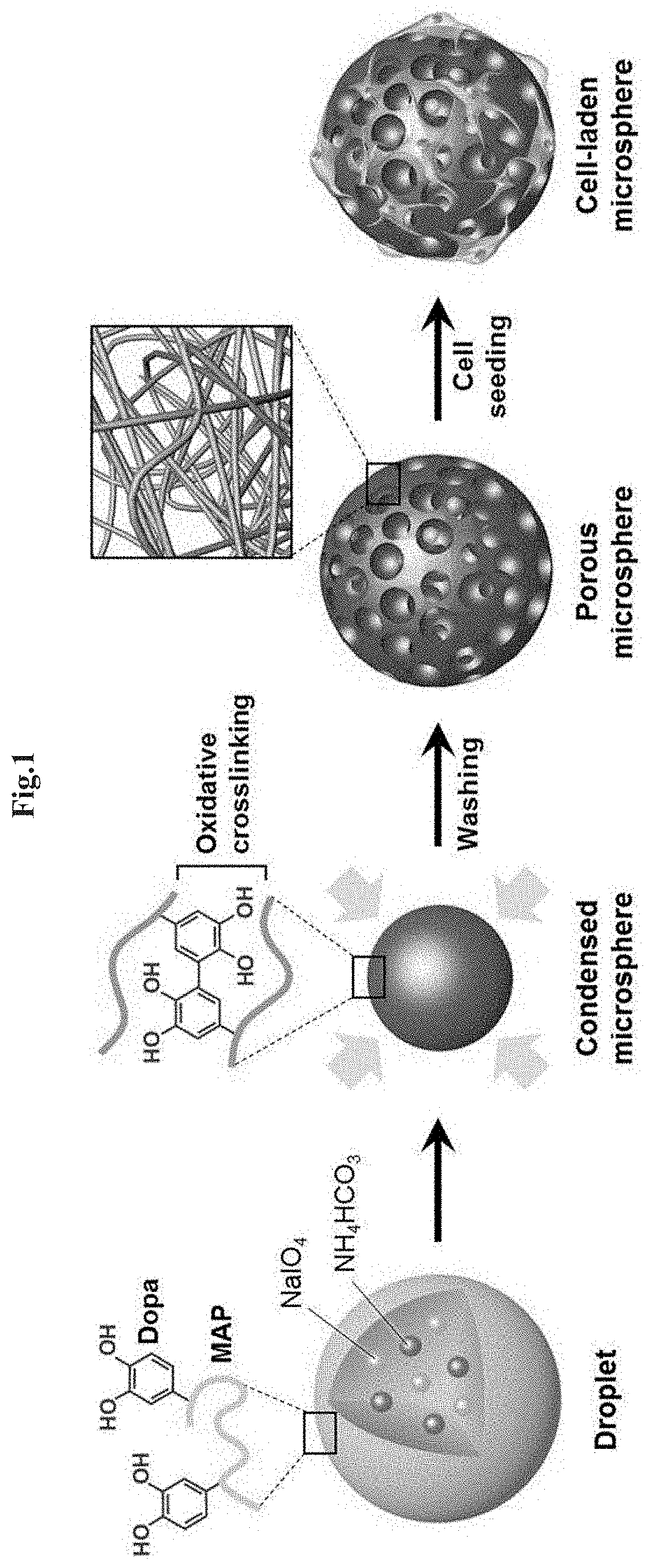
[0092]In order to prepare porous microspheres based on mussel adhesive protein, the following experiments were carried out sequentially. A scheme of the overall experiment for preparing the porous microsphere of the present invention is shown in FIG. 1.

[0093]1-1. Preparation of Mussel Adhesive Protein fp-151

[0094]For decapeptide which repeats about 80 times in natural mussel adhesive protein fp-1 and consists of 10 amino acids to be expressed in E. coli, the mussel adhesive protein fp-1 variant composed of 6 decapeptides was prepared. Mgfp-5 gene (Genbank No. AAS00463 or AY521220) was inserted between two fp-1 variants to allow successful expression in E. coli. Subsequently, the mussel adhesive protein fp-151 was produced through purification process using acetic acid (D. S. Hwang et. al, Biomaterials 28, 3560-3568, 2007).

[0095]Specifically, in the amino acid sequence of fp-1 (Genbank No. Q27409 or S23760), a fp-1 variant (herein...

example 2

of Physical and Chemical Characterization of Mussel Adhesive Protein fp-151-Based Porous Microspheres

[0107]2-1. Analysis of FT-IR Spectroscopy of Porous Microsphere Surfaces

[0108]FT-IR spectroscopy analysis was performed to confirm the chemical bonding of the surface of the mussel adhesive protein-based porous microspheres (MAP-PMS) prepared in Example 1, and the results are shown in FIG. 8. As a comparison group, the modified fp-151 (MAP) and NaIO4-treated fp-151 (MAP+NaIO4) coated surfaces were used.

[0109]As shown in FIG. 8, it was confirmed that the porous microsphere had a peak similar to that of the fp-151 coated surface treated with NaIO4 at a wavelength of about 1240 cm−1 indicating C—O bonds in the catechol. As a result, it was confirmed that DOPA was changed into the DOPA-quinone by the oxidizing agent, allowing sufficient crosslinking progress to form microspheres.

[0110]2-2. Charge Measurement on Porous Microsphere Surfaces

[0111]In order to determine the charge of the surf...

example 3

ion of Applicability of Mussel Adhesive Protein fp-15-Based Porous Microspheres

[0116]In order to confirm the applicability of the porous microspheres, the porous microspheres were introduced into the syringe, and the release pattern of the porous microspheres was observed. In addition, a mold was used to prepare the porous microspheres in a cylindrical structure. The adhesion was confirmed on the skin surface of the animal tissue with alginate microspheres. The results are shown in FIGS. 12 to 15.

[0117]As shown in FIGS. 12 and 13, it was confirmed that the microspheres can be injected to the outside through the syringe while maintaining the form. In addition, as shown in FIG. 14, it was confirmed that the microspheres can be produced in a cylindrical structure by using a mold. Further, as shown in FIG. 15, when the microspheres are applied to the surface of the skin tissue of the animal through a syringe, it was confirmed that they are attached to the application site even after sev...


PUM

PropertyMeasurementUnit
porosityaaaaaaaaaa
diameteraaaaaaaaaa
diameteraaaaaaaaaa

Abstract

The present invention relates to a porous microsphere comprising a mussel adhesive protein and a method of preparing the same. The porous microsphere comprising the mussel adhesive protein according to the present invention is capable of minimally invasive bio-injection through syringes to efficiently deliver therapeutic stem cells to the sites of tissue defects as cell carriers. Further, the present invention may be widely applied to scaffolds for tissue engineering, drug carriers, or the like, which may be suitably applied to the size of the defected site of tissue.

Description

TECHNICAL FIELD[0001]The present invention relates to a porous microsphere comprising a mussel adhesive protein and method for preparing the same.BACKGROUND ART[0002]Stem cells have a differentiation ability to develop into various tissues. In the tissue engineering and regenerative medicine field, studies have been actively conducted to transplant stem cells, thereby restoring human tissues or organs and to treat diseases. In particular, direct injection of stem cells into biological tissues can be applied in a minimally invasive manner so that it has been attracting attention in the clinical field. However, there are the limitations of low cell delivery efficiency such as damage or death of cells easily caused during the injection.[0003]Microspheres (MS) are three-dimensional structures with a diameter of 1 μm to 1000 μm and have a high surface area-volume ratio, allowing efficient cell loading. Therefore, their applicability is increasing as an injection-type cell carrier capable...

Claims


Application Information

Patent Timeline
Patent Type & Authority Applications(United States)
IPC IPC(8): C07K14/435C12M1/12A61K47/64A61K9/00
CPCC12M25/14A61K9/0019A61K47/64C07K14/43504A61K47/6927C12M25/16C12M23/20A61L27/56A61L27/227C07K2319/35A61K9/5052A61K9/5089A61K35/28A61K35/32A61K35/44A61K35/30A61K35/13C12N15/63A61L27/22C07K2319/00
Inventor CHA, HYUNG JOONKIM, DONG PYOJO, YUN KEEMIN, KYOUNG IK
Owner POSTECH ACAD IND FOUND
Why Eureka
Try Eureka