Preservation and transport of an ex vivo biological sample comprising ultrasound application

a biological sample and ex vivo technology, applied in the field of ex vivo biological sample transport and preservation devices, can solve the problems of major, relevant problems, injury to the graft during the period, etc., and achieve the effects of reducing the injury to the biological sample, increasing the viability of the sample, and improving the quality of life of the transplant patien

Pending Publication Date: 2020-11-19
INST DINVESTIGACIONS BIOMEDIQUES AUGUST PI I SUNYER (IDIBAPS)
View PDF0 Cites 1 Cited by
  • Summary
  • Abstract
  • Description
  • Claims
  • Application Information

AI Technical Summary

Benefits of technology

[0018]It is an object of the invention to provide a device for transport and preservation of an ex vivo biological sample and a method of the type indicated above that reduces injury to the biological sample, and accordingly increases viability thereof in comparison to what could be obtained with known devices. The invention also considers, in the case of transplants, the problem of improving the quality of life of transplant patients, and in the worst cases, of significantly reducing the need for another transplant.
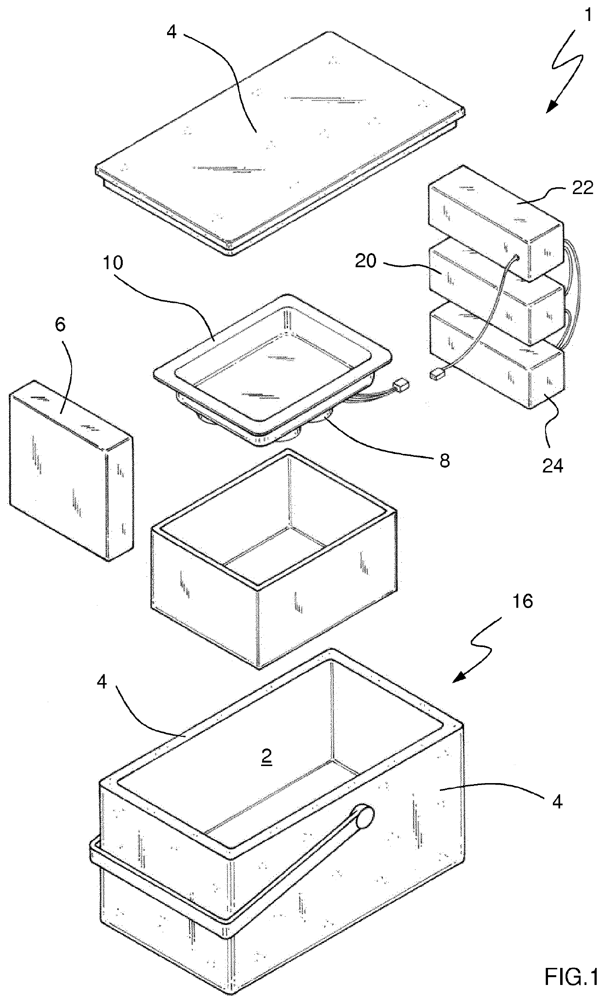
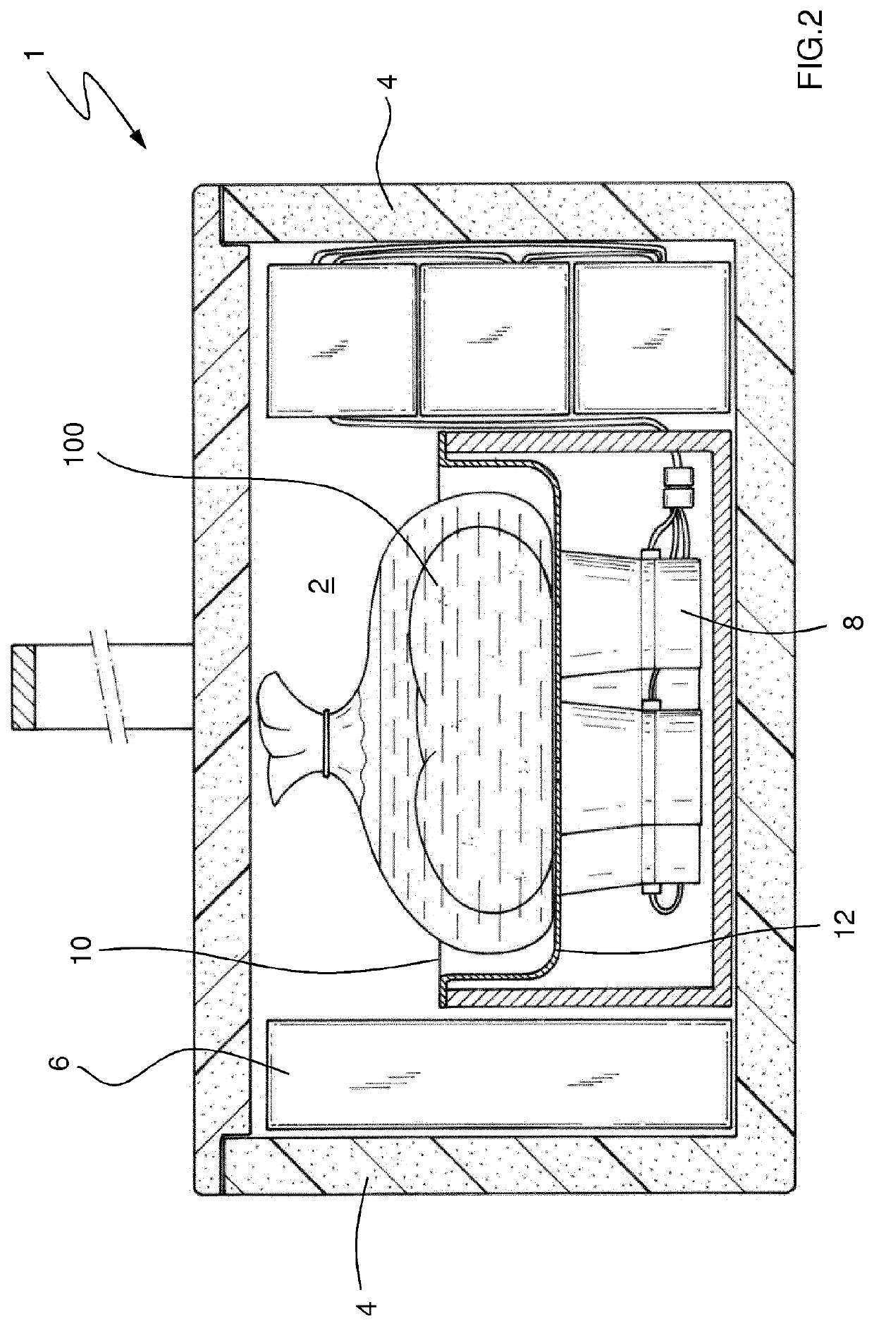
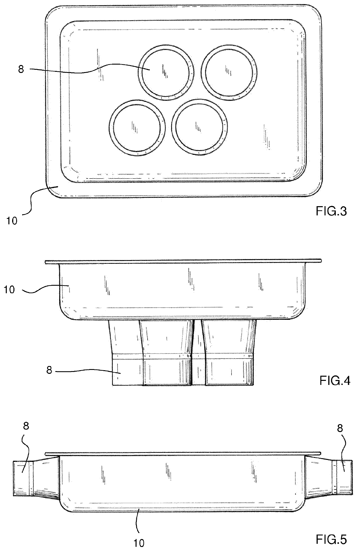
[0019]This objective is achieved by means of a device for ex vivo transport of the type indicated above, characterized in that it further comprises at least an ultrasound system suitable for generating and applying ultrasound on said biological sample. Applying ultrasound combined with cooling the inner chamber can therefore be carried out such that local heating of the biological sample, and the subsequent increase in temperature that would cause damage therein, are prevented. Particularly, the device also comprises an auxiliary container containing a preservation solution, without external oxygen delivery and for containing said biological sample immersed in said preservation solution.

Problems solved by technology

Despite the advances achieved in surgical techniques in the past few decades, the injury the graft sustains during the period in which the donor organ is transported until it is implanted in the recipient (cold ischemia time) is still a major socially and clinically relevant problem yet to be solved.
This all negatively results in a poor quality of life for the patient, the need for transplanting again, and more problems in the availability of organs for transplant.
This factor is responsible for PGD and PGF and negatively affects post-operative results, the quality of life and survival of the patient.
Such organs are extremely vulnerable to the injury they sustain during transport, before being implanted in the recipient, and have a higher risk of experiencing PGD and PGF.
They are therefore not viable for transplant.
There are no means for transmitting ultrasound between the inner vessel and the outer container, so the system cannot work as well as if the ultrasound was applied directly on a wound for the purpose of wound healing.
Patent document U.S. Pat. No. 4,767,402 A, relating to the field of transdermal drug delivery by means of applying ultrasound, also relates to the risks associated with the use thereof.
The patent document particularly advises against the prolonged use of ultrasound due to the risks associated with the increase in skin temperature and the possible damage this technique can cause.
As a result, applying ultrasound for organ and tissue preservation is not advisable due to the mentioned factors of: local heating of the tissue, difficulty in penetrating considerable tissue or organ thicknesses, and transmission difficulty in gaseous media.
When the temperature exceeds 38° C., exposure to ultrasound is usually interrupted because it damages tissues.

Method used


Image

  • Preservation and transport of an ex vivo biological sample comprising ultrasound application
  • Preservation and transport of an ex vivo biological sample comprising ultrasound application
  • Preservation and transport of an ex vivo biological sample comprising ultrasound application

Examples


experimental examples

[0070]Different tests conducted to put the method according to the invention into practice are described below.

Methodology

[0071]The experimental protocol was carried out from livers and kidneys of Landrace pigs. The kidney and liver were perfused with preservation solution at 4° C. to remove blood contained in the organ, and the organ was kept under cold conditions, bathed in ice until the organs were immersed in different preservation solutions. Next the biological samples were placed in the cooler and kept in the cooler with or without ultrasound for 8 hours (liver) and 24 hours (kidney). All the procedures were performed under anesthesia and the study respected the European Union regulations concerning experiments using animals (Directive 86 / 609 / EEC).

Experimental Design

[0072]Protocol 1

[0073]The experimental groups formed were the following:[0074]GROUP 1: cold ischemia group in the conventional transport system and with preservation solution. This group was divided into different ...


PUM


Abstract

The present invention relates to a device for transport and preservation of an ex vivo biological sample and corresponding method. The device comprises a chamber for containing the biological sample, delimitated by walls made of a thermal insulating material. The device, furthermore, incorporates cooling means that keep the temperature inside the chamber below the temperature outside the device. Finally, an ultrasound system suitable for generating and applying ultrasound on the biological sample is provided. The invention also proposes a method for transport and preservation which combines applying cooling and ultrasound to reduce cell damage in the biological sample.

Description

CROSS-REFERENCE TO RELATED APPLICATIONS[0001]The present application is a divisional of U.S. patent application Ser. No. 15 / 625,611, filed Jun. 5, 2017, which is a U.S. National Stage of International Patent Application No. PCT / EP2015 / 080688, filed Dec. 18, 2015, which claims the benefit of European Patent Application No. 14382548.7, filed Dec. 19, 2014. The entirety of each of these application is hereby incorporated by reference.FIELD OF THE INVENTION[0002]The invention relates to a device for transport and preservation of an ex vivo biological sample comprising a chamber for containing the biological sample, delimitated by walls made of a thermal insulating material and cooling means for keeping the temperature inside the chamber below the temperature outside the device, as well as to a corresponding method for preservation of the biological sample.[0003]The invention also relates to the use of the device according to the invention for transport and preservation of an ex vivo bio...

Claims


Application Information

Patent Timeline
Patent Type & Authority Applications(United States)
IPC IPC(8): A01N1/02A61N7/00
CPCA61N2007/0004A01N1/0273A61N7/00A61F2007/108A01N1/0278A01N1/02
Inventor PERALTA, CARMEN
Owner INST DINVESTIGACIONS BIOMEDIQUES AUGUST PI I SUNYER (IDIBAPS)
Why Eureka
Try Eureka