Wireless and noninvasive epidermal electronics

a technology of epidermal electronics and wires, applied in the field of wireless and noninvasive epidermal electronics, can solve the problems of high cost, high cost, inaccurate, harmful or invasive existing diagnostics, etc., and achieve the effect of preserving a high level of accuracy, convenient and rapid operation, and avoiding unnecessary admissions

Active Publication Date: 2021-01-28
RHAEOS INC
View PDF0 Cites 3 Cited by
  • Summary
  • Abstract
  • Description
  • Claims
  • Application Information

AI Technical Summary

Benefits of technology

[0006]The devices presented herein provide a platform for measuring flow in subdermal conduits and are advantageously non-invasive and rapid, while preserving a high level of accuracy. The devices may be conformal to skin and wireless without a need for hard-wire connection to bulky external components, such as controllers, digital monitors and power supplies. In this manner, the device is painless and non-obtrusive to a patient, akin to wearing an adhesive bandage.
[0007]One particular application for any of the devices and methods described herein is detection of shunt malfunction, specifically ventricular shunts. Extended use can capture occult malfunction, akin to a holter monitor for cerebrospinal fluid.
[0008]Provided herein is a soft, wireless, noninvasive, non-surgical, skin-mounted device for the continuous measurement of fluid flow in a subdermal conduit, such as shunt-based CSF flow. The epidermal device exploits the precise measurement of thermal transport to characterize CSF flow in underlaid shunts. The device platform is ultrathin (<100 μm), soft (70 kPa), flexible resulting in a continuously wearable device mechanically invisible to the wearer. Similar in size to a Band-Aid® adhesive bandage, the device is composed primarily of soft, silicone rubber (no hard edges) and transmits recorded data wirelessly via Bluetooth to a companion mobile app. Patient data and in vitro tests confirm device efficacy in producing clinical-quality data suitable for shunt malfunction diagnostics. To assist in handling durability and device placement, a carrier substrate having an open passage through a central portion of the carrier substrate, may be provided around the active sensors region, where intimate conformal contact is desired. The carrier substrate may have a relatively larger mechanical parameter compared to the substrate that supports the sensors, such as being less flexible, elastic and / or soft, so that the device can be handled in a manner similar to an adhesive bandage (e.g., does not tear during application and use) but without sacrificing conformability and patient comfort.
[0011]Provided herein are various conformable devices capable of reliably, accurately, and continuously measuring subdermal fluid flow, including in a conduit. Various active components are supported by a substrate, such as a substrate that is characterized as soft, stretchable and flexible. A thermal actuator, an upstream temperature sensor and a downstream temperature sensor is supported by the substrate. A microprocessor is in electronic communication with the temperature sensors and other relevant components, such as the actuator, to calculate subdermal fluid flow from the measured upstream and downstream temperatures. Such a configuration maximizes patient comfort during use, facilitating long-term monitoring of fluid flow. Any of the devices may be wireless, further facilitating low patient impact monitoring, including without a need for hard-wire connections. In this manner, the patient may even return to home, without adversely impacting monitoring.
[0012]To further increase the accuracy and reliability of fluid flow measurement, any of the devices described herein may have additional temperature sensors. The position of those sensors may be described relative to a notional line (e.g., imaginary) line that is formed by drawing a line over spatially aligned upstream sensor, actuator, and downstream sensor. The additional sensors function as reference sensors and assist with determining various skin properties and related convection-type properties, for example, useful in determining fluid flow rate and the effect of the actuator independent of flow. Preferably, at least one sensor is positioned so that a temperature reading is obtained that is independent of whether or not the thermal actuator is actuated.
[0015]Additionally, various methods for increasing the signal to noise ratio of thermal measurements are also provided. For example, additional layers that partially or fully encapsulate and insulate various components may enhance the ability to isolate thermal energy provided by the various actuation means and increase the accuracy and reliability of sensing. Conductive layers may also be provided to increase the efficacy of thermal actuation and thermal sensing by providing a conduit for thermal energy to be directed to or received from the skin. Various discontinuous thermal conductive layers may further increase the signal to noise ratio for a variety of measurements, including thermal sensing.

Problems solved by technology

Existing diagnostics are expensive, inaccurate, and often harmful or invasive, and can lead to unnecessary admissions, further testing, or needless surgery.
Hydrocephalus is a common and costly condition caused by the accumulation of cerebrospinal fluid in the brain.
Cerebrospinal fluid (CSF) is produced within the ventricles of the brain and is responsible for its nourishment and protection, but dysfunction in its drainage or reabsorption can lead to devastating neurological complications.
Unfortunately, shunts have extremely high failure rates; most children undergo 2-3 surgical revisions before adulthood and 30% are expected to have at least 1 revision operation in the first year, with rates approaching 98% by 10 years in some studies.
That technology, however, is cumbersome and time-consuming.
The device's cumbersome, multi-step protocol; equivocal or negative past clinical studies; and need for ice-pack cooling have limited its acceptance.
Additionally, patient discomfort due to prolonged skin cooling (detrimental for pediatric diagnostics) and absence of chronic monitoring further limits its diagnostic relevance.

Method used


Image

  • Wireless and noninvasive epidermal electronics
  • Wireless and noninvasive epidermal electronics
  • Wireless and noninvasive epidermal electronics

Examples


example 1

Epidermal Electronics for the Noninvasive, Wireless, Quantitative Assessment of Ventricular Shunt Function

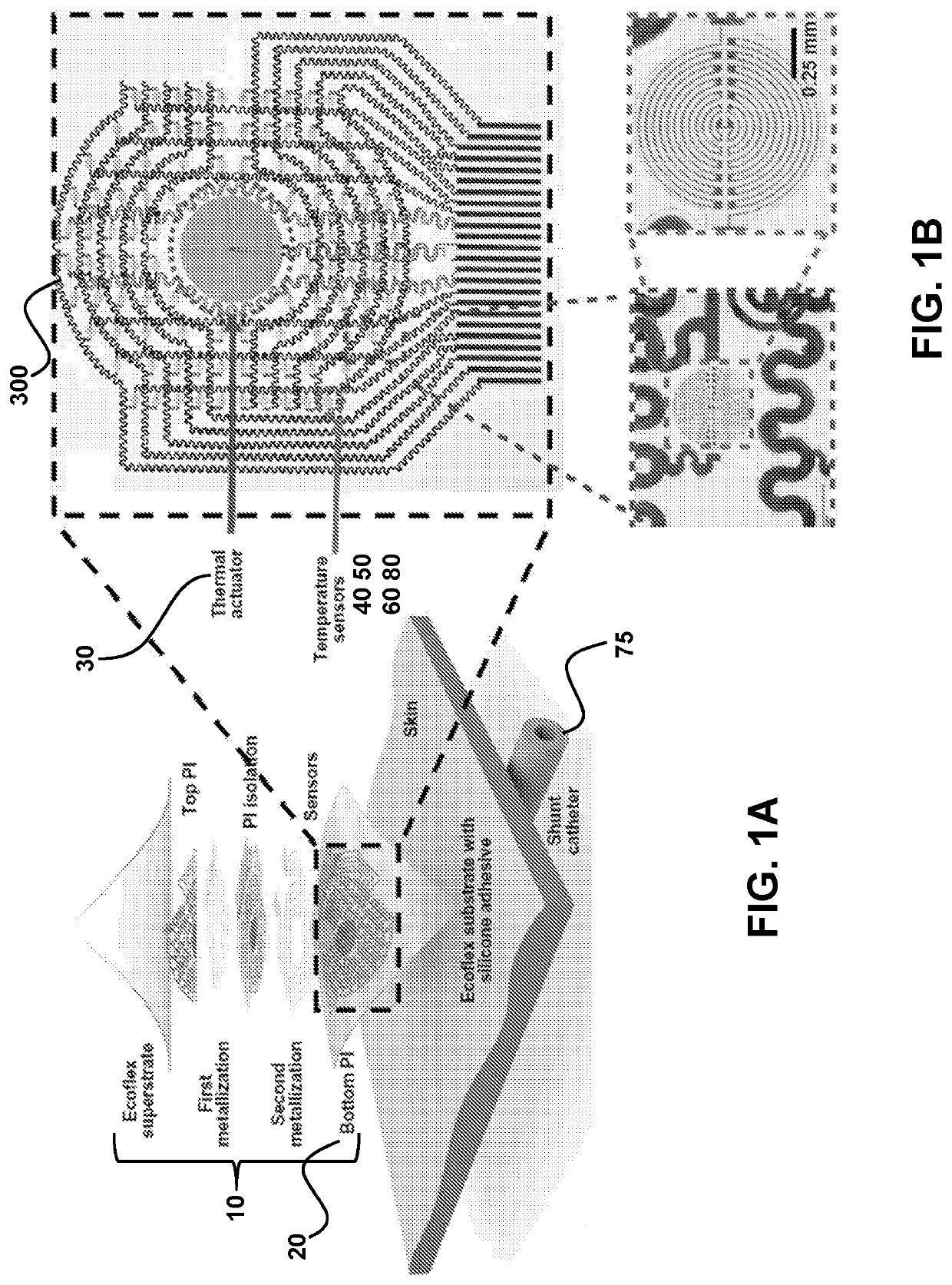
[0106]Ventricular shunts represent an essential component of clinical treatment for hydrocephalus, a common and debilitating neurological disorder that results from the overproduction and / or impaired reabsorption of cerebrospinal fluid (CSF) produced in the ventricular system of the brain [Rachel]. Hydrocephalus arises from a number of causes, including but not limited to cancer, hemorrhage, trauma, and congenital malformations. This condition affects an estimated 750,000 patients in the United States alone, and it is responsible for ˜3.1% of all pediatric acute care costs [Lam, Patwardhan, Shannon, Stone]. 125,000 pediatric hydrocephalus patients in the US account for 400,000 days spent in the hospital each year [Simon]. Shunts assemblies typically involve two silicone catheters, connected upstream and downstream of a regulating valve, to drain excess CSF from the ventricle to ...

example 2

System Characterization and Use

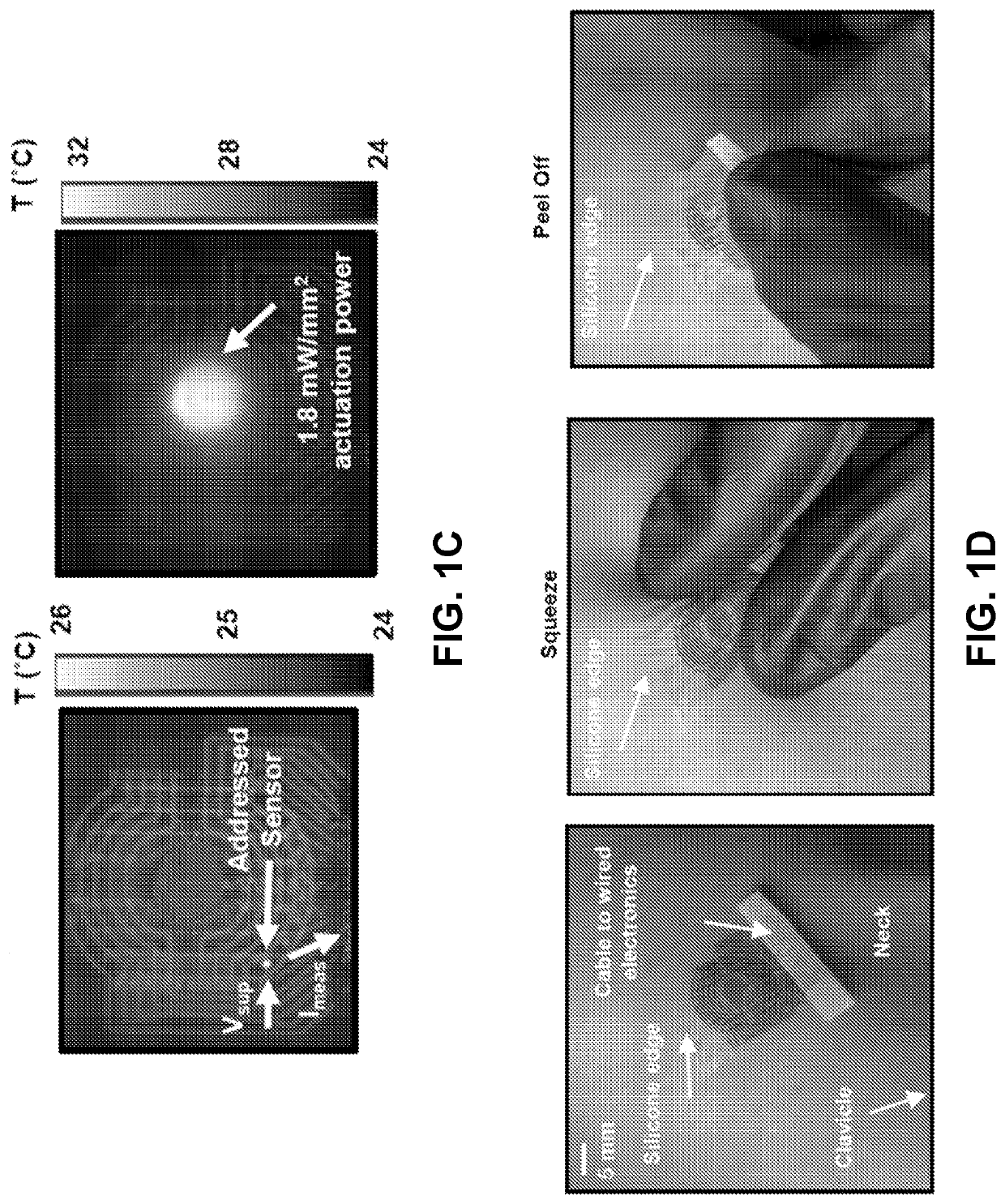
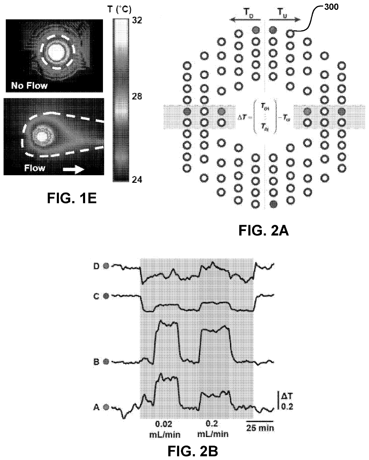
[0165]FIGS. 24-28 illustrate various features of the devices and methods described herein. FIG. 24 illustrates the position of the device and temperature sensors and various parameters used to calculate temperature and flow-rate in a subdermal conduit (described in FIG. 24 as “catheter”). Unknown quantities include skin-related parameters, such as depth of the conduit from the skin surface (hskin), as well as thermal conductivity (k) and diffusivity (α) of the skin. Known parameters include the thermal conductivity (k) and diffusivity (α) of the conduit and fluid in the conduit, outer and inner diameters of the conduit, and upstream, downstream distance between the sensors and the actuator (labeled “heater”), and temperature measured by the sensor and applied by the heater. In controlled conditions, the flow-rate (Q) of the fluid in the conduit may be known. From these parameters, flow-rate may be determined, as summarized in the flow-chart of FIG. 25....

example 3

REFERENCES RELATED TO EXAMPLE 3

[0222]1. Gao, L., et al., Epidermal photonic devices for quantitative imaging of temperature and thermal transport characteristics of the skin. Nat Commun, 2014. 5: p. 4938.


PUM


Abstract

Provided are conformable devices to measure subdermal fluid flow and related methods. A soft, stretchable and flexible substrate supports a thermal actuator and various specially positioned temperature sensors. A microprocessor in electronic communication with sensors calculates subdermal fluid flow from the measured upstream and downstream temperatures, as well as various application-dependent parameters. Devices and methods provided herein are particularly useful for measuring cerebral spinal fluid in a ventricular shunt placed for treatment of hydrocephalus.

Description

CROSS-REFERENCE TO RELATED APPLICATIONS[0001]This application claims the benefit of and priority to U.S. Provisional Patent Application No. 62 / 650,826 filed Mar. 30, 2018, and U.S. Provisional Patent Application No. 62 / 791,390, filed Jan. 11, 2019 each of which are specifically incorporated by reference in their entirety to the extent not inconsistent herewith.STATEMENT REGARDING FEDERALLY SPONSORED RESEARCH OR DEVELOPMENT[0002]Not applicable.BACKGROUND OF INVENTION[0003]Hydrocephalus and shunt-related expenditures cost the US system over $2 billion dollars in annual expenses, with 125,000 shunt surgeries per year and an untreated mortality rate estimated at 50-60%. Existing diagnostics are expensive, inaccurate, and often harmful or invasive, and can lead to unnecessary admissions, further testing, or needless surgery. To address these issues, provided herein is a noninvasive, thermal biosensor capable of diagnosing ventricular shunt malfunction.[0004]Hydrocephalus is a common and ...

Claims


Application Information

Patent Timeline
Patent Type & Authority Applications(United States)
IPC IPC(8): A61B5/00A61B5/01
CPCA61B5/7278A61B5/01A61B2562/18A61B2562/164A61B2562/0271A61B5/00A61B2562/028A61B2562/046A61B5/145A61B5/14507A61B5/4851A61B5/031A61B5/026
Inventor ROGERS, JOHN A.KRISHNAN, SIDDHARTHRAY, TYLER R.AYER, AMIT B.GUTRUF, PHILIPPREEDER, JONATHAN T.LEE, KUN HYUCKSU, CHUN-JU
Owner RHAEOS INC
Why Eureka
Try Eureka