Biosensor based on trititanium dicarbide two-dimensional metal carbide catalyzed luminol electrogenerated chemiluminescence probe and preparation method

Pending Publication Date: 2021-04-08
QINGDAO UNIV
View PDF0 Cites 6 Cited by
  • Summary
  • Abstract
  • Description
  • Claims
  • Application Information

AI Technical Summary

Benefits of technology

[0015]Advantageous effects of the present invention are:
[0016]It is found for the first time in the present invention that Ti3C2 MXenes can improve the ECL signals of luminol, and using this property to prepare Ti3C2 MXenes as probes, then the electrode of the biosensor

Problems solved by technology

These methods have the disadvantages of requiring expensive instruments, complex technical skills and time-consuming operations, etc.
Therefore, it is a huge challenge to develop a simple, sensitive and reliabl

Method used


Image

  • Biosensor based on trititanium dicarbide two-dimensional metal carbide catalyzed luminol electrogenerated chemiluminescence probe and preparation method
  • Biosensor based on trititanium dicarbide two-dimensional metal carbide catalyzed luminol electrogenerated chemiluminescence probe and preparation method

Examples


embodiment 1

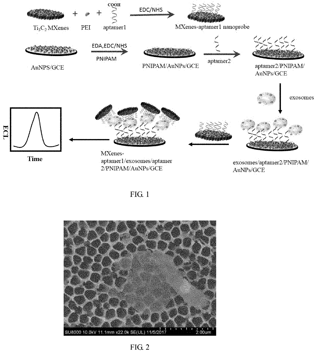
Synthesis of MXenes-Aptamer1 Nanoprobe

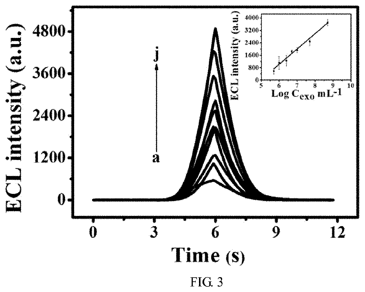
[0049]Ti3AlC2 (1.0 g) powder was immersed in 15 mL of 48% (by mass) HF, and was stirred for 24 h at 45° C. Then, the suspensions were centrifuged to separate solids from the supernatant. After that the solid products were washed several times at 5000 rpm for 5 minutes each time, and were dried at room temperature. The layered Ti3C2Tx was obtained and stored at 4° C. until use The demixed Ti3C2 (0.05 g) powder was immersed in 1 mL of DMSO, stirred at room temperature for 24 h, centrifuged at 12,000 rpm and washed five times for 5 min each time, then the supernatant was discarded and DI water was added for smashing in a cell lysis instrument for 2 h. Finally, the solution was centrifuged at 3500 rpm for 60 min, and the supernatant (i.e., Ti3C2 MXenes nanosheets dispersion) was retained and stored at 4° C. for later use. Its structural characteristic is shown in FIG. 2.

[0050]200 μL of (0.005 g / mL) PEI, 3 ml Ti3C2 MXenes nanosheets and 2 mL DI water...

embodiment 2

[0057]This embodiment is the same as Embodiment 1, and the difference lies in that:

Assembly of Electrode

[0058]GCE after AuNPs modification: Drop 6 μL of AuNPs (18 nm) solution on pretreated GCE, the electrode was incubated to be dry at 37° C., and then the electrode was immersed in 120 μL of mixture solution containing 2 mg mL−1 EDA, 400 μM EDC and 100 μM NHS at 37° C. for 2 h. At the same time, 1 mg mL−1 carboxyl-terminated PNIPAM was activated by 400 μM EDC and 100 μM NHS at 37° C. for 1 h. The GCE incubated in the EDA was further immersed in the PNIPAM solution activated for 1 h and was incubated for 1 h. The immobilization of aptamer2 was finished by incubating the above electrode in 40 μL of 0.8 μM aptamer2 solution at 37° C. for 2 h, washed and blown dry to obtain a the electrode of the biosensor, which was recorded as aptamer2 / PNIPAM / AuNPs / GCE.

Assembly of Sensor

[0059]The aptamer2 / PNIPAM / AuNPs / GCE was immersed in exosomes with different concentrations at 25° C. for 1 h. And th...

embodiment 3

[0061]This embodiment is the same as Embodiment 1, and the difference lies in that:

Assembly of Electrode

[0062]GCE after AuNPs modification: Drop 6 μL of AuNPs solution on pretreated GCE, the electrode was incubated to be dry at 37° C., and then the electrode was immersed in 120 μL of mixture solution containing 2 mg mL−1 EDA, 400 μM EDC and 100 μM NHS at 37° C. for 2 h. At the same time, 1 mg mL−1 carboxyl-terminated PNIPAM was activated by 400 μM EDC and 100 μM NHS at 37° C. for 1 h. The GCE incubated in the EDA was further immersed in the PNIPAM solution activated for 1 h and was incubated for 1 h. The immobilization of aptamer2 was finished by incubating the above electrode in 40 μL of 1.2 μM aptamer2 solution at 37° C. for 1.5 h, washed and blown dry to obtain the electrode of the biosensor, which was recorded as aptamer2 / PNIPAM / AuNPs / GCE.

Assembly of Sensor

[0063]The aptamer2 / PNIPAM / AuNPs / GCE was immersed in exosomes with different concentrations at 50° C. for 30 min. And then, t...


PUM

PropertyMeasurementUnit
Temperatureaaaaaaaaaa
Timeaaaaaaaaaa

Abstract

An electrogenerated chemiluminescence (ECL) probe is based on trititanium dicarbide two-dimensional (2D) metal carbide catalyzed luminol and a preparation method. The biosensor includes the probe and the electrode of the biosensor, wherein the probe includes the Ti3C2 MXenes nanosheets, a linker molecule and a bio-recognition molecule 1; the Ti3C2 MXenes nanosheets are linked with the linker molecule by electrostatic adsorption; the linker molecule is linked with the bio-recognition molecule 1 by an amide group, contains a primary or secondary amine group, and presents positive potential in water; the bio-recognition molecule 1 is a single-stranded DNA sequence 1 having a carboxyl group at the 5′ end, and a CD63 protein on exosomes is recognized by the single-stranded DNA sequence 1. It was found for the first time that Ti3C2 MXenes can improve the ECL signal of luminol, the Ti3C2 MXenes could be applicable to the ECL probe.

Description

FIELD OF THE INVENTION[0001]The present invention relates to the field of materials and analytical chemistry, in particular to a novel two-dimensional (2D) nanomaterial, i.e., Ti3C2 MXenes material which catalyzes electrogenerated chemiluminescence (ECL) signal of luminol, and a method for constructing an ECL biosensor by using the carboxyl-terminated poly(N-isopropylacrylamide) (carboxyl-terminated PNIPAM) polymer molecules to expose more active sites at a suitable temperature so as to detect exosomes.BACKGROUND OF THE INVENTION[0002]Exosomes are nanoscale extracellular vesicles (30˜100 nm) released from multivesicular bodies by an endolysosomal pathway. Exosomes carry abundant cellular genetic materials, including transmembrane proteins and cytoplasmic proteins, mRNA, DNA, and microRNA, and thereby act as mediators between mediate cells. They play an important role. And experiments have shown that they are related to diseases, especially related to the pathogenesis of cancer. Exos...

Claims


Application Information

Patent Timeline
IPC IPC(8): G01N21/76G01N33/543G01N33/551C09K11/07B01J31/22G01N27/30G01N27/327
CPCG01N21/76G01N33/5438G01N33/551G01N2021/757B01J31/2295G01N27/308G01N27/3278C09K11/07C09K11/06C09K2211/1044G01N21/763G01N33/582G01N27/4146B01J2231/40B01J2531/0211B01J2531/46C09K2211/1018
Inventor WANG, ZONGHUAZHANG, HUIXINLIU, YANG
Owner QINGDAO UNIV
Why Eureka
Try Eureka