
Detection of immune checkpoint molecules by deglycosylation

a technology of glycosylation and immune checkpoint, which is applied in the field of molecular biology, can solve the problems of inability to use pd-l1 as a predictive biomarker in nsclc, and the inability to direct biomarkers, so as to increase the expression and increase the level of said immune checkpoint protein

Pending Publication Date: 2021-05-27
BOARD OF RGT THE UNIV OF TEXAS SYST
View PDF0 Cites 1 Cited by
  • Summary
  • Abstract
  • Description
  • Claims
  • Application Information

AI Technical Summary

Benefits of technology

The present patent provides an in vitro method for detecting the level of an immune checkpoint protein in a sample, such as PD-L1, PD-L2, TIM-3, B7-H3, B7-H4, VISTA, CD40, PD-1, CTLA-4, or OX-40L. The method involves obtaining a fixed sample, such as saliva, blood, urine, normal tissue, or tumor tissue, and contacting it with an anti-immune checkpoint protein antibody. The sample can be a formalin fixed sample or paraformaldehyde fixed sample. The fixed sample can be isolated from saliva, blood, urine, normal tissue, or tumor tissue. The method can be performed using immunoblotting, immunohistochemistry, ELISA, or immunofluorescence. The immune checkpoint inhibitor can be administered to a subject with an increased level of the immune checkpoint protein to treat cancer or other immune checkpoint-associated diseases. The patent also provides a pharmaceutical composition comprising an immune checkpoint inhibitor for use in treating cancer in a subject with an increased level of the immune checkpoint protein.

Problems solved by technology

Despite significant improvements in survival, the majority of NSCLC patients fail to respond to checkpoint inhibitors, notably antibodies targeting programmed death-1 (PD-1) and programmed death ligand-1 (PD-L1), and uncertainties remain regarding how best to use these therapies in clinical practice.
However, there are limitations to using PD-L1 as a predictive biomarker in NSCLC due to a lack of accurate diagnostic assays that can measure PD-L1 expression.
However, there is no direct biomarker available for anti-PD-L1 therapy since PD-L1 IHC staining readout is not associated with patient response after anti-PD-L1 therapy, where it remains unclear if this discrepancy comes from certain unknown biological mechanism or technical issue for detection (Garon et al.

Method used


Image

  • Detection of immune checkpoint molecules by deglycosylation
  • Detection of immune checkpoint molecules by deglycosylation
  • Detection of immune checkpoint molecules by deglycosylation

Examples


example 1

ection Method

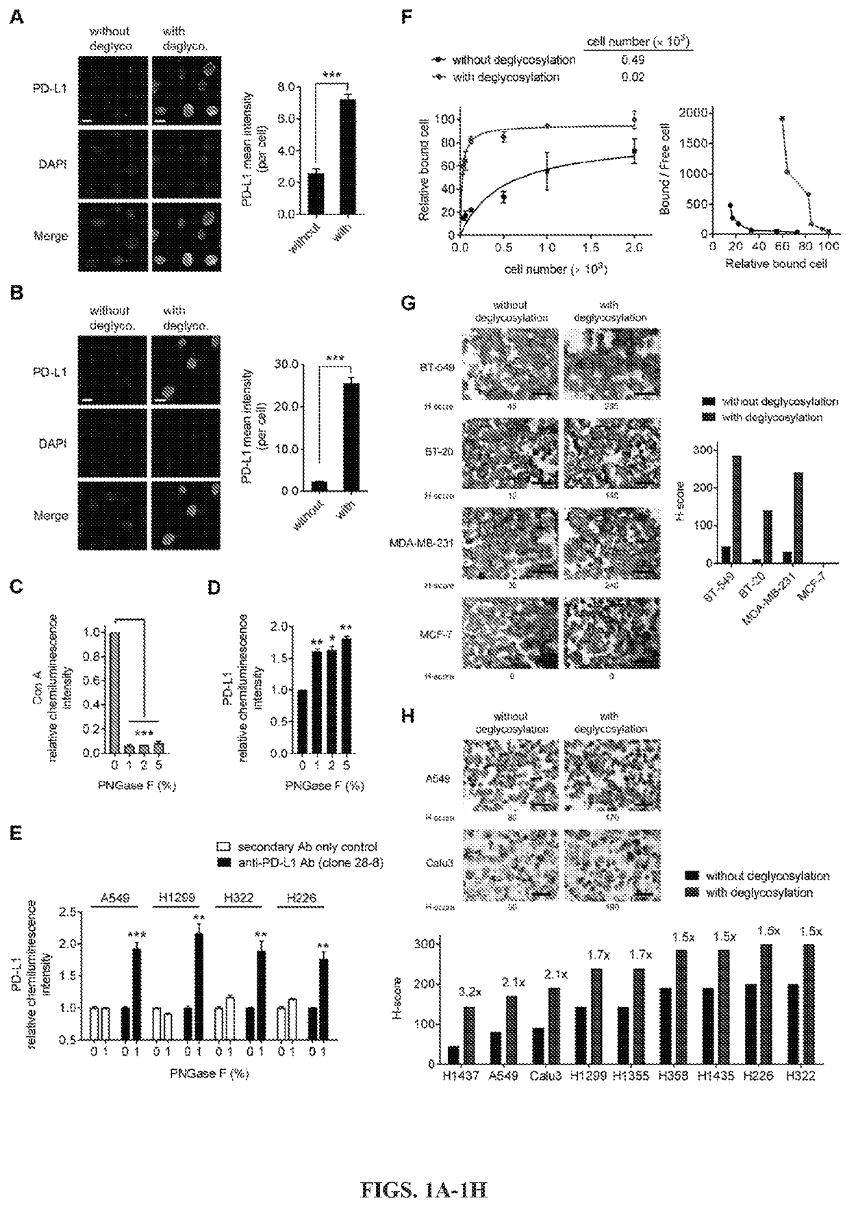
[0125]Removal of N-linked glycosylation enhances anti-PD-L1 signal in human cancer cells: The migration pattern of PD-L1 on gel electrophoresis was heterogeneous as illustrated by a range of bands at ˜50 kDa with heavy glycosylation in a panel of human lung and basal-like breast cancer (BLBC) but not non-BLBC cell lines (FIGS. 6A and 6B). Treatment with recombinant glycosidase (peptide-N-glycosidase F; PNGase F) to remove the entire N-linked glycosylation (deglycosylation, herein after) resulted in a homogenous pattern of PD-L1 at ˜33 kDa (FIGS. 6A and 6C). To determine whether the N-linked glycan structure of PD-L1 hinders antibody-based detection targeting the PD-L1 antigen, cells were first pretreated with or without PNGase F followed by immunofluorescence confocal microscopy analysis. The fluorescent intensity of PD-L1 was significantly enhanced after PNGase F treatment in lung cancer and BT-549 BLBC cells compared with no treatment (FIGS. 1A, 1B, and 6D). The resul...

example 2

and Methods

[0139]Cell culture: All human cells lines cultured at 37° C. under 5% CO2 were obtained from the American Type Culture Collection (Manassas, Va., USA), including breast cancer (BT-549, BT-20, MDA-MB-231, MCF-7), lung cancer (H1437, A549, Calu3, H1299, H1355, H358, H1435, H226, H322), and immune (Jurkat T lymphocytes, THP1 monocytes) cell lines. Human breast cancer cell lines (BT-549, BT-20, MDA-MB-231, MCF-7) and H1435 cells are female-derived cell lines; other cell lines used are male-derived cells. All cell lines were independently validated by STR DNA fingerprinting at The University of Texas MD Anderson Cancer Center and characterized as mycoplasma negative. BT-549, BT-20, MDA-MB-231, MCF-7, and A549 cells were maintained in Dulbecco's modified Eagle's medium (DMEM) / F12, supplemented with 10% fetal bovine serum (FBS) and 1% antibiotic mixture. Calu3 cells were cultured in Eagle's Minimum Essential Medium, supplemented with 10% FBS and 1% antibiotic mixture. Other cell...


PUM

PropertyMeasurementUnit
Fractionaaaaaaaaaa
Fractionaaaaaaaaaa
Timeaaaaaaaaaa

Abstract

Provided herein are methods of detecting glycosylated proteins, such as immune checkpoint proteins. Further provided are methods of treating cancer, such as by administering immune checkpoint inhibitors.

Description

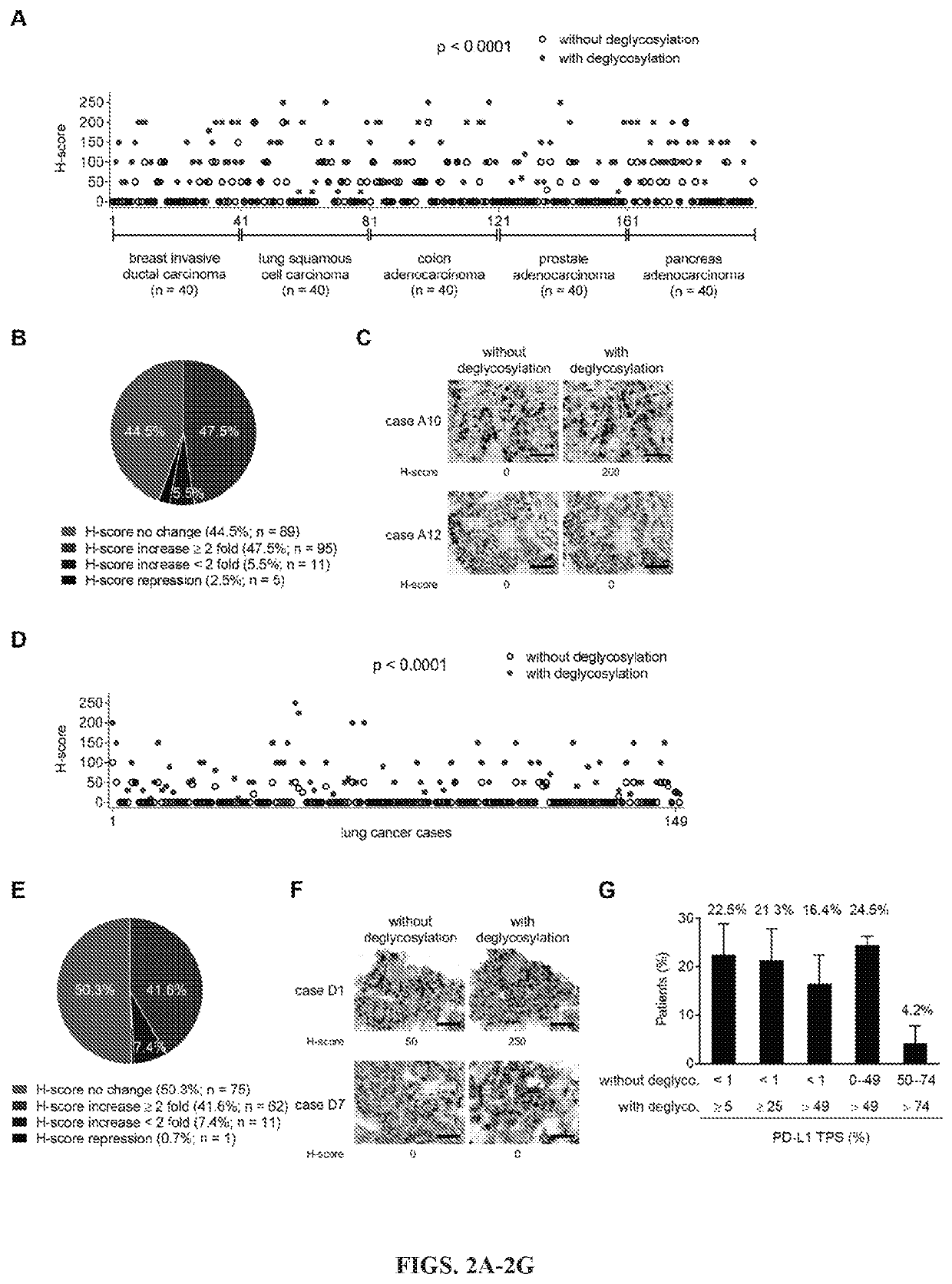
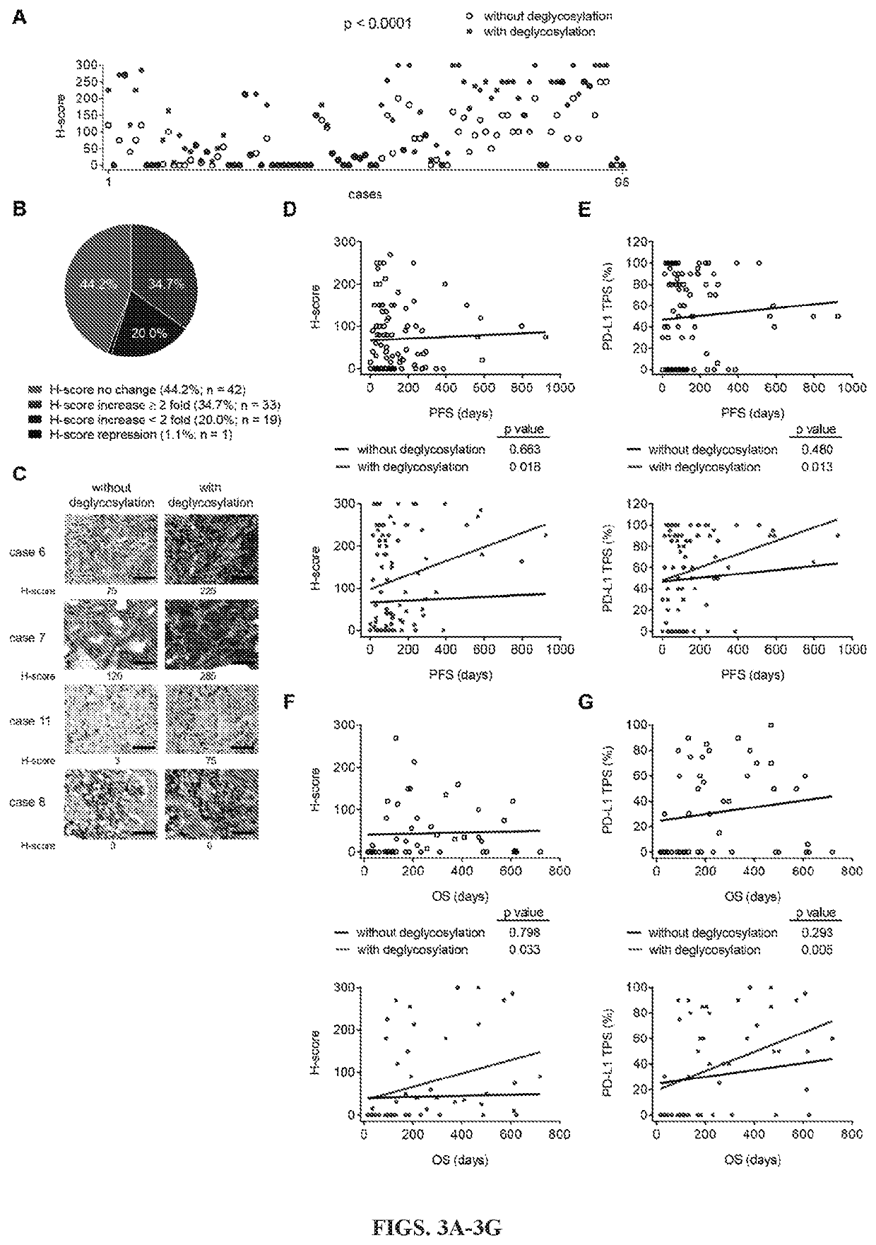
[0001]This application claims the benefit of U.S. Provisional Patent Application No. 62 / 681,929 filed Jun. 7, 2018, the entirety of which is incorporated herein by reference.BACKGROUND1. Field[0002]The present invention relates generally to the field of molecular biology. More particularly, it concerns the detection of glycosylated proteins, such as immune checkpoint molecules.2. Description of Related Art[0003]The Food and Drug Administration (FDA) approval of immune checkpoint inhibitors has dramatically changed treatment paradigms for patients with advanced-stage or metastatic non-small cell lung cancer (NSCLC). Despite significant improvements in survival, the majority of NSCLC patients fail to respond to checkpoint inhibitors, notably antibodies targeting programmed death-1 (PD-1) and programmed death ligand-1 (PD-L1), and uncertainties remain regarding how best to use these therapies in clinical practice. Given the risk of immune-related and other adverse effects associated wi...

Claims


Application Information

Patent Timeline
IPC IPC(8): G01N33/574C12N9/80A61K45/06C07K16/28G01N33/563
CPCG01N33/57492C12N9/80A61K45/06G01N2800/52C07K16/2827G01N33/563C12Y305/01052C07K16/2818G01N2333/70532G01N2333/70521C07K2317/92
Inventor LEE, HENG-HUANWANG, YING-NAIHUNG, MIEN-CHIE
Owner BOARD OF RGT THE UNIV OF TEXAS SYST
Learn More