
Probing mechanical properties of biological matter

Pending Publication Date: 2021-08-12
LUMICKS CA HLDG BV
View PDF0 Cites 0 Cited by
  • Summary
  • Abstract
  • Description
  • Claims
  • Application Information

AI Technical Summary

Benefits of technology

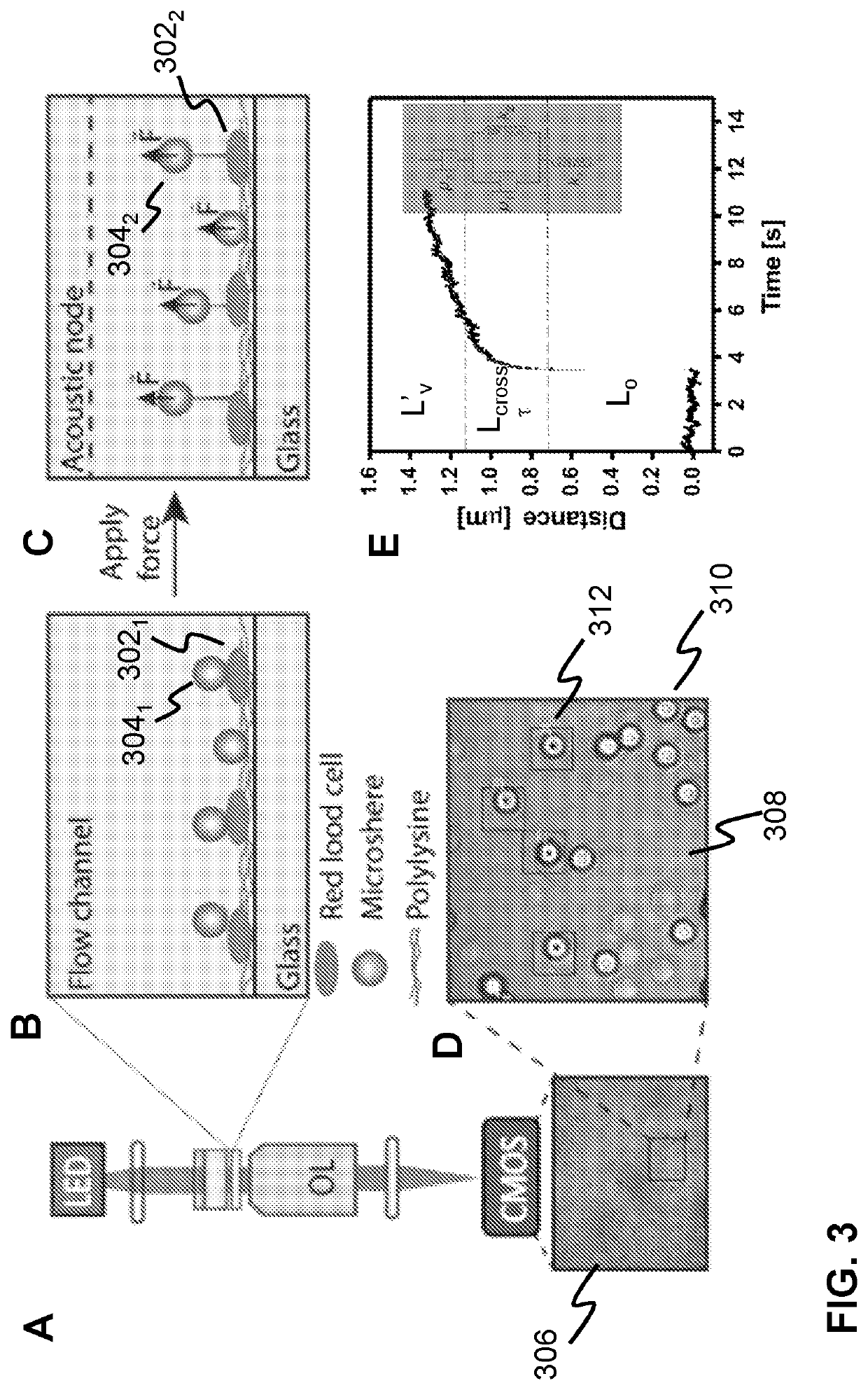
The invention relates to a method for studying the mechanical properties of living cells and biological matter. It uses particles of known size and material that produce strong acoustic contrast with the cells, allowing for the probing of mechanical properties without affecting the cells. The particles used should also have a high optical contrast, allowing for accurate tracking of their displacement due to the acoustic force. This method is easy to use and can be used for biomedical or diagnostic applications. It can be done in a fast and efficient way, providing large statistics on the mechanics of cells and biological matter.

Problems solved by technology

Although cell mechanics can be probed, this technique has the disadvantage that it entails a single cell manipulation tool requiring an acoustic trap with a small focus (small enough to manipulate a microbead) which can be moved around.
More in general, current state of the art acoustic probing techniques are single cell probing techniques which are not suitable for simultaneously determining individual cell mechanics parameters of a large number of cellular bodies and / or for different parts of a biological soft matter layer.
Additionally, many of these techniques suffer from low signal-to-noise ratio, slow detection, small sample size and / or only localised and indirect interaction with the cellular membranes of probed cells.
When trying to apply this technique to study mechanical properties of biological matter, such as cellular bodies and / or biological soft matter layers problems arise.
Large microbeads may have a large contact area and thus do not allow to controllably probe the mechanical response of specific parts of a cellular body or soft matter layer.
However, the force on the microbeads scales with the third power of the volume of the microbeads so possibilities to reduce the size of conventional microbeads will be limited.
Additionally, when trying to track microbeads in the z-direction, the image of the cellular body or soft matter layer may interfere with the optical signal of the microbead, thereby deteriorating the accuracy of the measurements.

Method used


Image

  • Probing mechanical properties of biological matter
  • Probing mechanical properties of biological matter
  • Probing mechanical properties of biological matter

Examples


Embodiment Construction

[0090]It is noted that the drawings are schematic, not necessarily to scale and that details that are not required for understanding the present invention may have been omitted. The terms “upward”, “downward”, “below”, “above”, and the like relate to the embodiments as oriented in the drawings, unless otherwise specified. Further, elements that are at least substantially identical or that perform an at least substantially identical function are denoted by the same numeral, where helpful individualised with alphabetic or subscript numeric suffixes.

[0091]While the embodiments and examples hereunder are described with reference to cellular bodies, it is appreciated that these embodiments and examples are not limited thereto and also include systems and methods for probing mechanical properties of biological soft matter layers such as a tissue layers, lipid bilayers, organ on chip, etc.

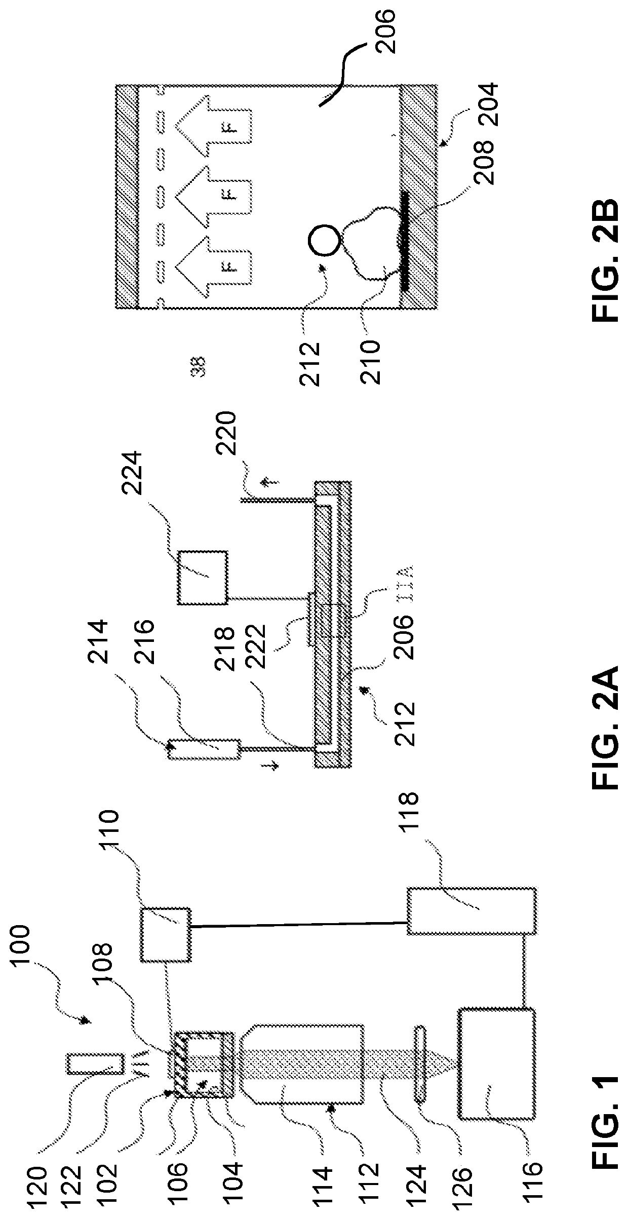
[0092]FIG. 1 schematically depicts an acoustic force spectroscopy system according to an embodiment of...


PUM


Abstract

A method for probing mechanical properties of cellular bodies includes: providing a plurality of particles in a fluid medium contained in a holding space of a sample holder, each of the plurality of particle being attached to a cellular body; generating a resonant bulk acoustic wave in the holding space, the resonant bulk acoustic wave exerting an acoustic force on each of the plurality of particles, each of the plurality of particles having an acoustic contrast factor and a size, the acoustic contrast factor and the size being selected such that the force exerted on a particle is larger than the force exerted on the cellular body to which the particle is attached; measuring a displacement of a particle in response to the exertion of the force on the particle, the measured displacement being associated with a mechanical property of the cellular body attached to the particle.

Description

TECHNICAL FIELD[0001]The present disclosure relates to probing mechanical properties of biological matter, and, in particular, though not exclusively, to methods and systems for probing mechanical properties of biological matter using acoustic waves, and a computer program product for executing such methods.BACKGROUND OF THE INVENTION[0002]The measurement of mechanics of biological matter, e.g. cellular bodies and / or biological soft matter layers such as a tissue layers, lipid bilayers, organ on chip, etc., is crucial for a better understanding of biological processes, e.g. cellular responses during the progression of many diseases, including malaria, anaemia and cancer. A variety of biophysical techniques, including atomic force microscopy, optical tweezers, and micro-pipette aspiration, have been developed to spatially manipulate a cell in order to probe its mechanics.[0003]Hwang et a., describe in their article Cell Membrane Deformation Induced by a Fibronectin-Coated Polystyrene...

Claims


Application Information

Patent Timeline
IPC IPC(8): G01N15/14B01L3/00
CPCG01N15/14G01N2015/0065B01L2400/0436B01L3/508B01L3/502761G01N29/222G01N2291/02416G01N2291/02466G01N2291/015G01N29/032G01N29/348B01L2400/0457B01L2200/0647B01L2300/0654G01N33/4833G01N15/01
Inventor WUITE, GIJS JAN LODEWIJKSORKIN, RAYAKAMSMA, DOUWESITTERS, GERRITPETERMAN, ERWIN JOHANNES GERARDBERGAMASCHI, GIULIADE GROOT, MATTIJS
Owner LUMICKS CA HLDG BV
Learn More