Ultrasound blood-flow monitoring

a technology of ultrasonic and blood flow, which is applied in the field of ultrasonic blood flow monitoring, can solve the problems of high equipment cost and inability to carry out such analyses, and the techniques are not well suited to the unattended monitoring of patients in hospital wards or at home, so as to optimize cerebral flow, reduce stress, pain and discomfort, and reduce the effect of large variations in cerebral blood flow

Pending Publication Date: 2021-08-19
CIMON MEDICAL AS
View PDF4 Cites 10 Cited by
  • Summary
  • Abstract
  • Description
  • Claims
  • Application Information

AI Technical Summary

Benefits of technology

[0017]Thus it will be seen that, in accordance with these aspects, rather than a skilled operator having to manually hold an ultrasound transducer against the subject, an ultrasound transducer is fastened to the subject. This can facilitate the monitoring of blood flow over an extended period of time, without requiring the expense of a human operator attending the subject continually during the data collection process. Preferably the ultrasound transducer will be fastened to the subject on an external surface of the subject and thus will be non-invasive (i.e. fastening will preferably not involve a surgical procedure).
[0432]In some embodiments of any of the aspects disclosed herein the ultrasound transducer may comprise a force sensor. The ultrasound transducer or a separate controller may comprise a detector configured to process signals from the force sensor to determine when a contact force between the ultrasound transducer and the subject exceeds a threshold level. This can be useful to prevent restricting blood flow due to excessive pressure from the ultrasound transducer, and therefore provide more accurate or consistent measurements of the characteristic of blood flow. Small vessels close to the skin are especially vulnerable to compression.

Problems solved by technology

The equipment for carrying out such analyses can also be very expensive (e.g., over one million U.S. dollars for a 3D ultrasound imaging system).
Such techniques are therefore not well suited to the unattended monitoring of patients in settings such as hospital wards or at home.

Method used


Image

  • Ultrasound blood-flow monitoring
  • Ultrasound blood-flow monitoring
  • Ultrasound blood-flow monitoring

Examples


example 1

Analysis of Cerebral Blood Flow in Neonatal Preterm Humans with Unfocused Doppler Ultrasound

[0603]The test subject was an infant of gestational age 32, birth weight: 1830 gram receiving no respiratory support. Ultrasound apparatus as described herein was used to obtain continuous measurements from the cerebral circulation via the anterior fontanelle for 7 seconds with 10 second pauses in between. FIGS. 30a, 30b and 30c show the same recording, but present Doppler curves from different depth ranges (represented by white rectangle). In FIG. 30a, the Doppler curve was obtained from a depth of 10-15 mm. In FIG. 30b, the Doppler curve was obtained from a depth of around 20 mm. In FIG. 30c, the Doppler curve was obtained from a depth of around 25 mm. Safety measurements were visualized continuously for each recording (right upper corner of FIGS. 30a-c).

[0604]A trend curve was visualized based on multiple recordings as represented in FIG. 30 (FIG. 31). Each small circle represents one 7 se...

example 2

Monitoring of Cerebral Blood Flow in Neonatal Humans with Unfocused Doppler Ultrasound—Comparison with Conventional Ultrasound

[0605]Background

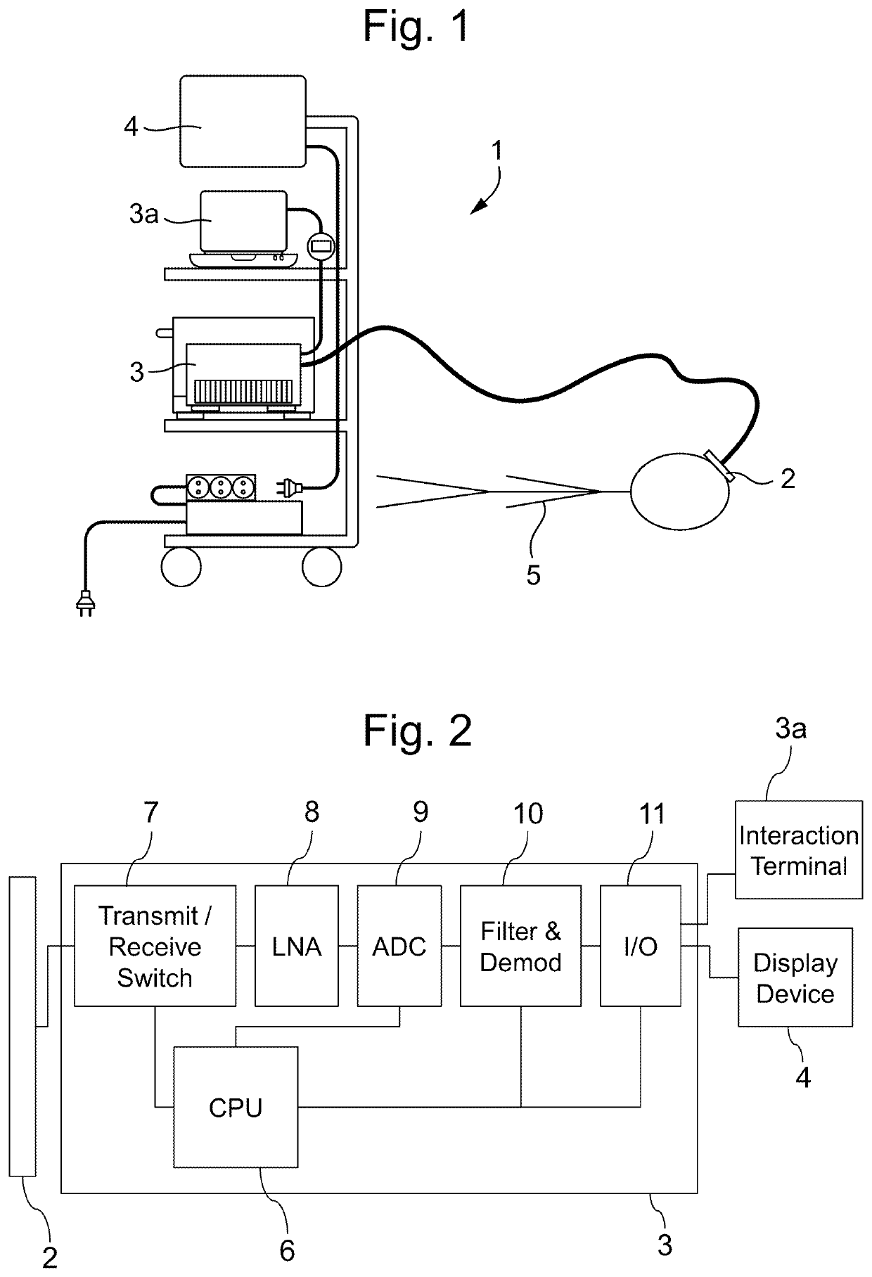
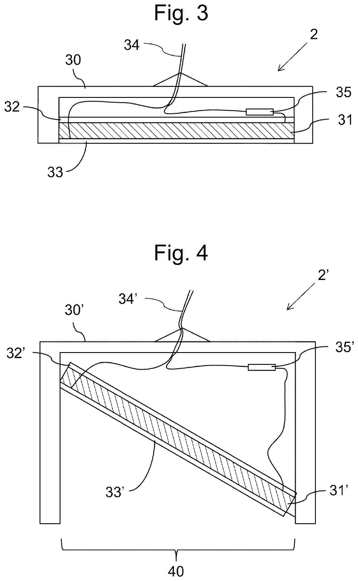
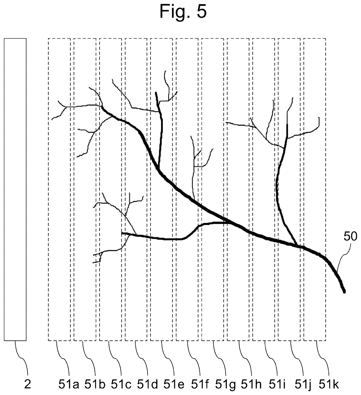
[0606]There is a strong need for continuous cerebral circulation monitoring in neonatal care, because brain injury due to low or variable blood flow frequently complicates prematurity and critical illnesses in neonates. NeoDoppler is a novel, non-invasive method based on unfocused Doppler ultrasound (as described herein) which is designed to monitor cerebral blood flow continuously. By recording and analysing the cerebral circulation over time in different depths of the brain simultaneously, the timing of medical interventions can be optimised. The NeoDoppler probe is operator independent and can be gently fixed to the fontanel by a specially designed housing.

[0607]Objective

[0608]In this feasibility study, the general quality of the NeoDoppler measurements and the fluctuations of cerebral blood flow in neonates over time were investigated. Com...

example 3

of Microvascular Circulatory Chancres

[0615]Background

[0616]Microvascular physiological responses or endothelial functions as vaso-constriction or -dilatation and vasomotion, are well studied in healthy as well as in diabetic subjects. A range of non-invasive methods has been developed and is shown to adequately assess vasomotor responses. There are a number of potential devices and techniques that are in use to evaluate microcirculatory function, i.e. transcutaneous oxygen tension (TcPO), skin pulp blood flow (i.e. laser Doppler fluxometry), iontophoresis or capillaroscopy. These techniques, as of today, need further development to optimally cover their clinical purposes due to lack of standardization and official guidelines which results in large differences in methodology and reduces reproducibility and comparability between studies performed.

[0617]The present study was performed to compare and validate a novel flat unfocused ultrasound probe in accordance with at least some aspec...


PUM


Abstract

The invention provides a method of monitoring blood flow in a vertebrate animal subject. Unfocussed plane-wave ultrasound pulses are transmitted into the subject, along a transmission axis, from a single-element ultrasound transducer (2) fastened to the subject (5). Reflections of the ultrasound pulses are received, generating a succession of pulse-Doppler response signals over time. Each pulse-Doppler response signal is processed to determine a first respective spatial-maximum velocity value for blood flowing towards the single transducer element (2), and a second respective spatial-maximum velocity value for blood flowing away. Heartbeats are identified from said spatial-maximum velocity values and a quality metric is assigned to each identified heartbeat. A subset of the spatial-maximum velocity values is identified for which the assigned quality metric exceeds a threshold level. The values from the subset are monitored, and, when a set of values from the subset satisfies a predetermined alert criterion an audible or visual alert is signalled.

Description

BACKGROUND OF THE INVENTION[0001]This invention relates to apparatus and methods for characterising or monitoring blood flow using ultrasound.[0002]Various techniques have been used to analyse blood flow in human or animal subjects. These include laser Doppler scanning, near-infrared spectroscopy, and Doppler ultrasound imaging. However, such analyses must typically be performed by a skilled technician, who must be present with the patient throughout. The equipment for carrying out such analyses can also be very expensive (e.g., over one million U.S. dollars for a 3D ultrasound imaging system). Such techniques are therefore not well suited to the unattended monitoring of patients in settings such as hospital wards or at home.[0003]The present invention seeks to provide a better approach.SUMMARY OF THE INVENTION[0004]From a first aspect, the invention provides a method for determining a characteristic of blood flow in a vertebrate animal subject, the method comprising:[0005]transmitt...

Claims


Application Information

Patent Timeline
Patent Type & Authority Applications(United States)
IPC IPC(8): A61B8/06A61B8/08A61B8/00
CPCA61B8/06A61B8/488A61B8/5207A61B8/4483A61B8/461A61B8/4227A61B8/0883A61B8/0866
Inventor TORP, HANSHERGUM, TORBJØRN
Owner CIMON MEDICAL AS
Why Eureka
Try Eureka