
Method and device for protein sequence analysis

a protein sequence and protein technology, applied in the field of protein sequence analysis, can solve the problems of enzymes often failing to cleave all scissile bonds, time-consuming and laborious, and inconvenient, and achieve the effects of simple and fast protein digestion, short time, and acceleration of protein digestion

Pending Publication Date: 2022-04-21
FUDAN UNIV
View PDF0 Cites 0 Cited by
  • Summary
  • Abstract
  • Description
  • Claims
  • Application Information

AI Technical Summary

Benefits of technology

The present invention relates to a method and device for protein sequence analysis using microdroplet chemistry. The method involves forming a solution containing protein into small droplets, which are then introduced into a mass spectrometer for real-time detection. The microdroplets accelerate the protein digestion process, resulting in a higher sequence coverage without the need for any pre-treatment steps. The method and device are suitable for analyzing a wide variety of proteins and do not require any pre-treatment steps for enzymatic digestion. Overall, the invention provides a convenient and efficient interface for protein sequencing with high accuracy.

Problems solved by technology

In bottom-up proteomics, enzymatic digestion of proteins is an essential and critical approach for breaking down proteins into smaller polypeptides prior to analysis for protein structure elucidation by mass spectrometry (MS).1 In a typical enzymatic digestion process, the protein solution is mixed with a proper amount of enzyme, such as trypsin, and incubated overnight at 37° C. However, such process is time-consuming.
To further accelerate protein digestion, various attempts have been taken to reduce the digestion time from overnight to several minutes, including increasing the digestion temperature, using columns or porous materials for trypsin immobilization, addition of organic solvents, applying microwave energy or focused ultrasonic field, or a combination of any thereof.2 Still, the conventional methods for sequence analysis of protein require pre-treatment steps prior to enzymatic digestion, which is relatively time consuming and inconvenient.
The most ideal case in enzymatic digestion for proteomics study is achieved when all cleavage sites are digested, but in practice, enzymes often fail to cleave all scissile bonds, even though the reaction time is sufficiently long.
This failure to achieve complete coverage is mainly attributed to neighboring amino acids around the cleavage sites.
However, the application of microdroplet to biochemical analysis has seldom been investigated.

Method used


Image

  • Method and device for protein sequence analysis
  • Method and device for protein sequence analysis
  • Method and device for protein sequence analysis

Examples


example 1

[0073]Optimization of the Performance of Microdroplet-MS.

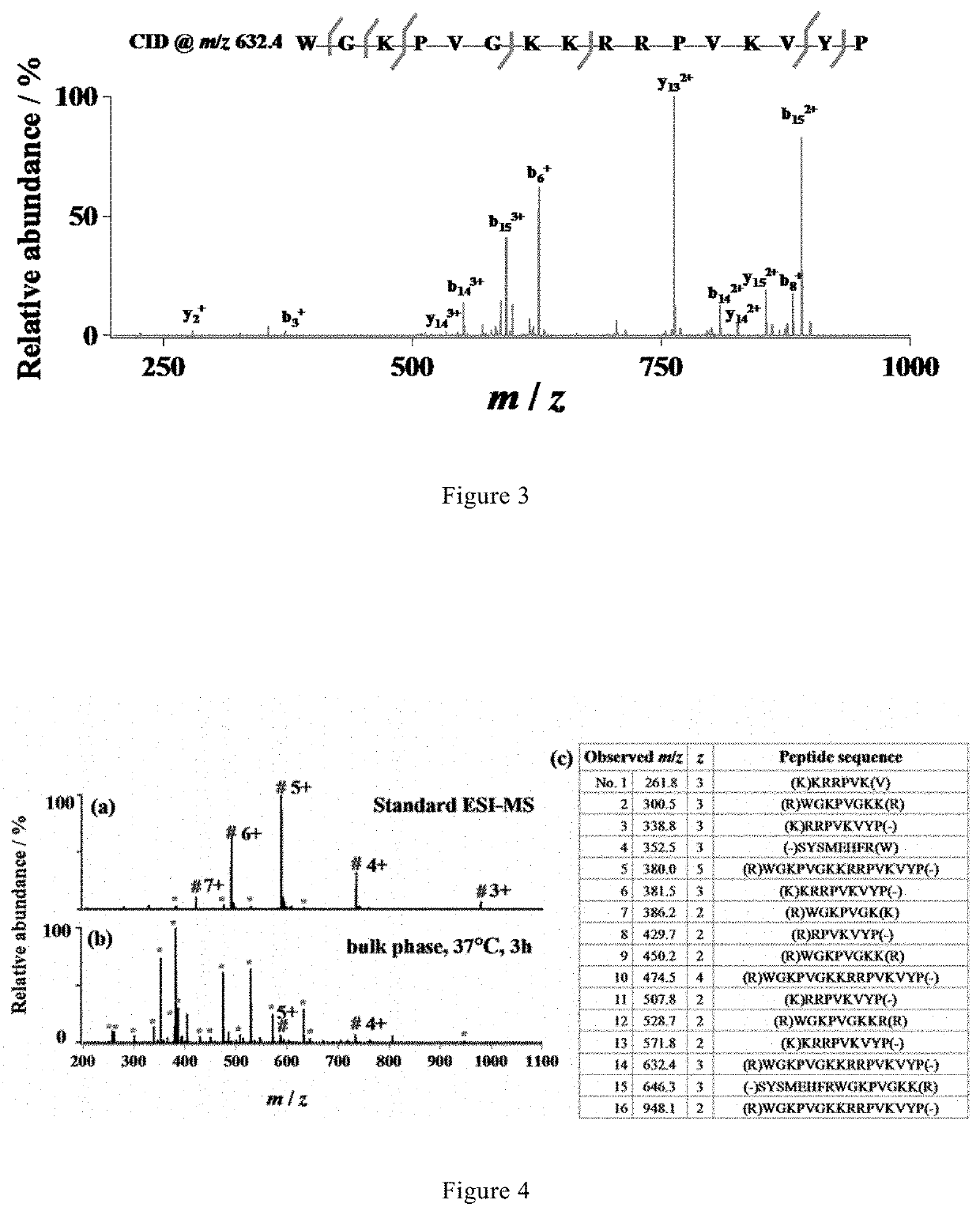
[0074]To optimize of the performance of microdroplet-MS, tryptic digestion of ACTH was used as a simple model system.

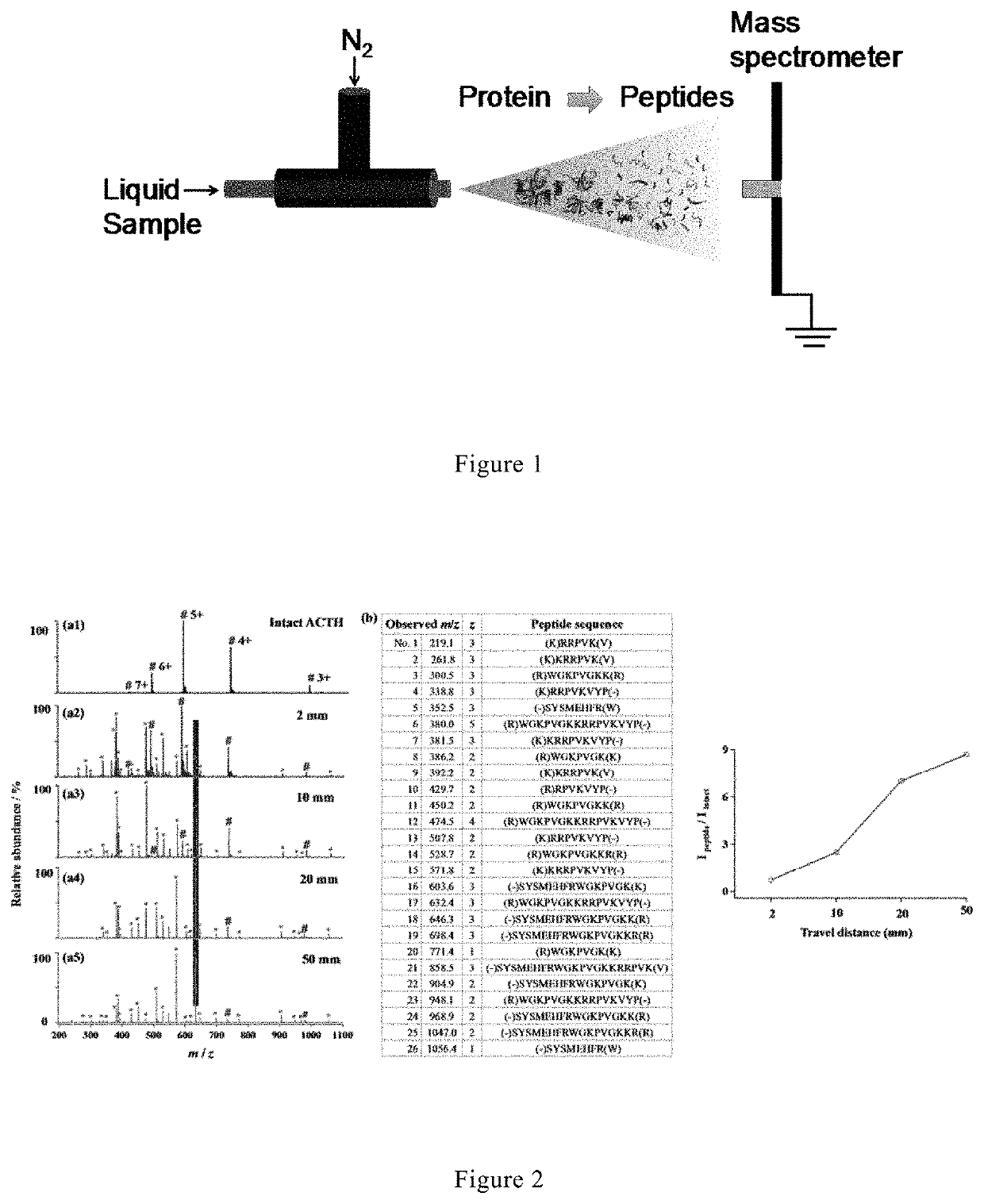
[0075]A stream of microdroplets was generated by infusing an aqueous sample solution containing 10 μM adrenocorticotropic hormone from human (ACTH, 1-24, Genscript, China) and 5 μg / mL trypsin in 5 mM ammonia bicarbonate (NH4HCO3, pH 8) with a syringe at a flow rate of 10 μL / min into a homemade sprayer (with a capillary of 50 μm i.d and 148 μm o.d, as shown in FIG. 1).

[0076]The sample solution was sprayed from the tip of the fused silica capillary (148 μm o.d., 50 μm i.d., Polymicro Technologies, China) and assisted by a nebulizing gas of dry N2 with a pressure of 120 psi. By placing the sprayer in front of a high-resolution mass spectrometer (LTQ Orbitrap Elite, Thermo Scientific, San Jose, Calif.) at a proper position, the microdroplets were directed into MS for real-time detection. The MS inlet capillary was m...

example 2

[0085]Microdroplet-MS for Digestion and Analysis of Protein that Particularly Recalcitrant to Tryptic Digestion.

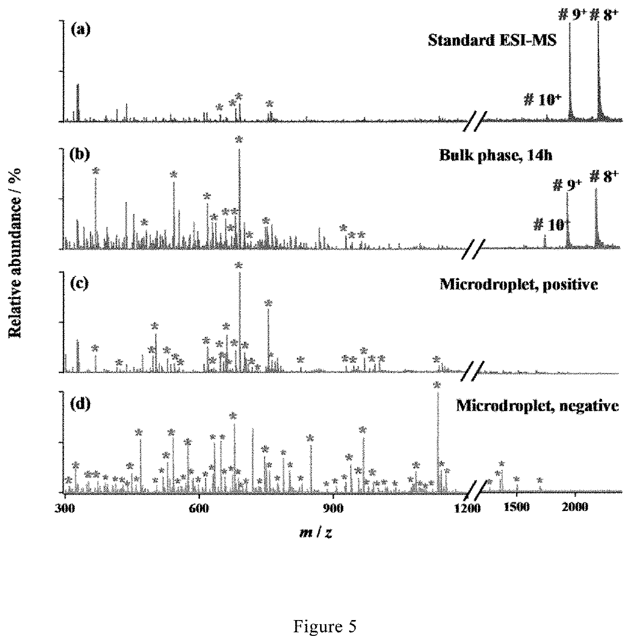
[0086]A stream of microdroplets was generated by infusing an aqueous sample solution containing myoglobin (10 μM) and trypsin (5 μg / mL) in 5 mM ammonia bicarbonate (NH4HCO3, pH 8) with a syringe at a flow rate of 10 μL / min into a homemade sprayer (with a capillary of 50 μm i.d and 148 μm o.d, as shown in FIG. 1).

[0087]The sample solution was sprayed from the tip of a fused silica capillary (148 μm o.d., 50 μm i.d., Polymicro Technologies, China) and assisted by a nebulizing gas of dry N2 with a pressure of 120 psi. By placing the sprayer in front of a high-resolution mass spectrometer (LTQ Orbitrap Elite, Thermo Scientific, San Jose, Calif.) at a proper position, the microdroplets were directed into MS for real-time detection when applying a positive high voltage of +3 kV (BOHER H V, Genvolt, U.K.) to the sprayer. The MS inlet capillary was maintained at 275° C. and capill...

example 3

[0097]Microdroplet-MS for Digestion and Analysis of Cytochrome c.

[0098]According to the same method as described in Example 1 and Example 2, microdroplet-MS was further used for the digestion and analysis of cytochrome c under a positive voltage of +3 kV.

[0099]As shown in FIG. 8, 33 peptide fragments corresponding to 83% sequence coverage of cytochrome c was successfully identified. The results demonstrate that microdroplet-MS is a universal tool for protein digestion.


PUM

PropertyMeasurementUnit
pressureaaaaaaaaaa
voltageaaaaaaaaaa
distanceaaaaaaaaaa

Abstract

A method and a device for protein sequence analysis, and the use of microdroplets for improving protein sequencing by accelerating enzymatic digestion, wherein the method comprises the following steps: a) forming a solution containing protein into microdroplets having a size small enough to result in acceleration of protein digestion; b) introducing the microdroplets into a mass spectrometer (MS) for real-time detection; c) obtaining analysis result of the protein from the mass spectrometer (MS); wherein the protein is fully digested in the microdroplets before entering the mass spectrometer (MS). The method and device can achieve simple and nearly complete protein digestion in a very short time and obtain high sequence coverage.

Description

TECHNICAL FIELD[0001]The present invention relates to a method and a device for protein sequence analysis. More particularly, the present invention relates to a method of protein sequence analysis and a microdroplet-MS (microdroplet-mass spectrometer) device for protein sequence analysis. The present invention also relates to the use of microdroplets for improving protein sequencing by accelerating enzymatic digestion, wherein the microdroplets are formed from protein-containing solution and have a size small enough to result in acceleration of protein digestion.BACKGROUND ART[0002]In bottom-up proteomics, enzymatic digestion of proteins is an essential and critical approach for breaking down proteins into smaller polypeptides prior to analysis for protein structure elucidation by mass spectrometry (MS).1 In a typical enzymatic digestion process, the protein solution is mixed with a proper amount of enzyme, such as trypsin, and incubated overnight at 37° C. However, such process is ...

Claims


Application Information

Patent Timeline
Patent Type & Authority Applications(United States)
IPC IPC(8): G01N33/68
CPCG01N33/6848G01N33/6821G01N33/6818
Inventor ZARE, RICHARDZHONG, XIAOQIN
Owner FUDAN UNIV
Learn More