Cell-matrix microspheres, methods for preparation and applications

a cell-matrix and microsphere technology, applied in the field of cell-matrix microspheres, can solve the problems of preventing their use in regenerative medicine and tissue engineering, unable to allow cell migration and penetration, and no microencapsulation system for these materials, so as to increase cell density, reduce matrix concentration, and high surface tension

Active Publication Date: 2014-03-25
VERSITECH LTD
View PDF7 Cites 11 Cited by
  • Summary
  • Abstract
  • Description
  • Claims
  • Application Information

AI Technical Summary

Benefits of technology

[0010]A method has been developed to produce stable cell-matrix microspheres with up to 100% encapsulation efficiency and high cell viability, using matrix or biomaterial systems with poor shape and mechanical stability for applications including cell therapeutics via microinjection or surgical implantation, 3D culture for in vitro expansion without repeated cell splitting using enzymatic digestion or mechanical dissociation and for enhanced production of therapeutic biomolecules, and in vitro modelling for morphogenesis studies. The modified droplet generation method is simple and scalable and enables the production of cell-matrix microspheres when the matrix or biomaterial system used has low concentration, with slow phase transition, with poor shape and mechanical stability.
[0012]The method of forming microspheres include the steps of mixing and dispensing the composition into liquid droplets in the right order and the right time with a dispensing unit. The pH of the first matrix is adjusted to be suitable for cell survival. The cell suspension and other biomolecules are mixed thoroughly with the matrix components as soon as possible to evenly distribute cells throughout the solution form of the matrix before accelerating phase transition. The liquid droplets are collected with a dry collection platform without mechanical disturbance. The platform has a surface property for maintaining the spherical shape of the liquid droplets, such as high surface tension. By maintaining the temperature low, the rate of matrix phase transition can be controlled as low as possible at a range between 2 minutes to 10 hours depending on the matrix concentration, preferably 30 minutes. The volume of the microspheres dispensed is preferably about 2.5 μl. The diameter of the dispensed liquid droplets is preferably 2 mm. In addition, the method comprises accelerating the rate of matrix phase transition after dispensing by raising the temperature at the collection platform preferably to 37° C. The matrix components of the liquid droplets are allowed to undergo phase transition to form cell-matrix microspheres for a period of time, sufficient for phase transition to reach equilibrium.
[0013]The microspheres are stabilized by collecting the cell-matrix microspheres from the collection platform with minimal mechanical disturbance; maintaining the microspheres free-floating in a first medium for an extended period of time until the size of the microspheres becomes substantially constant; and releasing the microspheres from the first medium. The microspheres can be maintained free-floating either by keeping in suspension status in non-adhesive culture dish such as a Petri dish, or by culturing the microspheres in spinner flask or rotating vessel bioreactors. The size and mechanical strength of the microspheres can be controlled by controlled by at least one of the following parameters: cell density, collagen concentration, serum concentration, composition of the ECM, ratio of the first and second ECM, volume of the liquid droplets, duration of the free-floating status of microspheres. Increasing cell density, decreasing matrix concentration or decreasing volume of liquid droplets decreases size and increases stability of cell-matrix microspheres.
[0016]The cell matrix microspheres are also useful for 3D cultures. These have numerous advantages over the existing microcarrier systems. The system can be used to culture protein-secreting cells in physiologically relevant microenvironment with significantly increased protein productivity via matrix-induced proliferation control as compared to monolayer cultures with unrestricted proliferation.

Problems solved by technology

However, technological challenges associated with the localization, long term viability, host tissue-integration and functional remodeling (Tatard, et al., Curr.
Furthermore, since these systems avoid direct contact of the delivered cells with the host tissue, they do not allow cell migration and penetration.
This prevents their use in regenerative medicine and tissue engineering, which entails host-implant integration at cellular level.
However, there is no microencapsulation system for these materials because of their poor mechanical and shape stability (Yannas, Natural Materials, Ratner, et al. editors, Biomaterials Sciences—An introduction to materials in medicine, California, Academic Press, pp.
However, these methods encounter problems when the matrix materials are low in concentration, or with poor shape and mechanical stability such as collagen gel or hyaluoronic acid gel.
First, the cell-matrix droplets or emulsions formed barely survive the shear stress generated upon stirring during emulsification or stirring in the liquid collection bath.
The does not allow sufficient time for the formation of cell-matrix microspheres if the phase transition of the matrix takes a longer time and fragmentizes the microspheres, leading to low encapsulation efficiency.
However, not all cells grow successfully in suspension, only cells such as hybridomas and tumor cells.
However, these systems employ technologically demanding fabrication process for the bead preparations, making the commercial preparations costly.
The bead preparation has to be separated from the cell attachment procedure since most of the bead fabrication system employ harsh conditions such as high temperature, freeze-drying, organic solvent extraction and chemical crosslinking treatment that cells do not survive.
Moreover, the cell attachment procedure is a rate-limiting step of the microcarrier culture system (Sun, et al., J. Biosci. Bioeng., 90(1):32-6 (2000)) requiring prolonged culture for cell attachment to the solid surfaces or cell penetration into the porous beads (Bancel and Hu, Biotechnol. Prog., 12(3):398-402 (1996)).
However, the productivity of actively and unrestrictedly proliferating cells is usually low because these cells may not synthesize proteins at a maximal rate outside their tissue-specific microenvironment and in actively proliferating cells, most of the metabolic energy is activated to reproduction rather than synthetic activities (Sanches-Bustamante, et al., Biotechnol. Bioeng., 93(1):169-180 (2005)).
However, these proliferation controlling strategies lead to reduced cell viability (Mercille, et al., Cytotechnology, 15(1-3):117-28 (1994)) and increased apoptosis (Ko and Prives, Genes Dev., 10(9):1054-72 (1996)).
However, this system requires complicated designs for the vector systems and complicated genetic manipulation that interfaces the cell metabolism internally.
The advantages of this system include the use of external agents that do not interfere with the overall metabolism of cells but the problems are the downstream purification procedures eliminating this antibiotic, the genetic instability introduced by genetic manipulation as well as the instability of tetracycline in cultures.

Method used


Image

  • Cell-matrix microspheres, methods for preparation and applications
  • Cell-matrix microspheres, methods for preparation and applications
  • Cell-matrix microspheres, methods for preparation and applications

Examples


example 1

Production of hMSC-Collagen Microspheres

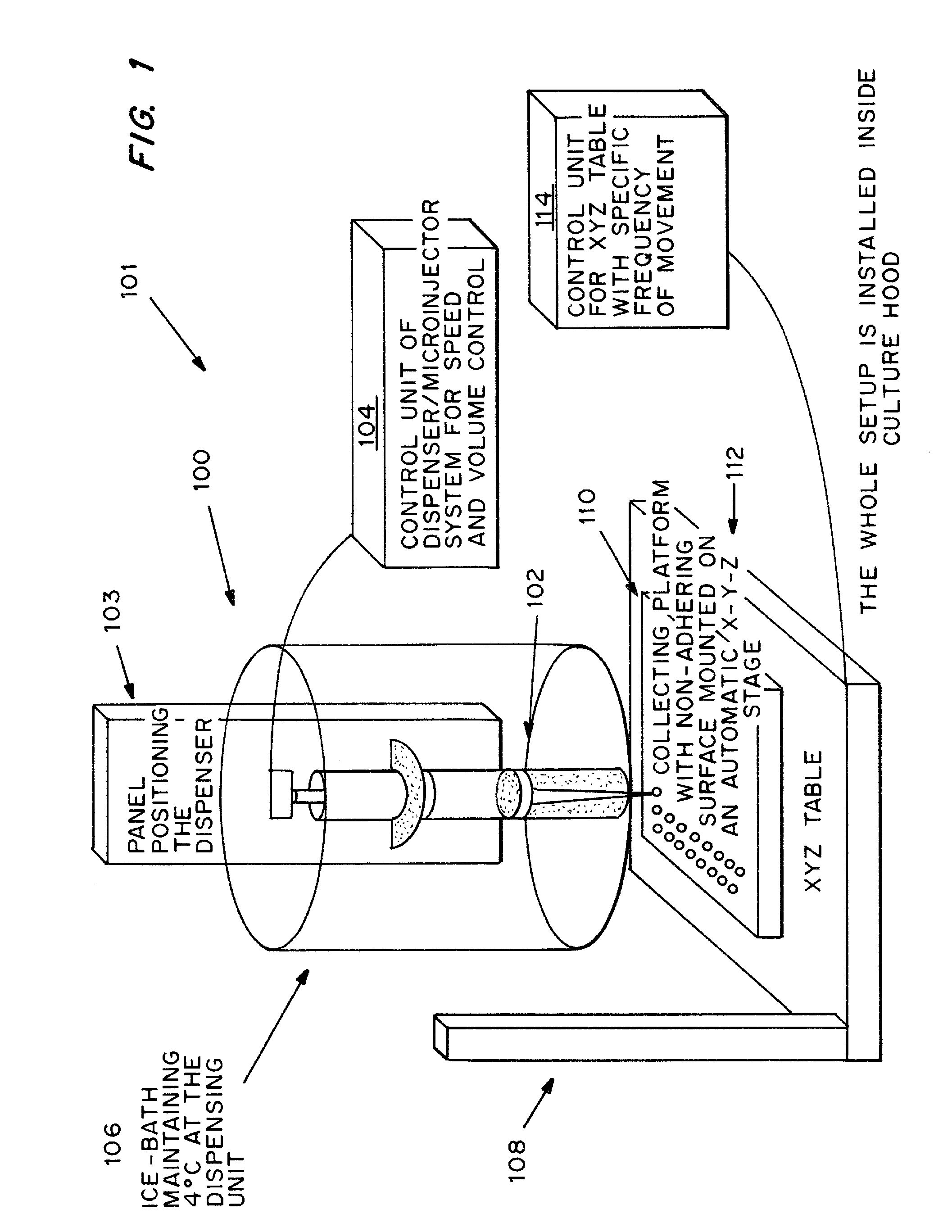
[0090]Rat-tail collagen solution type I in acetic acid, which mainly consists of triple helical monomers, was neutralized by NaOH and diluted into a final concentration of 0.5 mg / ml. All procedures were done in an ice-bath to prevent collagen gel formation. Human bone marrow derived mesenchymal stem cells (MSCs) in full medium, DMEM-LG with 10% FBS and 1% P / S, were suspended thoroughly in the neutralized collagen solution as soon as possible. A dispenser was then loaded with the ice-cold cell mixture and a small volume of 2.5 μl was dispensed at a time onto a bacterial culture dish covered with UV-irradiated parafilm. To prevent air-bubble formation in the liquid droplets, the dispenser was moved upwards or the collection platform downwards after dispensing the liquid droplets. The liquid droplets were thermally induced to reconstitute into a gel meshwork of organized collagen fibrils, interacting with the encapsulated cells, to form solid mic...

example 2

Production of Mechanically Stable Cell-Matrix Microspheres and Controlling Parameters of the Microsphere Size

[0091]Materials and Methods

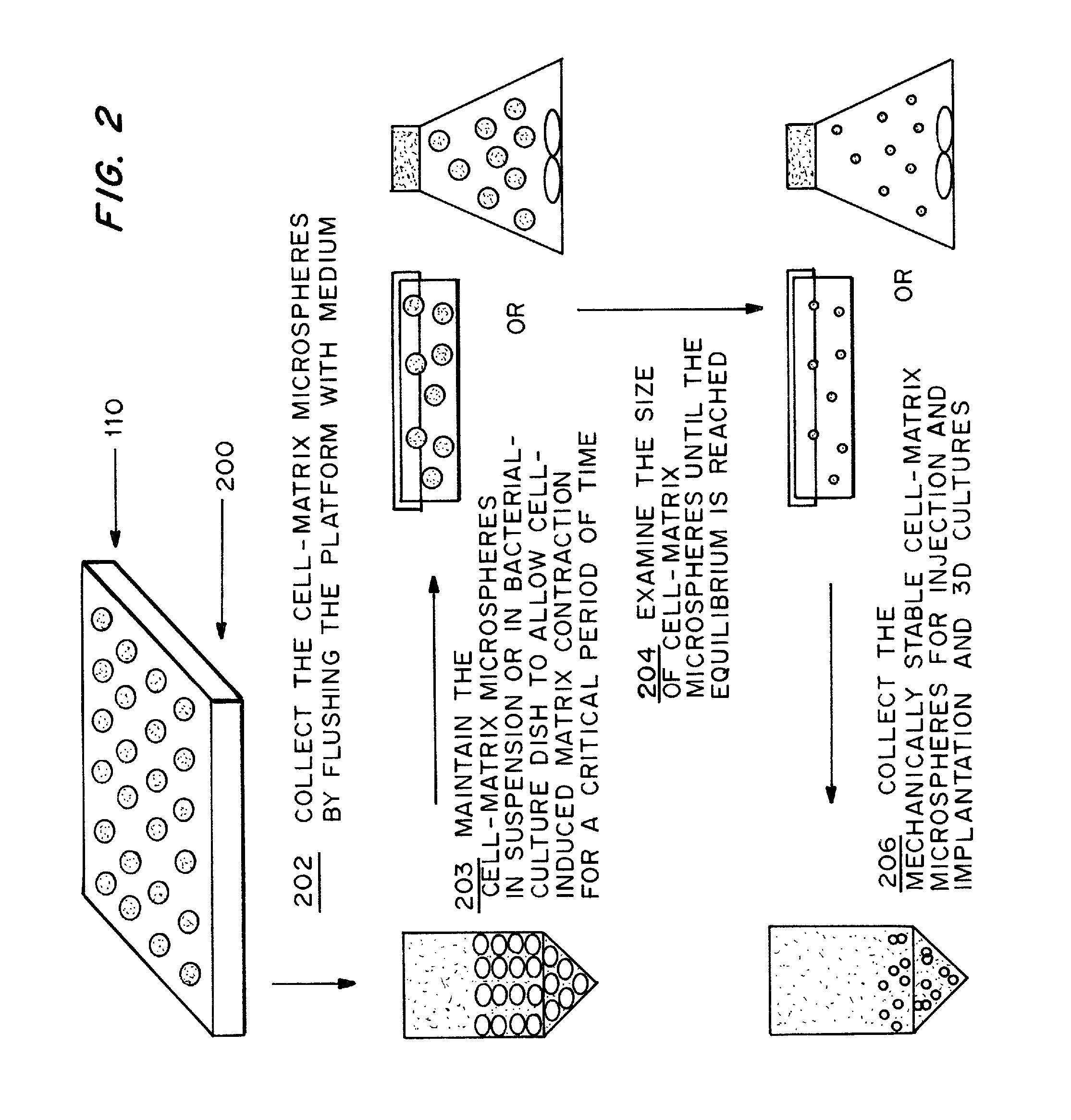
[0092]Following the steps shown in FIG. 2, rat-tail collagen solution type I was neutralized and diluted into different concentrations (0.5, 1.0, 2.0 and 3.0 mg / ml) in the presence of different concentrations (2×10⁴, 1×10⁵ and 5×10⁵ cells / ml) of human bone marrow derived MSCs in DMEM medium with 10% FBS as described in Example 1. The cell-matrix microspheres were collected into a DMEM medium containing bacterial culture dish. The collected microspheres were maintained at their free-floating state in the culture vessel at 37° C. for 2 to 7 days until the equilibrium is reached as characterized by a constant microsphere size.

[0093]Results

[0094]The temporal morphological change of the cell-matrix microspheres with different cell densities and collagen matrix densities were recorded. Microspheres at day 0 showed individual cells embedding in the collage...

example 3

Controlling the Growth Rate of Encapsulated hMSCs Inside the Collagen Microspheres

[0095]Materials and Methods

[0096]hMSCs were isolated from bone marrow aspirates from donors with informed consent in compliance with the Institute human ethics regulations. hMSCs were cultured as described by Li et al. 2004. Cells harvested from passage 2 and 3 by traditional monolayer culture were cryopreserved and used for production of cell-matrix microspheres with different collagen density: 0.5 and 2 mg / ml. Fully contracted mechanically stable hMSCs-collagen microspheres were obtained as described in Examples 1 and 2. These microspheres were incubated with 2 μM Calcein AM and 4 μM Ethidium homodimer-1 for 45 minutes for simultaneous staining of live and dead cells. Stained microspheres were fixed in 4% paraformaldehyde for 1 hour and examined using a laser confocal scanning microscope for stacked images. In a separate experiment, microspheres, with 100 microspheres per plate in triplicates, were c...


PUM

PropertyMeasurementUnit
temperatureaaaaaaaaaa
volumeaaaaaaaaaa
diameteraaaaaaaaaa

Abstract

A method has been developed to produce stable cell-matrix microspheres with up to 100% encapsulation efficiency and high cell viability, using matrix or biomaterial systems with poor shape and mechanical stability for applications including cell therapeutics via microinjection or surgical implantation, 3D culture for in vitro expansion without repeated cell splitting using enzymatic digestion or mechanical dissociation and for enhanced production of therapeutic biomolecules, and in vitro modeling for morphogenesis studies. The modified droplet generation method is simple and scalable and enables the production of cell-matrix microspheres when the matrix or biomaterial system used has low concentration, with slow phase transition, with poor shape and mechanical stability.

Description

CROSS-REFERENCE TO RELATED APPLICATIONS[0001]This application claims priority to U.S. Ser. No. 60 / 801,975 filed May 19, 2006.FIELD OF INVENTION[0002]The present invention relates generally to cell-matrix microspheres, associated products, methods for preparation and applications. More specifically, it related to methods producing cell-matrix microspheres and immobilizing living cells, methods and processes for culturing these cells; methods and processes for using these microspheres as therapeutics, as three dimensional (“3D”) microcarriers, and for production of biomolecules such as therapeutic proteins, in a more efficient and economical manner.BACKGROUND OF INVENTION[0003]Cell-based therapy provides a minimally invasive approach by local injection at the site of defect via microsyringe needles and presents a promising approach for tissue repair and regenerative medicine. However, technological challenges associated with the localization, long term viability, host tissue-integrati...

Claims


Application Information

Patent Timeline
Patent Type & Authority Patents(United States)
IPC IPC(8): C12N11/04C12N5/0775C12N5/077
CPCA61K35/12C12N5/0602C12N5/0012C12N5/0655C12N5/0663C12N2533/90C12N2533/54C12N2510/02C12M25/14A61P19/00A61P21/00A61P43/00A61P9/10
Inventor CHAN, BARBARA PUICHAN, GODFREY CHI-FUNGWONG, HOI LINGCHEUNG, PIK TOCHEAH, SONG-ENG KATHRYNCHAN, DANNY
Owner VERSITECH LTD
Why Eureka
Try Eureka