Supercharge Your Innovation With Domain-Expert AI Agents!

Contrast solution for the characterisation of biological samples by electron or correlative microscopy

a technology of contrast solution, which is applied in the field of contrast solution for the characterisation of biological samples by electron or correlative microscopy, can solve the problems of severe restrictions, inability to produce satisfactory results, and high cost of delivery and disposal of uranyl acetate derivatives

Pending Publication Date: 2022-06-16
FOND INST ITAL DI TECH +1
View PDF0 Cites 0 Cited by
  • Summary
  • Abstract
  • Description
  • Claims
  • Application Information

AI Technical Summary

Benefits of technology

The present invention is a solution that contains a mixture of a heteropolyacid and a lanthanide salt in a solvent that consists of water / organic solvent or an organic solvent. This solution can also contain additional components such as an organic or inorganic buffer with a specific pH or a strong base or acid. The invention also includes a method for preparing biological samples using this solution for analysis using either correlative microscopy or conventional electron microscopy. The technical effects of this invention include improved accuracy and sensitivity for the analysis of biological samples, as well as a simplified and streamlined preparation process for such samples.

Problems solved by technology

However, this and other similar approaches presently used in the sector do not produce satisfactory results in the case of correlative microscopy, since they do not provide contrast that is sufficient to allow visualisation of the sample by electron microscopy (EM) and necessarily require the presence of contrast agents containing heavy atoms.
However, the radioactive nature of uranyl acetate and the derivatives thereof causes severe restrictions from a safety standpoint.
Furthermore, the delivery and disposal of uranyl acetate derivatives is extremely costly due to the increasingly strict regulations they are subject to.
Furthermore, this type of contrast agent is unsuitable for optical microscopy and thus also for correlative microscopy, due both to an ample background (auto)fluorescence in the red and green region of the spectrum and the quenching action of the majority of synthetic and protein-based fluorophores on the fluorescence signal.
On the basis of these considerations, it is clear that the use of traditional contrast agents based on uranyl acetate has two opposite effects on the sample: on the one hand, it enables better visualisation in the case of electron microscopy, on the other hand it negatively influences the optical detection of fluorescence, thus inhibiting the possibility of carrying out correlative microscopy experiments.
Nonetheless, to date uranyl acetate remains the contrast agent most commonly used also for correlative purposes, even though it is not optimal for this technique, as it must be used in relatively high concentrations which, because of the high background signal, are not fully compatible with fluorescence-based techniques.
In this regard, in the present state of the art, the most commonly used expedient is to maintain the concentration of the contrast agent as low as possible, to the detriment, however, of sample contrast, since it is known that uranyl acetate (UA) does not provide satisfactory results if used at low concentrations.
As regards the presently commercially available contrast media that are alternatives to uranyl acetate (such as, for example, “Uranyless”, “336 uranyl acetate alternative”, “NanoW” and “platinum blue”), although they enable the problem of handling radioactive materials to be overcome, they perform poorly in terms of contrast efficiency and their use is thus even more disadvantageous in the case of correlative microscopy.
Furthermore, the tested compounds demonstrated to be devoid of the fixative properties of uranyl acetate.
However, in the prior art it is difficult to identify substances which, when added to lanthanide salts, are capable of forming chemically stable solutions with a low chemical risk and which make it possible to obtain better images, at least as far as electron microscopy is concerned, or at least ones that are comparable to those obtainable with uranyl acetate.
In particular, PTA provided low quality images in tests with both Gram-positive and Gram-negative bacteria.
None of these alternatives, however, have yet been thoroughly tested in the field of correlative microscopy, since, as mentioned previously, the contrast agents presently available for electron microscopy are not always suitable for an optimal implementation of the CLEM technique.

Method used


Image

  • Contrast solution for the characterisation of biological samples by electron or correlative microscopy
  • Contrast solution for the characterisation of biological samples by electron or correlative microscopy
  • Contrast solution for the characterisation of biological samples by electron or correlative microscopy

Examples


example 1

[0054]Preparation of the PTA-YbCl3 Solution (Stock Solution)

[0055]A solution of phosphotungstic acid (concentration 3.2 mM) in 10 ml of water / ethanol containing 20% (v / v) ethanol was prepared. The pH of this solution was brought to around 5 with sodium hydroxide 1 M. This solution had added to it an equal volume of an ytterbium chloride hexahydrate 48 mM (final concentration) solution in ethanol / water containing 20% (v / v) ethanol. The pH was again adjusted to 5 with sodium hydroxide 1 M. The mixture was kept under stirring overnight at room temperature. The solution thus obtained was called “X solution 1.5” and manifested a precipitate. The precipitate was removed by filtration and the pH was again adjusted to around 5 by adding a 20 mM solution of MES. The solution was then again kept under stirring overnight and again filtered, this time through a membrane with a pore size of 200 nm so as to obtain a solution free of precipitate and called “stock solution”. The stock solution was ...

example 2

[0056]Use of the Solution According to the Present Invention as a Contrast Agent for Characterisation by Correlative Microscopy (CLEM) Using the HPF / FS Protocol for Biological Samples.

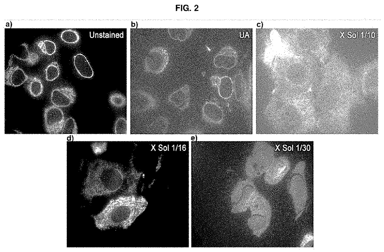
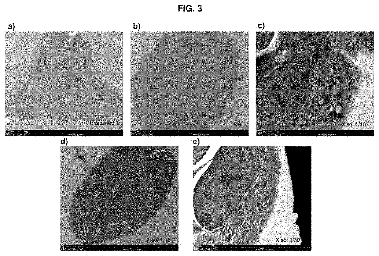
[0057]The stock solution obtained as per example 1 (0.2 ml) was freeze-dried and reconstituted in 2 ml (“X Sol 1 / 10”), 3.2 ml (“X Sol 1 / 16”) or 6 ml (“X Sol 1 / 30”) of a water / acetone mixture containing 95% (v / v) acetone in order to obtain the solution according to the present invention, in three different concentrations, respectively.

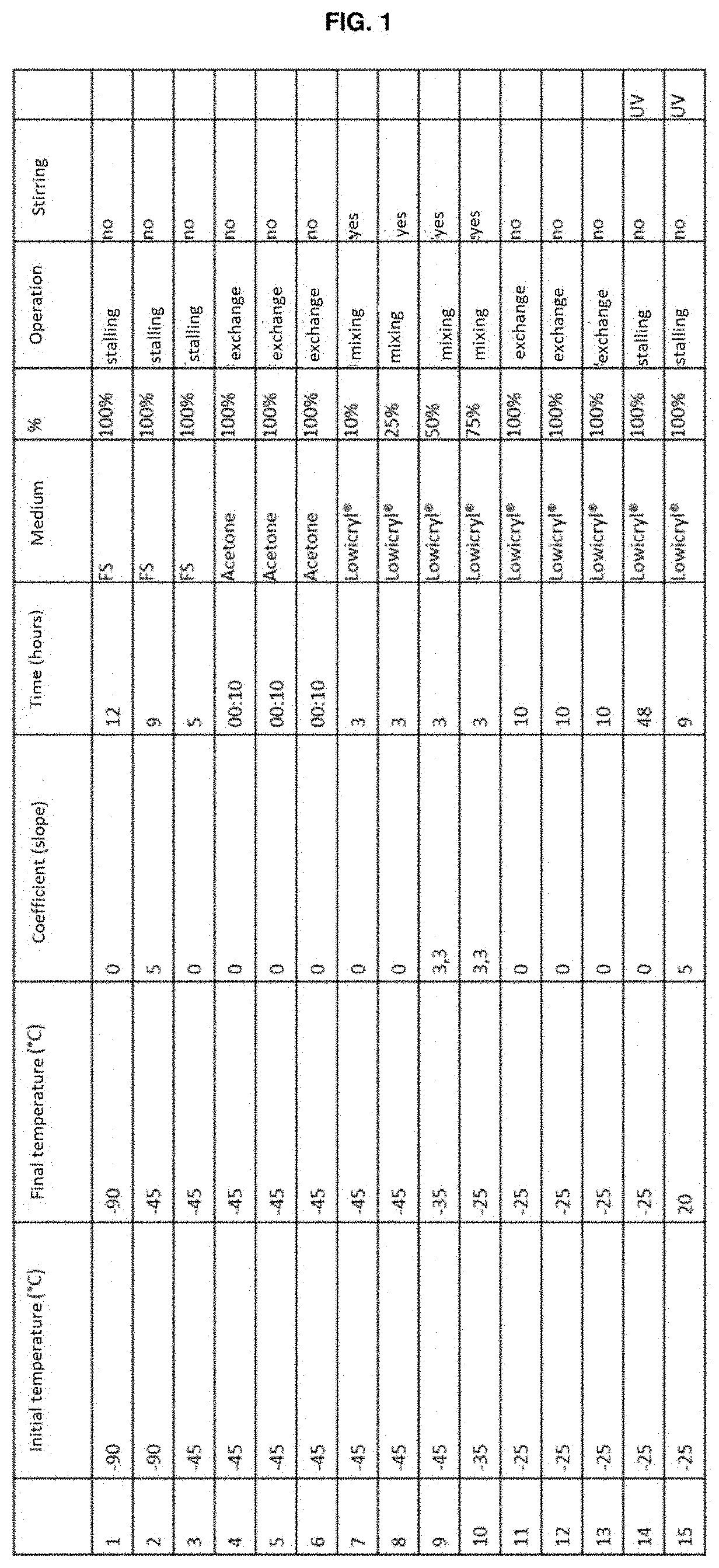
[0058]Each solution was centrifuged and the supernatant was used as the substitution medium in the standard HPF / FS protocol (illustrated in FIG. 1) on line cell stably transfected with a fluorescent protein conjugated to the endoplasmic reticulum. These were used as the standard sample for the correlative protocols. The cells embedded with this protocol were processed by means of the technique of high-pressure freezing / substitution of water with an organic solvent at cryog...

example 3

[0061]Use of the Solution According to the Present Invention as a Contrast Agent for Characterisation by Correlative Microscopy (CLEM) with the HPF / FS Protocol for Biological Samples (Drosophila Oocyte)

[0062]An experiment similar to the one described in Example 2 was carried out using Drosophila oocytes as the biological sample and using ethanol as an organic solvent in the place of acetone. The images obtained are shown in FIG. 5.

[0063]The images obtained thus show that the solution of the present invention enables comparable results to be obtained in terms of fluorescence emission, while at the same time ensuring, however, better contrast than when a solution containing uranyl acetate is used, thus proving more effective in the case of characterisation by correlative microscopy (CLEM).


PUM

PropertyMeasurementUnit
molar ratioaaaaaaaaaa

Abstract

The invention relates to a solution comprising: a heteropolyacid and a lanthanide salt in a solvent consisting of a water / organic solvent mixture or an organic solvent. Optionally, the solution can also comprise an organic or inorganic buffer with a pH comprised in the interval of 4-6 or a strong base or acid. The object of the present invention also includes a use of said solution and any products of the reaction between the components thereof as a contrast medium for biological samples and a method for preparing biological samples that uses said solution in at least one step, for analysis by either correlative microscopy (CLEM) and electron microscopy (EM).

Description

TECHNICAL FIELD[0001]The present invention relates to the field of substances used in the preparation of biological samples that must be characterised by means of electron or correlative microscopy. More precisely, the invention relates to a contrast solution that is alternative to uranyl acetate for the characterisation of biological samples by either correlative microscopy (CLEM) or electron microscopy (EM).PRIOR ART[0002]In recent years, correlative microscopic characterisation methods have attracted great interest in the field of diagnosis and research, thanks above all to the possibility of integrating information coming from different microscopic methods. One of the most interesting techniques is so-called correlative light-electron microscopy (CLEM), a technology that combines fluorescence microscopy (FM) or light microscopy (LM) and electron microscopy (EM). This technique enables the extrapolation, from a single sample, of data that are normally not correlatable with one an...

Claims


Application Information

Patent Timeline
Patent Type & Authority Applications(United States)
IPC IPC(8): G01N1/30
CPCG01N1/30G01N2001/302
Inventor CAPPELLO, VALENTINASIGNORE, GIOVANNIDI PIETRO, SEBASTIANOSANTI, MELISSAMOSCARDINI, ALDOGEMMI, MAURO
Owner FOND INST ITAL DI TECH
Features
  • R&D
  • Intellectual Property
  • Life Sciences
  • Materials
  • Tech Scout
  • Unparalleled Data Quality
  • Higher Quality Content
  • 60% Fewer Hallucinations
Learn More