Urothelial cell induction agent and method for inducing urothelial cells

a technology of urothelial cells and induction agents, which is applied in the direction of genetically modified cells, skeletal/connective tissue cells, drug compositions, etc., can solve the problems of insufficient use of methods, poor long-term results, and cancer formation

Pending Publication Date: 2022-09-22
CELLAXIA INC
View PDF0 Cites 0 Cited by
  • Summary
  • Abstract
  • Description
  • Claims
  • Application Information

AI Technical Summary

Benefits of technology

[0021]According to the present invention, urothelial cells can be generating from somatic cells in a short period of time by direct reprogramming. Since the urothelial cells can be easily derived from the somatic cells of the person who receives transplantation, problems such as immunological rejection and the like do not occur when the obtained urothelial cells are transplanted. In addition, since urothelial cells can be directly induced from somatic cells without going through pluripotent states such as iPS cells and ES cells, problems caused by pluripotent stem cells such as canceration and the like can be avoided.

Problems solved by technology

However, these gastrointestinal tissues easily resorb urine, causing many problems including cancer formation, metabolic acidosis, infection, stone formation, etc.
However, although short-term results are good, long-term results are poor, and there is no fundamental treatment method as the situation stands.
This achieved sufficient results for short-terms after the transplantation, but this method has not been widely used because the autologous bladder tissue may become dysfunctional after long-term follow-up.
However, these cells were hardly applicable to clinical use, because the procedures required autologous urothelial cells.
However, transplantation of cells derived from human pluripotent stem cells could cause teratoma formation.
However, conversion of somatic cells into urothelial cells has not been reported.

Method used


Image

  • Urothelial cell induction agent and method for inducing urothelial cells
  • Urothelial cell induction agent and method for inducing urothelial cells
  • Urothelial cell induction agent and method for inducing urothelial cells

Examples


example 1

[0132]Adult Human Skin Fibroblasts (aHDFs) were Induced to Express Urothelial Cell-Like Phenotype by Introducing Specific Factor.

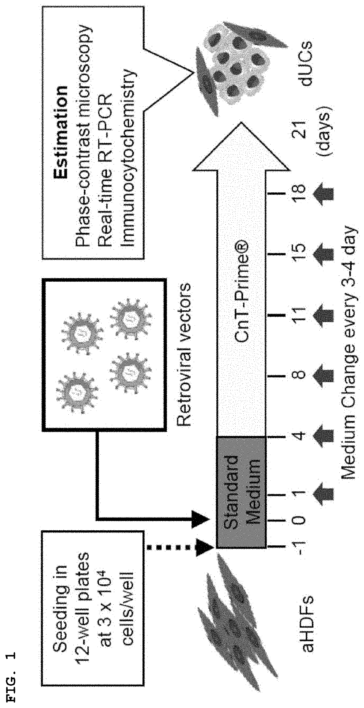
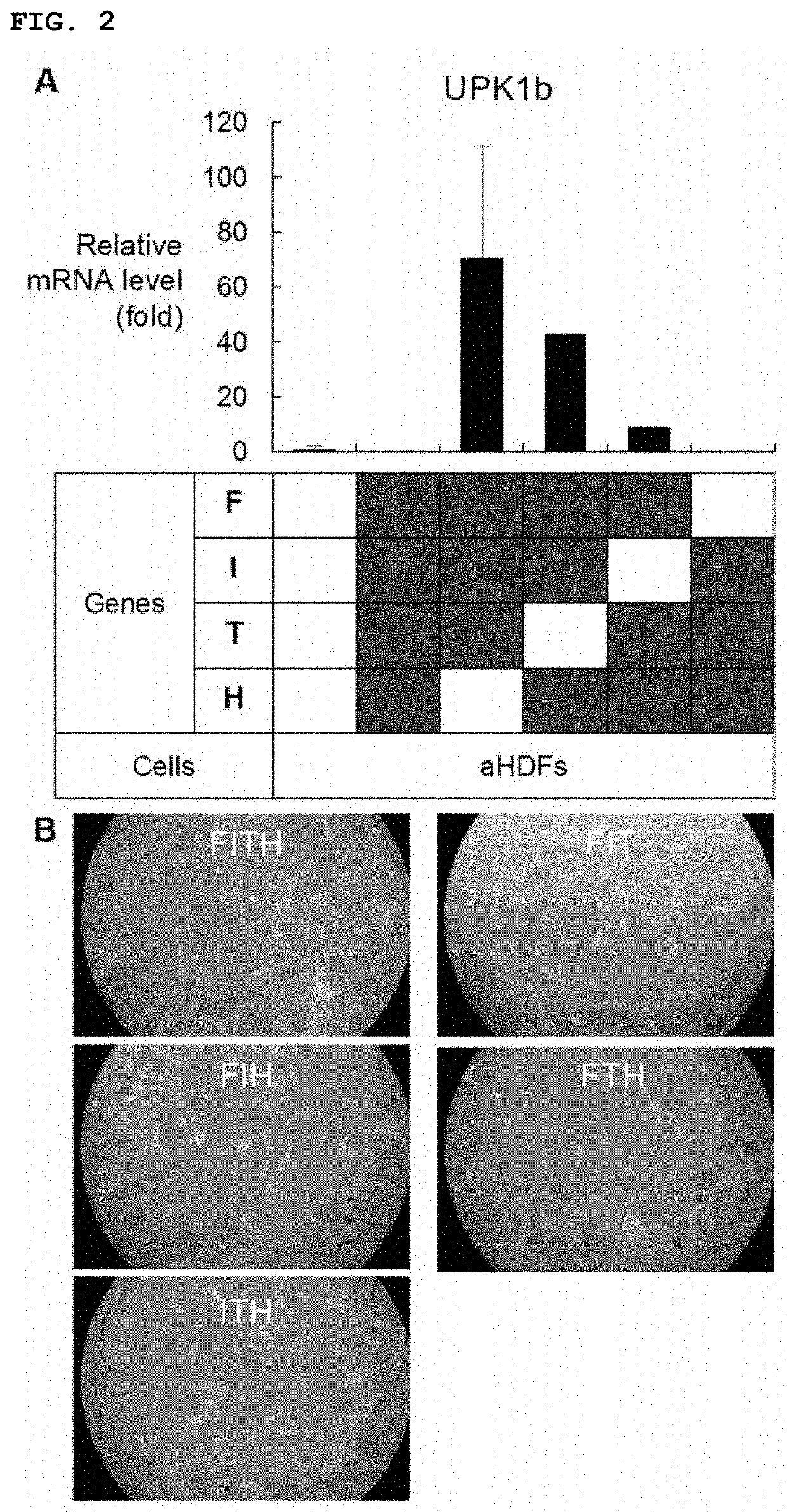
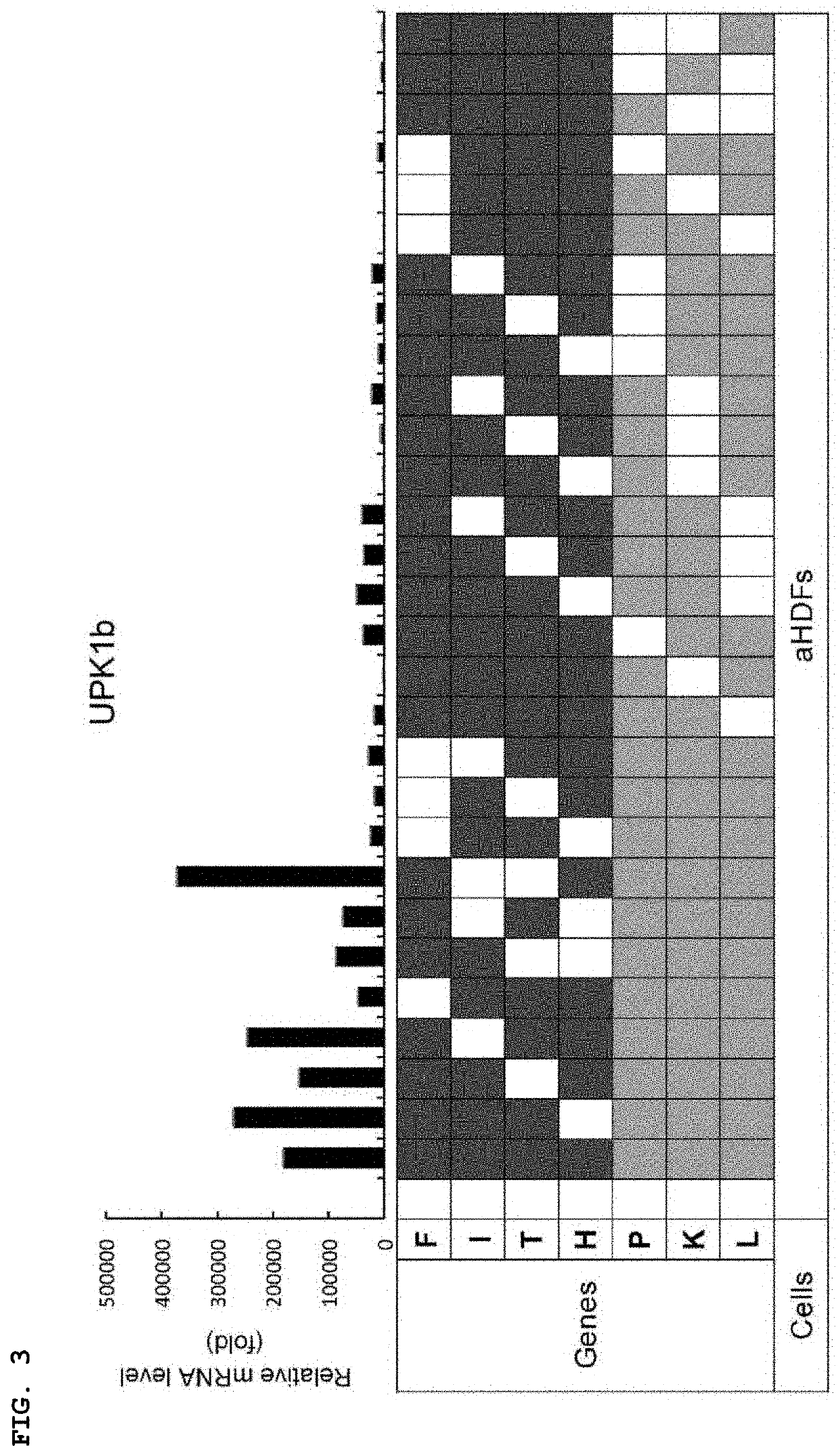
[0133]First, three transcription factors were focused on. FOXA1(F) and IRF1(I) are important in the development of urothelial cells. On the other hand, TP63 (T) is a transcription factor that is expressed in epithelial stem cells and is associated with the development and differentiation of urothelial cells. In addition, sonic hedgehog (SHH) gene (H), which is expressed in the basement membrane cells of urothelial cells and regulates signals important for cell proliferation during the regeneration process, was added. These genes were introduced into adult human skin fibroblasts (aHDFs) using a retroviral vector and the cells were cultured in CnT-Prime medium for a period considered to be suitable for cell maintenance (21 days) (FIG. 1). In cells into which FIT or FIH was introduced, uroprakin (UPK1b), which is a development marker for urothelial cells, was...

example 2

[0137]Characteristics of Directly-Converted Urothelial Cells (dUCs).

[0138]The cells transduced by the method of Example 1 are indicated as directly-converted urothelial cells (dUC). Then, the detailed characteristics of the cells were investigated. In the time-course analysis, cells transduced by F, T, L, and K markedly expressed UPK1b 8 days after gene introduction, and the expression peaked 15 days later (FIG. 8a). The UPK2 mRNA expression began to rise after 11 days in the FTLK-transduced cells and peaked 21 days later. The formation of epithelial cell colony was observed after 11 days and increased thereafter (FIG. 8b).

[0139]In addition to the expression of UPK1b and UPK2, the expression of E-cadherin, which is a transmembrane type protein expressed in general epithelial cells, and keratin 8 / 18, which is a cytoskeleton, was confirmed. Immunocytochemical analysis revealed that about 42% of the cells expressed UPK1b, whereas 23%, 26%, and 26% of UPK2-positive, E-cadherin-positive,...

example 3

[0141]Pluripotency, Liver, Small Intestine Markers were not Expressed in dUCs.

[0142]To exclude the possibility that fibroblasts were temporarily converted to iPS cells or endodermal progenitor cells that subsequently produced urothelial cells, expression of pluripotent markers and non-urothelial endodermal markers, and the expression of non-urothelial endodermal markers in cells into which F, T, L, and K were introduced during the conversion process were examined. As a result, gene expressions of NANOG, POU5F1, LIN28, and SOX2 were not observed during the culture period in cells into which F, T, L, and K were introduced. NANOG protein was not observed, either (FIG. 10). Similarly, cells into which F, T, L, and K had been introduced did not express mRNA of small intestinal epithelial cell marker CDX2 and hepatocyte marker albumin (ALB) at any observation time (FIG. 11). In addition, cells into which F, T, L, and K had been introduced did not express mRNA of CD31, which is a mesenchym...


PUM

PropertyMeasurementUnit
concentrationaaaaaaaaaa
phase contrast microscopeaaaaaaaaaa

Abstract

The present invention aims to provide a method for preparing urothelial cells that can be applied to the treatments of urologic diseases, particularly, diseases caused by urothelial cell damage, diseases caused by loss of urothelial cells and dysfunction, and the like, urothelial cells prepared by said method, and a medium for inducing (generating) urothelial cells. Urothelial cells prepared by a method for inducing a urothelial cell, including a step of introducing at least one member selected from the group consisting ofFOXA1 (Forkhead box A1) gene or an expression product thereof,TP63 (tumor protein P63) gene or an expression product thereof,MYCL (L-Myc) gene or an expression product thereof, andKLF4 (Kruppel-like factor 4) gene or an expression product thereofto a mammalian somatic cell as an exogeneous factor can be applied to the treatment of urologic diseases.

Description

TECHNICAL FIELD[0001]The present invention mainly relates to a urothelial cell induction method. More particularly, it relates to a method for inducing urothelial cells by direct reprogramming, and an agent for converting somatic cells to urothelial cells.BACKGROUND ART[0002]Urothelial cells form urothelium on the surface of the urinary tract that is composed of renal pelvis, ureter, urinary bladder and proximal urethra. The urothelium contracts or expands depending on the amount of urine stored and provides a strong barrier to urine exudation and bacterial invasion. Both or either of loss of urothelial cells and dysfunction are / is associated with urinary tract diseases such as bladder cancer, overactive bladder, congenital bladder anomalies, bladder injury, interstitial cystitis, neurogenic bladder, and contracted bladder. Damaged urothelium in patients could be treated by transplantation of autologous intestine or colon tissue segments (e.g., surgical method of removing intestinal...

Claims


Application Information

Patent Timeline
Patent Type & Authority Applications(United States)
IPC IPC(8): C12N5/071C12N15/86A61K35/22A61P13/02
CPCC12N5/0685C12N15/86A61K35/22A61P13/02C12N2506/1307C12N2740/10022C12N2740/10042C12N2501/11A61P13/00A61P13/10A61P35/00A61P43/00C12N2501/603C12N2501/604C12N2501/606C12N2501/60C12N2501/33C12N2740/13043C12N5/0684C12N15/85A61K48/00C12N2510/00C12N2501/999
Inventor INOUE, YUTAMAZDA, OSAMKISHIDA, TSUNAOUKIMURA, OSAMUSEKI, MAKOTO
Owner CELLAXIA INC
Why Eureka
Try Eureka