Anti-t-cell nanobody and nucleic acid encoding sequence thereof, and uses of the same
- Summary
- Abstract
- Description
- Claims
- Application Information
AI Technical Summary
Benefits of technology
Problems solved by technology
Method used
Image
Examples
example 1
Preparation of Anti-CD3 ε Nanobody
[0039]In this example, the preparation process of the anti-CD3 ε (CD3 epsilon) nanobody (NB) is as follows. The heavy chain variable domain (VHH) production protocol is as follows. The VHH gene was constructed in expression vector pET22b (Amp resistance) or pSB-init (CmR resistance); The plasmid was identified by restriction enzyme digestion and sequenced verification. 1 μL identified plasmid (about 50 ng) was added to BL21(DE3), and incubated overnight at 37° C. LB culture medium containing resistance was inoculated with a single colony and the cultures were incubated overnight at 37° C., 220 r / min Overnight culture was inoculated in a fresh LB medium (10 L-20 L) containing resistance at a ratio of 1:100, and cultured at 37° C. and 220 r / min. It was cooled to room temperature when the OD600 reaches 0.8. Isopropyl-β-D-thiogalactopyranoside (IPTG) was added with a final concentration of 0.1 mM and induced overnight at 20° C., 220 r / min. The cells and...
example 2
Result of T Cell Proliferation and Activation Assay of Anti-CD3 ε Nanobody
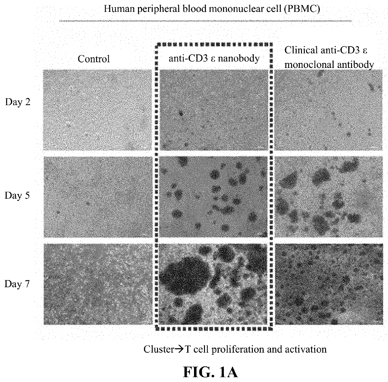
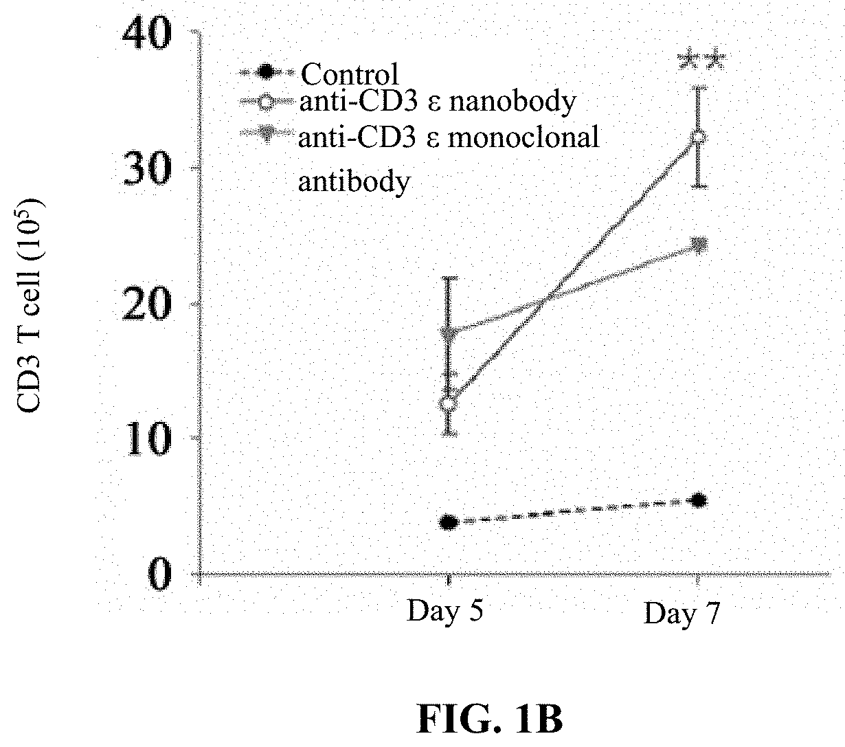
[0044]In this example, the procedures of T cell (i.e., peripheral blood mononuclear cell (PBMC)) proliferation and activation assay of the anti-CD3 ε nanobody are as follows. 1×10⁶ of PBMC cells were plating on 12-well plate presence with or without anti-CD3 ε nanobody (1 μg / ml) or clinical CD3 ε monoclonal antibody OKT3 (10 mg / ml, Invitrogen, Cat:MA1-10175). IL-2 50 IU / ml (Gibco, PHC0021) and IL-15 2 μg / ml (Sino Biological, Cat No:10360-H07E) were added. After 5 or 7 days, the total cell numbers were recorded, then stained with FITC-conjugated CD3 monoclonal antibody (OKT3, 11-0037-42, eBioscience) and then analyzed by flow cytometry. The pictures were taken by micorscope at 40×. The CD3 positive cells were calculated as % of CD3 cells×total cell number.
[0045]The result of T cell (i.e., PBMC) proliferation and activation assay of the anti-CD3 ε nanobody is shown in FIGS. 1A and 1B. As shown in FIGS. 1A and 1B...
example 3
Evaluation of Effect of Anti-CD3 ε Nanobody on Enhancing CD3+ T Cell Proliferation in PBMCs
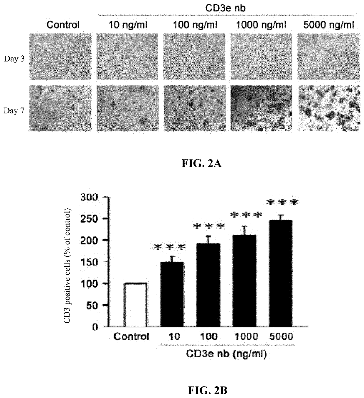
[0046]In this example, the procedures regarding evaluation of effect of the anti-CD3 ε nanobody on enhancing CD3+ T cell proliferation in PBMCs are as follows. 1×10⁶ of PBMC cells were plating on 12-well plate presence with or without anti-CD3 ε nanobody (10, 100, 1000, 5000 ng / ml). IL-2 50 IU / ml (Gibco, PHC0021) and IL-15 2 μg / ml (Sino Biological, Cat No:10360-H07E) were added. After 3 or 7 days, the total cell numbers were recorded, then stained with FITC-conjugated CD3 monoclonal antibody (OKT3, 11-0037-42, eBioscience) and then analyzed by flow cytometry. The pictures were taken by micorscope at 40×. The CD3 positive cells were calculated as % of CD3 cells×total cell number.
[0047]The result regarding evaluation of effect of the anti-CD3 ε nanobody on enhancing CD3 positive T cell proliferation in PBMCs is shown in FIGS. 2A-2C, wherein CD3ε nb represents anti-CD3 ε nanobody. As shown in FIG...
PUM
| Property | Measurement | Unit |
|---|---|---|
| Composition | aaaaa | aaaaa |
Abstract
Description
Claims
Application Information
 - R&D
- Intellectual Property
- Life Sciences
- Materials
- Tech Scout
- Unparalleled Data Quality
- Higher Quality Content
- 60% Fewer Hallucinations
Browse by: Latest US Patents, China's latest patents, Technical Efficacy Thesaurus, Application Domain, Technology Topic, Popular Technical Reports.
© 2025 PatSnap. All rights reserved.Legal|Privacy policy|Modern Slavery Act Transparency Statement|Sitemap|About US| Contact US: help@patsnap.com


