Repair of the
articular cartilage by surgically
transplanting autogenous or autologous osteochondral core grafts has had limited success, but is not always indicated.
Thus, these methods have limited scope and are generally confined to unique kinds of damage.
In either case, the resection involves considerable surgical skill, and results in prosthetic devices physically anchored into the
bone structure.
Not only is such reconstruction expensive major
surgery, but moreover, the mechanical means of attachment may fail as the patient grows older.
This ability to deform can also be a detriment, however, when it is desired to isolate portions of the
articular cartilage or bone surfaces from loads.
Moreover, such devices are prone to tearing or disintegration under repeated stress due to their low tensile strength and modulus.
Anchoring devices may create an area susceptible to fatigue fracture, causing
dislocation of the
prosthesis and further damage to the knee joint.
These devices have been found to cause pain in the knee joint.
This type of prosthetic device and the so-called "McKeever" device require very invasive
surgical procedures, require large arthrotomy, require bone and tissue resection, and are irreversible processes.
The device is a hard, self-centering meniscal device devoid of physical means that fix its location.
In many, if not most cases, a device will be inserted into one compartment only, generally the medial compartment, as the
meniscus and associated
articular surfaces in these compartments (left knee medial and
right knee medial compartments) are most subject to wear and damage.
However, it is possible to insert two separate devices into the medial and lateral compartments of the same knee, or to use two such devices that are mechanically but non-rigidly linked.
Thus, the device is devoid of means of physical attachment which limit its movement, for example, screws,
mating ridges and depressions, porous areas to accommodate tissue regrowth, and the like.
The continued load experienced at such points and the wear experienced as the knee flexes will substantially hinder the regeneration of
healthy tissue.
If a flexible, cushiony material is inserted within the knee compartment, the damaged area will still experience intimate contact with the damaged area under static loads, and will also experience continued wear and abrasion under non-static conditions.
However, more importantly, newly regenerated articular cartilage not having the necessary density or cohesiveness to withstand wear, will be rapidly eroded away.
If a soft and / or
low modulus elastomer or
thermoplastic is used for the entire device, not only is the load not concentrated on
healthy tissue, but moreover, damaged areas due to wear and / or degeneration will also be subjected to loading, decreasing the opportunity for the body's natural regenerative capability to function.
Movement of a device having a periphery with sharp corners would result in the potential for severe damage to the surrounding tissue and
articular surfaces, in addition to causing pain.
Any localized positions of higher loads are self-limiting due to the ability of the device to translate both rotationally and laterally which mimics the true motion of the natural
meniscus as described by Hollister.
Too large an angle will provide too high a centering force, and may accelerate wear of the femoral
condyle articular cartilage or the device itself.
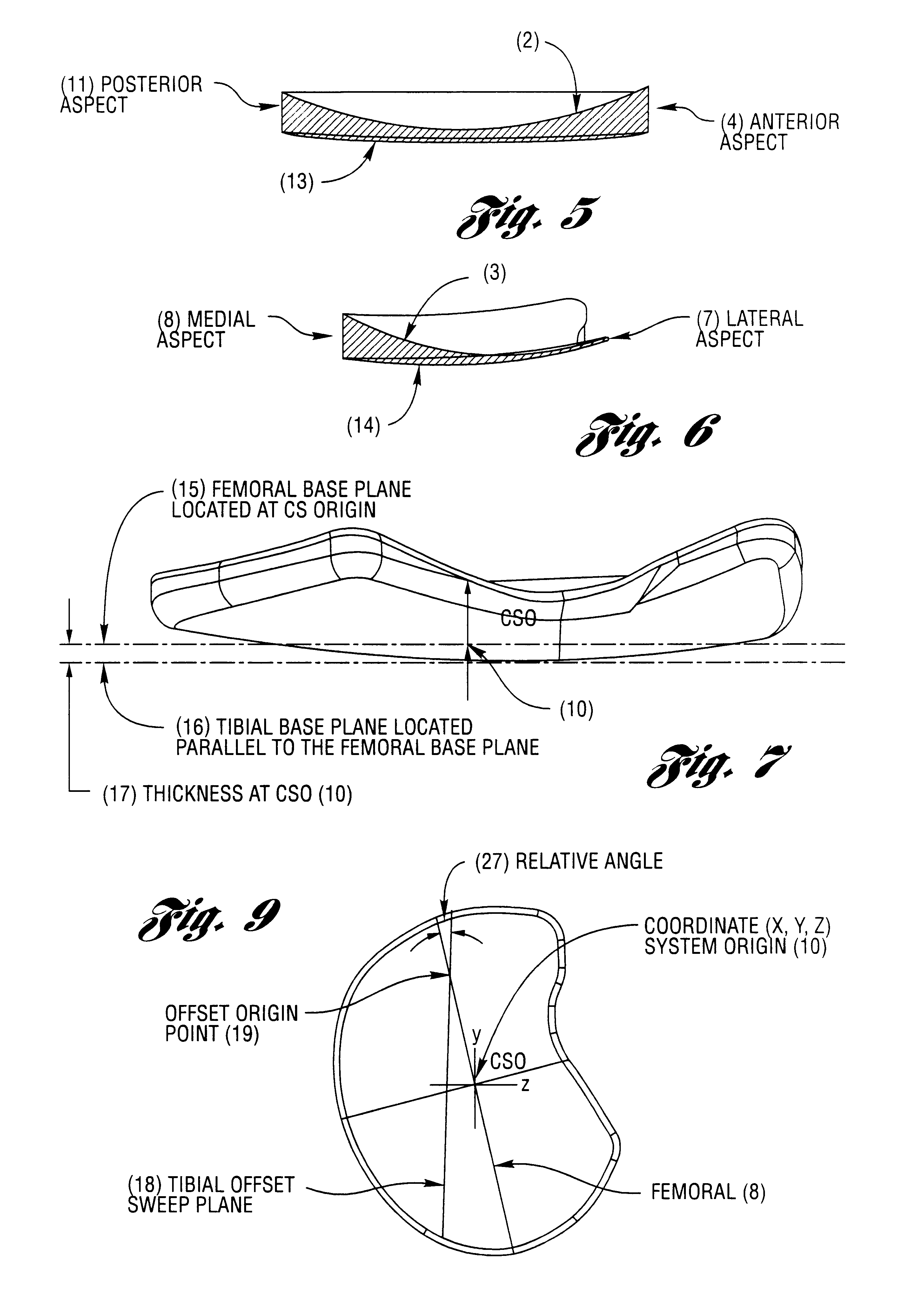
However, the contoured
mating surfaces of the femoral
condyle and femoral meniscal device surfaces can become increasingly dissimilar when the joint articulates, as the contour angles will not necessarily mate correctly throughout the entire articulation cycle.
The device is then introduced by arthroscopically assisted implantation, generally limited to extensive clean-up of existing damaged tissue, e.g., torn or particulate natural
meniscus damage.
Since the
surgical procedures used are not severe, and also not irreversible, an unsuitable device may be readily removed and replaced, either with a different device from a meniscal device
library, or by a custom device.
Such a device can never escape the sphere without lifting or otherwise dislocating the ball because the ever-increasing thickness of the device (from the
wedge shape) will cause increasing levels of interference with the ball as the device is moved in any lateral direction.
However, the device may move with the rotation of the ball if the
radius of the sphere is close to being concentric with the
radius of the ball.
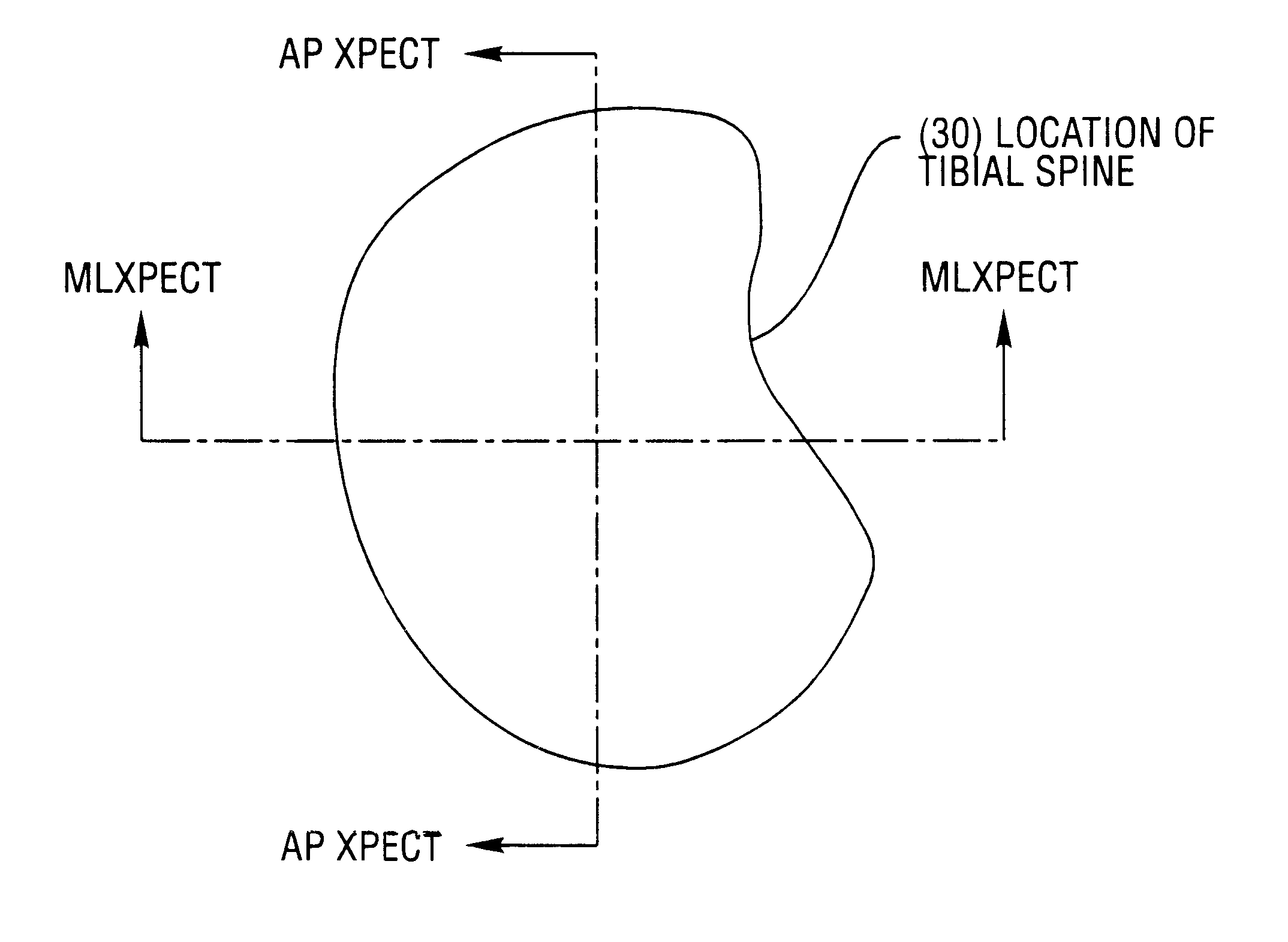
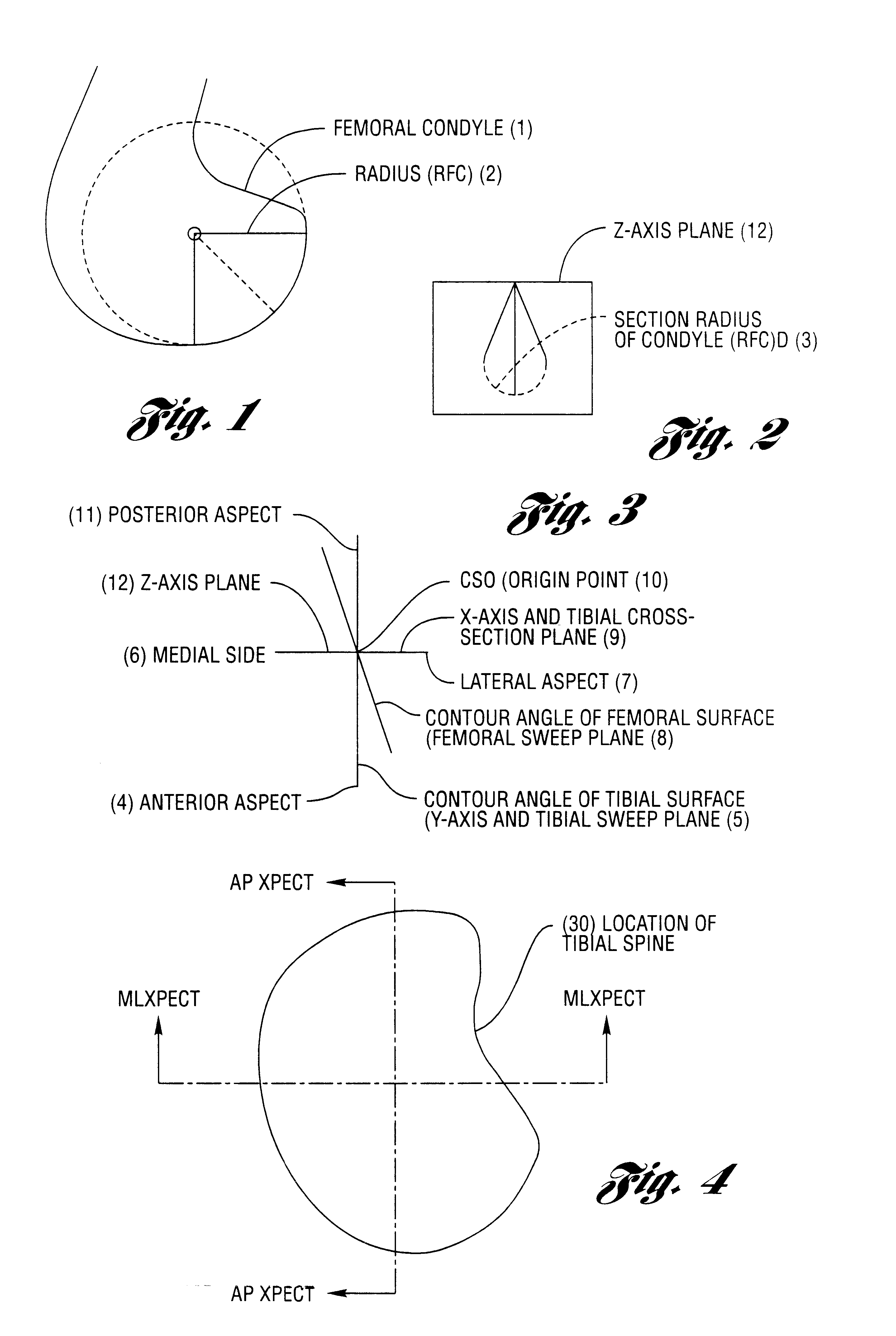
Although the meniscus is crescent shaped, the natural
anatomy of the knee completes the generally circular shape with the tibial spine, along the central axis of the knee, thus locating the femoral
condyle at all times in its
range of motion and limiting any potentially harmful positional excursions of the femoral condyle.
If the shape of the meniscus is damaged or not present then, it cannot perform this locating, load-bearing function.
Thus, the loads on the femoral condyle and tibial plateau become more concentrated leading to a gradual, arthritic degeneration of the articular
cartilage surface of the femoral condyle.

