
Biotinylated-chemokine antibody complexes

Inactive Publication Date: 2005-03-22
MILLENNIUM PHARMA INC
View PDF24 Cites 12 Cited by
  • Summary
  • Abstract
  • Description
  • Claims
  • Application Information

AI Technical Summary

Benefits of technology

[0007]The present invention overcomes these and other obstacles by providing a biotinylated-chemokine antibody complex that can be reversibly dissociated in the presence of free biotin. Accordingly, the invention provides a method by which the concentration of biotinylated-chemokine antibody complex can be adjusted by exposing the complex to varying amounts of free biotin in vivo or in vitro. The complexes of the invention are useful for a variety of applications, including modulating an immune response such as one which is mediated by the chemokine-induced recruitment of migratory cells to a site of inflammation, as well as for the targeted delivery of agents to cells which express a chemokine receptor. In a broader aspect, the invention embraces biotinylated peptide ligands that bind to G-protein coupled receptors. Thus, the invention permits the targeted delivery of agents (diagnostic and therapeutic agents) to pre-selected cells which express such receptors and, advantageously, both avoids the necessity for the synthesis of individual fusion proteins to accomplish these objectives and provides a mechanism for rapid down regulation of activity.
[0016]According to yet another aspect of the invention, a biotin composition comprising a therapeutically effective amount of a biotin of the invention and a pharmaceutically acceptable carrier is provided. The biotin composition is useful for administration to a patient who has or will receive a biotinylated-pharmacologically active agent antibody complex of the invention. Administration of the biotin composition facilitates dissociation of the complex in vivo and, thereby, allows one of ordinary skill in the art to further adjust the half-life of the complex in vivo. In general, the half-life of a biotinylated-chemokine of the invention is longer when the biotin conjugate is complexed with an anti-biotin antibody of the invention and is shorter when the biotin conjugate is present “free” (uncomplexed) in vivo. Accordingly, dissociation of the complex in vivo results in hastening the natural degradation of the biotin conjugate in vivo. The biotin composition also can be used to adjust the concentration in vitro.
[0020]According to still another aspect of the invention, a method for treating inflammation in a subject is provided. As used herein, the word treating embraces preventing, inhibiting, and ameliorating the symptoms of the particular condition which is being treated. The method of treatment involves: administering to a subject in need of such treatment a therapeutically effective amount of a biotin conjugate or of a complex comprising: (a) a biotin conjugate and an anti-biotin antibody. The biotin conjugate includes: (i) a biotin covalently coupled to (ii) an agent that selectively binds to a receptor expressed by a pre-selected cell associated with inflammation (e.g., a migratory cell). Administration of the biotin conjugate or the complex prevents or reduces inflammation in the subject. In certain embodiments, the biotin conjugate(s) and the anti-biotin antibody are simultaneously or sequentially administered to the subject. In the most preferred embodiments, the biotin conjugate is a biotinylated chemokine. Exemplary chemokines that can be used as the agent to which biotin is coupled in this aspect of the invention are provided in Table 1. In certain preferred embodiments, the pre-selected cell is a migratory cell, such as a T-cell or leukocyte, and the administration of a complex enhances recruitment of the migratory cell to a site of tumor cells in the subject.

Problems solved by technology

However, the half-life for chemokines in vivo is relatively short.
Unfortunately, each of the foregoing methods requires the creation of a chemokine fusion protein, thereby necessitating the development of tailored methods for the generation of each different chemokine construct.
Thus, despite the innovations advocated in connection with fusion protein technology, there exists no universal approach for the targeted delivery of chemokines to cells which express the cognate chemokine receptor.

Method used


Image

  • Biotinylated-chemokine antibody complexes
  • Biotinylated-chemokine antibody complexes
  • Biotinylated-chemokine antibody complexes

Examples


example 1

Preparation of a representative Biotinylated Chemokine

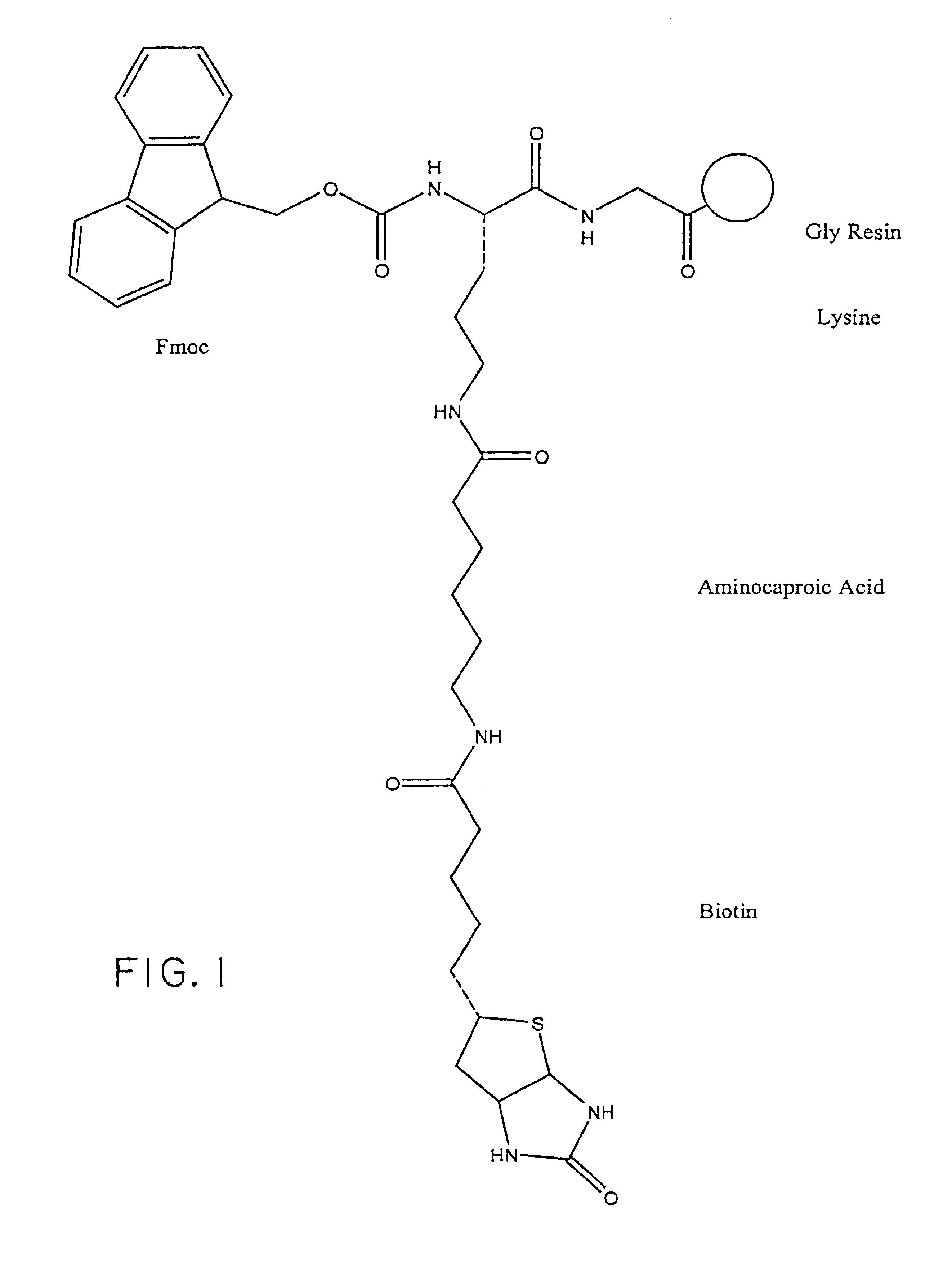
[0100]In general, the biotinylated pharmacologically active agents of the invention (“biotin conjugates”) are prepared in accordance with routine procedures known to those of ordinary skill in the art or are commercially available. Commercially-available biotinylated chemokines include: biotinylated human MCP-1, human MIP-1 alpha, and human MIP-1 beta (R&D Systems, Minneapolis, Minn.).

[0101]Specific protocols are provided below as exemplary methods for preparing a representative biotin conjugate; however, it is to be understood that alternative linker molecules and pharmacologically active agents can be substituted for the particular linker molecules and pharmacologically active agents described in the Examples to make a wide variety of biotin conjugates using no more than routine experimentation. In addition, the biotin conjugates embraced within the scope of the invention can be tested in high throughput screening assays, e.g.,...

example 2

Preparation of an Anti-biotin Antibody

[0104]In general, the anti-biotin antibodies of the invention are prepared in accordance with routine procedures known to those of ordinary skill in the art or are commercially available. An exemplary protocol for preparing a monoclonal antibody that selectively binds to biotin with a relatively high affinity (KA˜109M−1) is provided in H. Bagci, et al., FEBS 322(1): 47-50 (1993). See also, F. Kohen, et al., Meth. in Enzymol. 279:451463 (1997); Vincent, P., and Samuel, D., J. Immunol. Meth. 165:177-182 (1993); K. Dakshinamurti, et al., Meth. in Enzymol. 184:111-119 (1990); and Sigma Chemical Co. (St. Louis, Mo., cat. no. #B753, Murine IgG1 α-biotin clone BD-34). In addition, mice transgenic for human Vh and Vl genes are commercially available from Medarex, Annandale, N.J., or from phage display libraries of human Vh and Vl genes, prepared according to the procedure described in M.D. Sheets, et al., PNAS (USA) 95(11): 6157-62 (1998), entitled, “Ef...

example 3

Screening Assay for Binding to a Chemokine Receptor

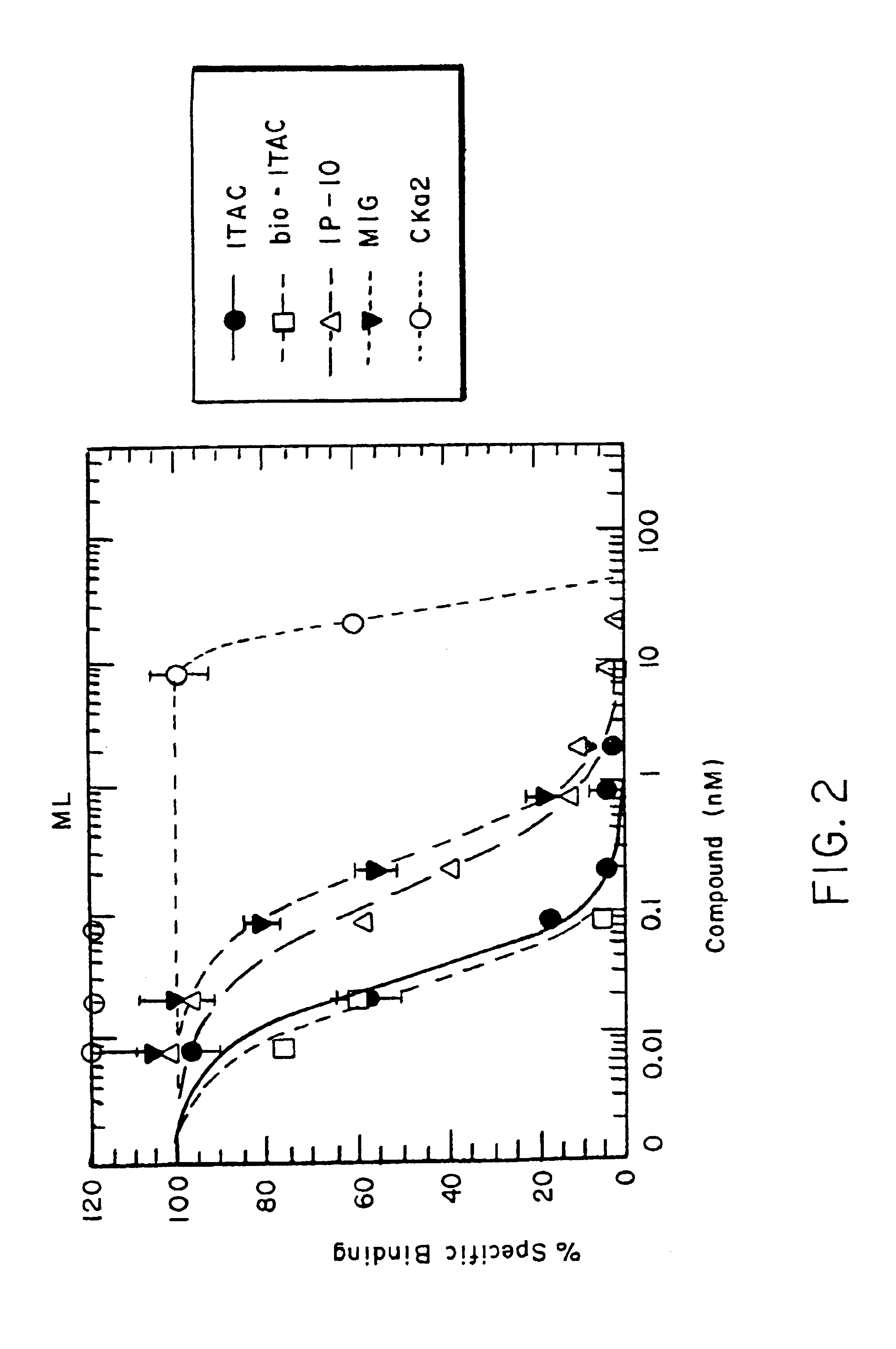
[0112]A general protocol for a screening assay is presented below (see FIG. 2 for the results of a representative screening assay).

[0113](a) Ligand Binding Assays.

[0114]Competition binding assays were performed with 1251-chemokine as the hot ligand and biotinylated or unbiotinylated chemokine as the cold competitor ligands. The binding reaction was performed with 500,000 receptor cell transfectants and 0.1 nM radiolabeled chemokine in binding buffer (50 mM HEPES12, 5 mM MgC12, 0.5% BSA and 0.05% azide) for 1 hour at room temperature in the presence or absence of cold chemokine in a range of 8 different concentrations from 0.0008-20 M. Non-specific binding is determined by residual binding obtained in the presence of 250 nM cold ligand. Cells are then washed with binding buffer containing 0.5 M NaCl and cell associated radioactivity counted with a top count radioactivity counter. Specific binding was calculated by subtracting non-spe...


PUM

PropertyMeasurementUnit
Timeaaaaaaaaaa
Timeaaaaaaaaaa
Timeaaaaaaaaaa

Abstract

Biotinylated pharmacologically active agents and complexes containing same are disclosed. In particular, biotinylated-chemokines are described. The complexes further include an anti-biotin antibody that selectively binds to biotin. The complex can be dissociated by contact with free biotin. The complexes are particularly useful for enhancing an immune response to tumor cells and virus-infected cells, in vivo or in vitro.

Description

[0002]This application claims priority under Title 35 § 119 (e) of United States Provisional Application No. 60 / 121,006, filed Feb. 22, 1999, and entitled BIOTINYLATED CHEMOKINE ANTIBODY COMPLEXES, the entire contents of which are incorporated herein by reference.FIELD OF THE INVENTION[0003]This invention relates to biotinylated compounds that, optionally, are complexed with an anti-biotin antibody or fragment thereof. More specifically, the invention relates to complexes comprising biotinylated-chemokines and an antibody or fragment thereof selectively bound to the biotin. The biotinylated-chemokines and complexes containing same are useful for modulating the mechanism underlying a variety of disease states, including a chemokine-mediated cellular response, and for the selective delivery of agents to sites of disease activity.BACKGROUND OF THE INVENTION[0004]Chemokines are a class of cytokine molecules that are involved in cell recruitment and activation in inflammation. These chem...

Claims


Application Information

Patent Timeline
IPC IPC(8): A61K47/48
CPCA61K47/48753B82Y5/00Y10S530/807A61K47/6898
Inventor NEWMAN, WALTERPICARELLA, DOMINICSOLER, DULCE
Owner MILLENNIUM PHARMA INC
Learn More