Method and apparatus for performing neuroimaging

a functional magneti resonance imaging and neuroimaging technology, applied in the field of magnetic resonance imaging, can solve the problems of affecting the application of fmri to the more physiologically relevant functions, impede the use of this technology in conscious animals, and most studies to date have been limited, so as to enhance mr signal mapping and eliminate movement artifacts

Inactive Publication Date: 2005-03-29
EKAM IMAGING
View PDF48 Cites 33 Cited by
  • Summary
  • Abstract
  • Description
  • Claims
  • Application Information

AI Technical Summary

Benefits of technology

Applicant's method and apparatus overcomes the difficulties of performing fMRI on conscious animals by utilizing a restraining assembly to eliminate movement artifacts in combination with RF resonator system to enhance MR signal for mapping changes in brain activity. The restraining assembly incorporates a coil design including a spatially adjustable volume coil for transmitting RF magnetic field pulses and a spatially adjustable dome shaped surface coil for receiving the RF response signals from the conscious animal. The significance of applicant's method of neuroimaging in conscious animals will change current imagery of the brain from either a static (as seen with most neurochemical measurements) or a low activation dynamic system in an anesthetized state (as seen with current fMRI or positron emission tomography (PET) measurements) to more physiologically relevant conditions.

Problems solved by technology

While most work on fMRI has been done in humans, it has been difficult to use this technology in conscious animals because of motion artifact.
As a result, most studies to date have been limited to animals which are typically anesthetized in order to minimize this problem of motion artifacts.
The low level of arousal during anesthesia either partially or completely suppresses the fMRI response and has impeded fMRI application to the more physiologically relevant functions that have been noted in humans.
However, signal-to-noise ratio is compromised because a large spatial domain contributes to the RF signal, resulting in additional noise and thereby obscuring the RF signal from the region of interest.
Unfortunately, a surface coil is ill suited for RF energy transmission owing to the fact that only a small proton area can be activated.
RF coil and tissue losses can significantly limit the potential SNR gains realized at high fields.
SNR (and reciprocal transmission efficiency) will suffer when the coil's ohmic resistance, radiation resistance, coupled tissue losses, RF magnetic field and angular frequency are not optimized.
These conductive and dielectic losses represented are limited in practice by using local surface coils, or volume coils efficiently coupled to a region of interest.
In addition to tissue loading, RF losses in the coils themselves become significant at higher frequencies.
From the above, it is apparent that radiative losses to the sample and environment, as well as conductive losses to the load of a coil become severe to the point of limiting and eventually degrading the SNR gains otherwise expected at higher magnetic field strengths.

Method used


Image

  • Method and apparatus for performing neuroimaging
  • Method and apparatus for performing neuroimaging
  • Method and apparatus for performing neuroimaging

Examples


Embodiment Construction

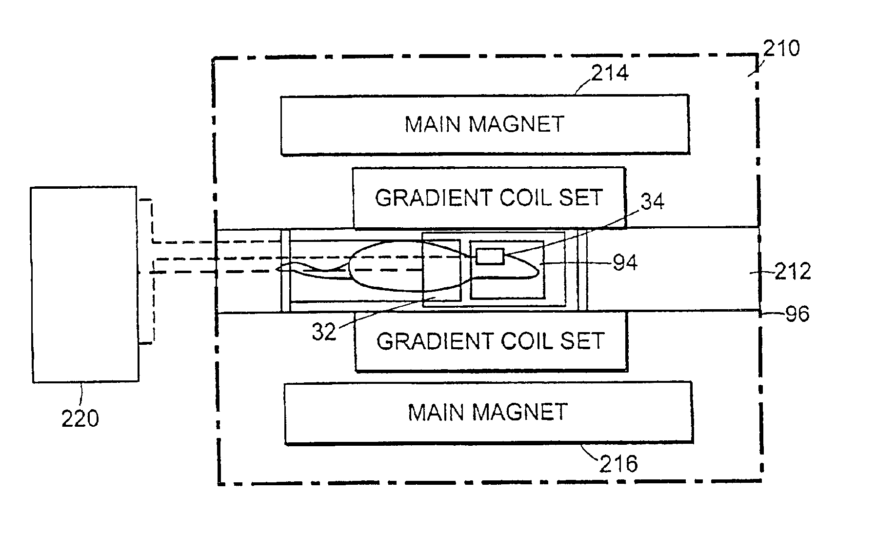
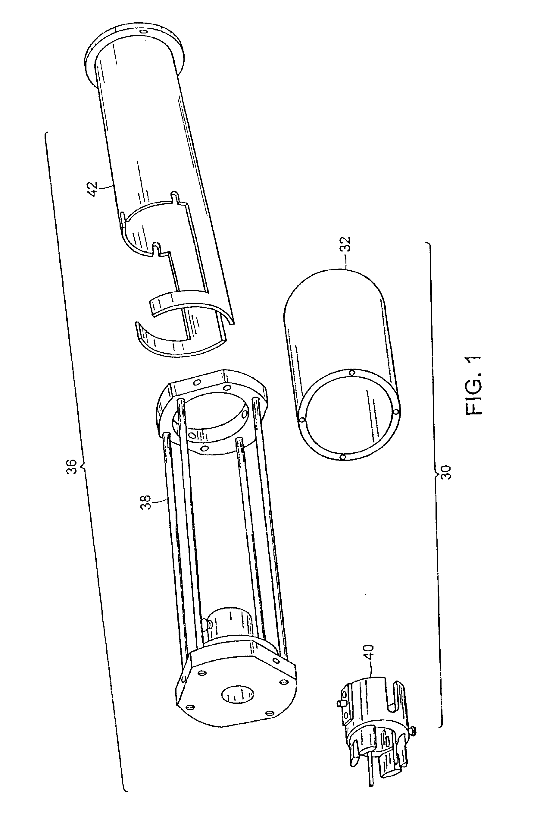
In the figures, like numbers are used to indicate like elements. FIG. 1 shows a multi-cylindrical, dual-coil animal restrainer 30 according to the invention. The multi-cylindrical, dual-coil animal restrainer 30 including a volume coil 32 and a surface coil 34, as seen in FIG. 8A, and the method described allow the functional magnetic resonance imaging (fMRI) of conscious animals.

Referring to FIG. 1, the multi-cylindrical, dual-coil animal restrainer 30 has the volume coil 32, which will be described further below in greater detail, and a restraining assembly 36. The restraining assembly 36 includes a support frame or chassis 38, a head retainer 40, and a body retainer 42.

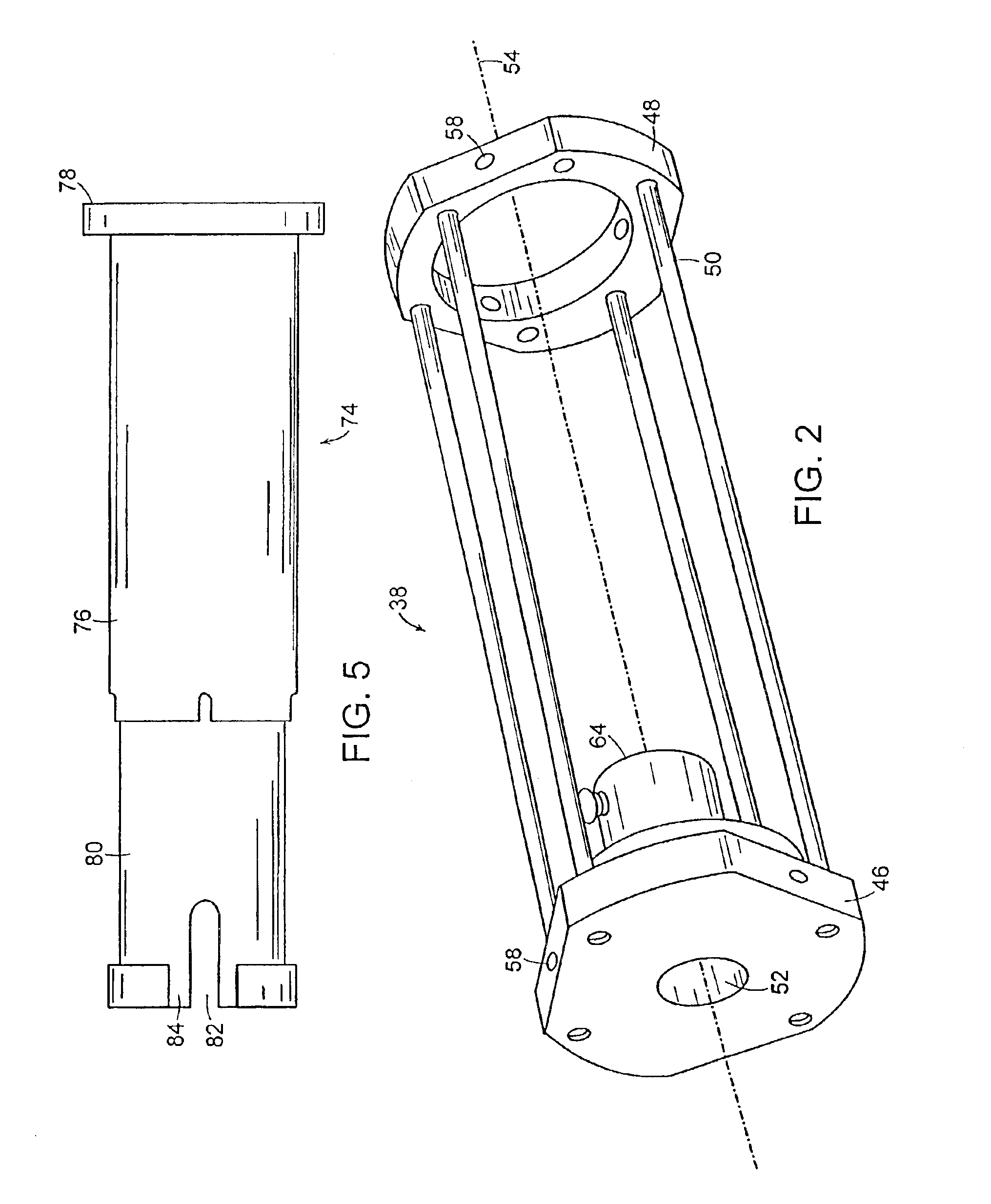
The frame 38 is shown in perspective in FIG. 2. The frame 38 has a first or front-end mounting plate 46 and a second or rear-end mounting plate 48 spaced apart by a plurality of support members or rods 50. The front-end mounting plate 46 has a hole 52 which is collinear with a longitudinal axis 54 of the frame 38. ...


PUM


Abstract

The present invention relates to systems and methods of performing magnetic resonance imaging (MRI) in awake animals. The invention utilizes head and body restrainers to position an awake animal relative to a radio frequency dual coil system operating in a high field magnetic resonance imaging system to provide images of high resolution without motion artifact.

Description

BACKGROUND OF THE INVENTIONThe present invention relates to magnetic resonance imaging, and more particularly to a method and apparatus for performing functional magneti resonance imaging (fMRI) in conscious animals.Human studies utilizing fMRI have advanced our understanding of the regional and functional interplay between populations of neurons serving sensory, integrative and motor functions. Changes in neuronal activity are accompanied by specific changes in hemodynamics such as cerebral blood flow, cerebral blood volume, and blood oxygenation. Functional MRI has been used to detect these changes in response to visual stimulation, somatosensory activation, motor tasks, and emotional and cognitive activity. When the brain is activated by any of these conditions, the blood flow and delivery of oxygen to the active regions the tissue oxygen uptake resulting in an increase in blood oxy-hemoglobin (Hb02) content. The susceptibility difference between diamagnetic oxy-hemoglobin and pa...

Claims


Application Information

Patent Timeline
Patent Type & Authority Patents(United States)
IPC IPC(8): A61B5/055G01R33/28
CPCA61B5/0555G01R33/341G01R33/34046G01R33/34007G01R33/3453A61B5/4094A61B5/055
Inventor FERRIS, CRAIG F.KING, JEAN AALLARD, ARTHUR C.LUDWIG, REINHOLDBOGDANOV, GENE
Owner EKAM IMAGING
Why Eureka
Try Eureka