[0010]The present invention is directed to tools and associated processing of mammographic images to enhance
image acquisition and review workflow. In particular, the present invention enables
rapid access to
digital image information for improved workflow management and identification of images, upon initial review, for substantially instant recall at a later time. The invention also allows for background processing of image information for enhanced functionality substantially without workflow
delay. The invention thus addresses certain perceived disadvantages of
digital mammography while providing improved digital processing functionality, thereby providing additional digital advantages.
[0011]According to one aspect of the present invention, digital images are processed in the background, i.e., they are automatically processed, free from specific task-orientated direction by a user, using resources that are not otherwise occupied addressing user-directed tasks. The associated
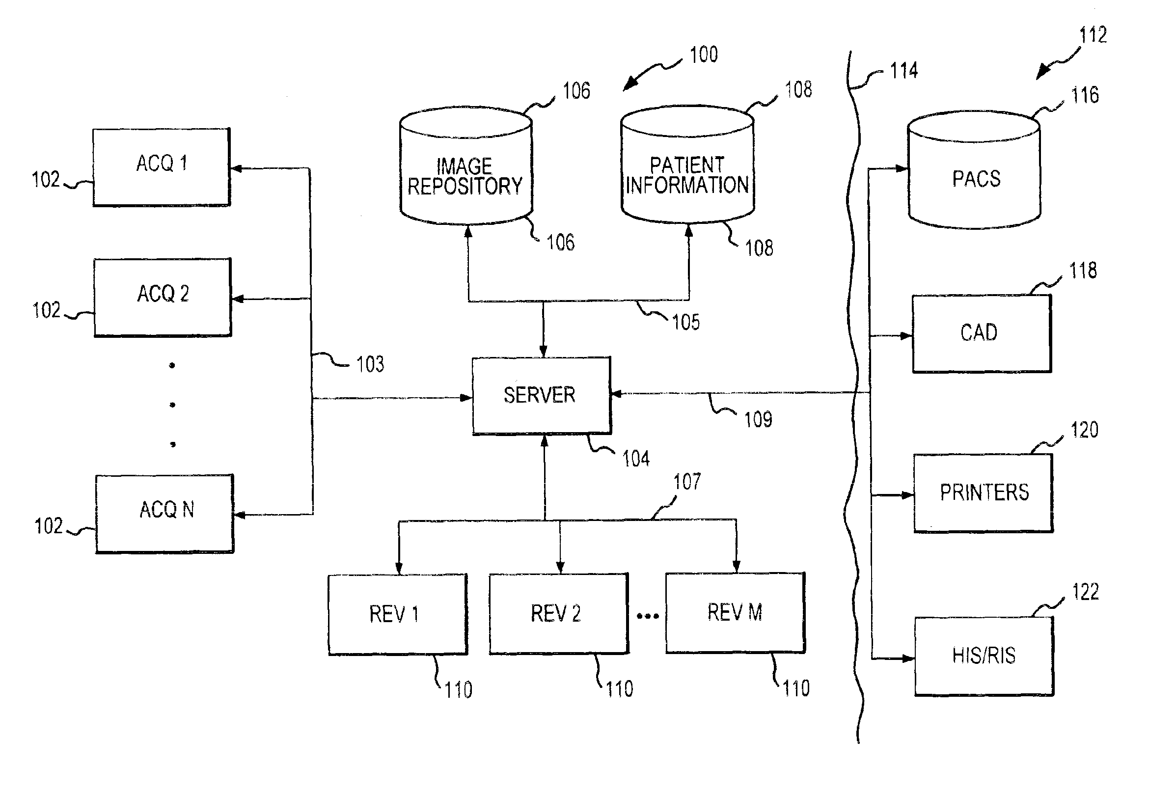
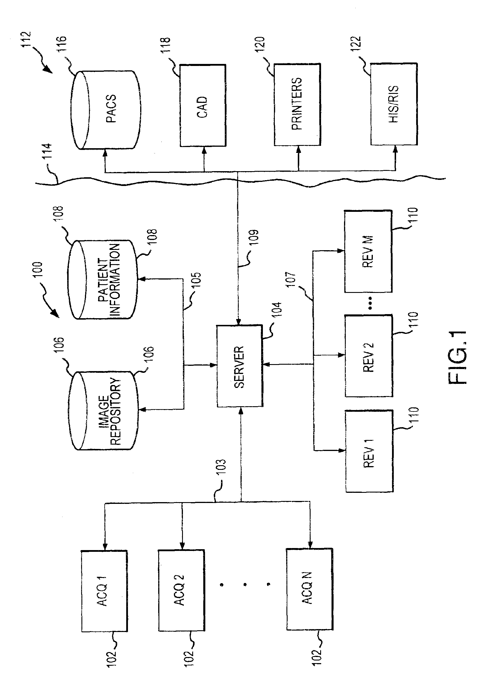
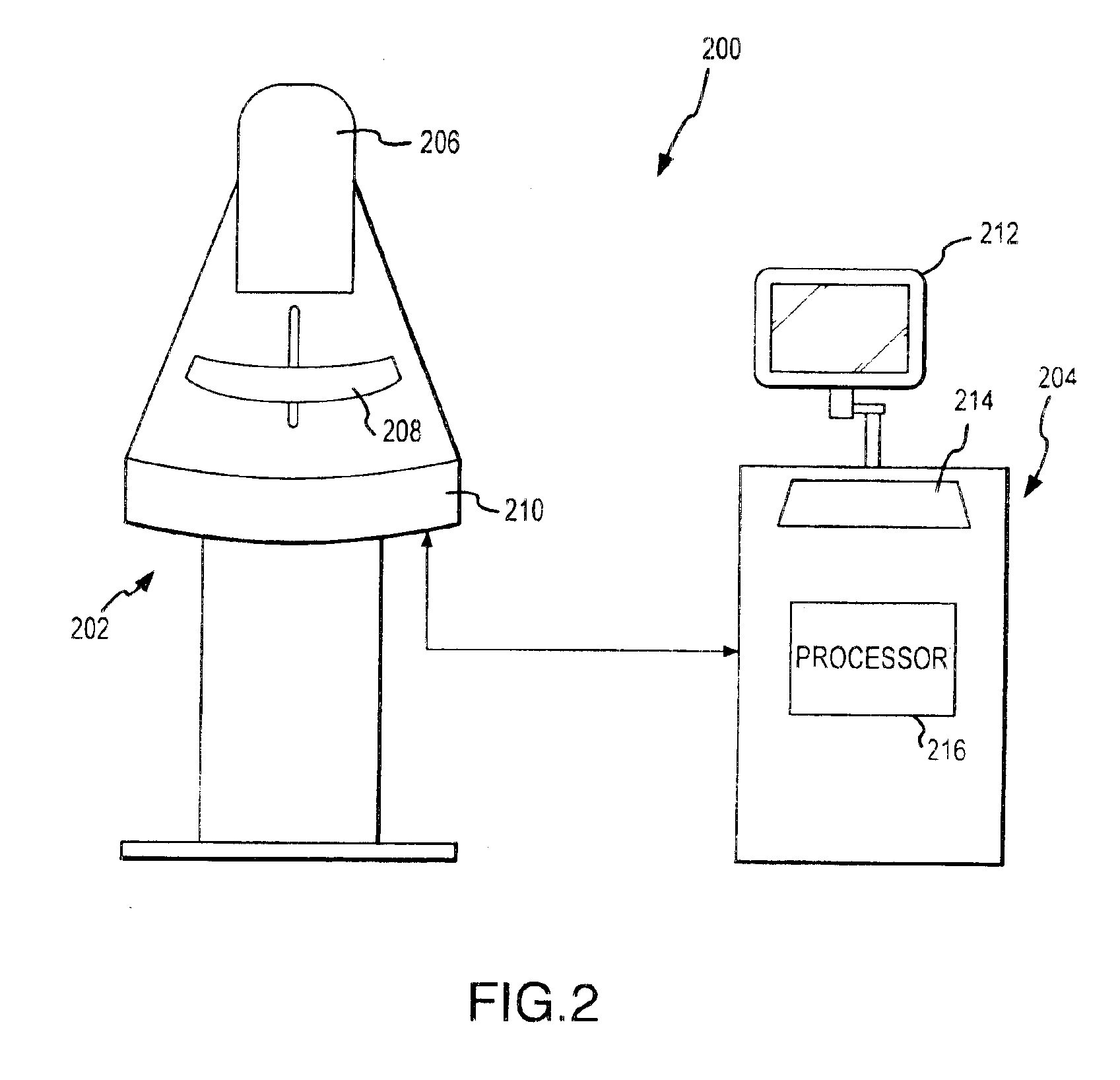
system includes image acquisition equipment for acquiring mammographic images from a patient, image review equipment for displaying images of the patient, and a processor for receiving imaging information from the acquisition equipment, processing the image information and making processed image information available to the image review equipment. The processor runs logic for identifying background tasks for particular images and executing the tasks in the background. The logic identifies at least one task and executes the task free from
concurrent user prompts. In this manner, certain processing can be accomplished without requiring significant
waiting time associated with processing from the perspective of the user.
[0013]Thus, in accordance with another aspect of the present invention, digital mammographic images are automatically preprocessed prior to initial review by a physician. An associated process involves operating image acquisition equipment to acquire
digital image information regarding a patient's breast and store the image information in an
image repository, operating a processor to process the image information in background to provide processed image information, and making the processed image information available for subsequent initial review by a physician. The background processing of the image information involves executing at least one task for processing the image information free from any concurrent prompts from a user. A variety of different types of tasks may be performed in this regard including, for example, performing a
diagnostic analysis of the image information, associating
diagnostic information with the imaging information, optimizing certain display parameters of the imaging information and preparing images for display in accordance with a desired review workflow. In this manner, certain enhancement tasks can be performed prior to initial review by a physician for improved efficiency.
[0014]In accordance with a further aspect of the present invention, a
mammographic image system is operated to prepare images for more rapid display during a
review process. An associated method involves determining an anticipated sequence for an image review session by a physician; accessing an
image repository to obtain image information corresponding to the anticipated sequence, and transferring the identified image information to a memory location for rapid display at a selected review
station. The anticipated sequence may be determined based on an image review protocol for a particular physician, based on the type of medical process (e.g., mammographic screening or diagnosis), based on an initial analysis of the images to identify areas of possible interest to a physician, or based on other potentially predictive criteria. The logic for determining the sequence may be resident on an image review platform, a separate
server or other location. The identified image information may be transferred to storage associated with the image review platform such as, for example, cache storage or may be retrieved from archive storage and stored in an active
image repository. The images thus transferred are available for rapid display on the display terminal or terminals of the review equipment so as to reduce loading
delay times otherwise associated with large image files.
[0015]According to another aspect of the present invention,
mammographic image information is automatically preprocessed for optimized display. An associated process involves accessing image information from an image repository, identifying a display parameter relative to an image
review process and optimizing the image information for display in connection with image review equipment based on the identified display parameter. Examples of optimization processes that may be performed in this regard include selecting and optimal monitor setting relative to a luminous range of a
display device, optimally filling the viewing area of a
display device, filling the available viewing area with a selected portion of the image and displaying the image in a known reference position or orientation to assist the physician in getting oriented to the image perspective. Such optimization logic may be executed by a processor resident at the image review site, at a central
server, or at another location. Preprocessing of
mammographic image information for optimal display increases the efficiency of the reviewing physician and reduces, eliminates or reverses a perceived
disadvantage of digital imaging relative to film-based mammography.
[0016]In accordance with a still further aspect of the present invention, annotated instances of a digital mammographic image can be stored in an image repository. The associated mammographic
image system includes an image repository; image review equipment for accessing images from the image repository; and logic for associating annotations with the image information to establish annotated image information and storing the annotated image information in the image repository. The annotations may be associated with the imaging information automatically or in response to user prompts. Thus, image information may be processed in background by a CAD utility to identify areas of interest and associate an
annotation with those areas of interest. For example, the
CAD system may perform a pixel-by-pixel or area-by-area analysis of an image to identify a potential
microcalcification or other area of interest and superimpose a symbol on the image to identify the area of interest and the nature of the potential diagnosis. Alternatively, a physician may tag an image for later review. Thus, for example, a physician may perform an initial review of a large number of images corresponding to all of the patients who were screened on a given day. A subset of these images may be tagged by the physician (e.g., by selecting an appropriate icon or providing another input) for later review. These images can be readily recalled by the physician based on a search for tagged images. It will thus be appreciated that the
annotation does not necessarily affect the visual display of the image. Additionally, images may be marked as reviewed, as may be desired.

